hidden

Atherosclerosis in Prehistorical Times

Since some of the risk factors for atherosclerosis such as eating fast food, lacking physical activity, and developing diabetes appeared with the modernization of our societies, it is natural to think that atherosclerosis is a disease of the modern world.

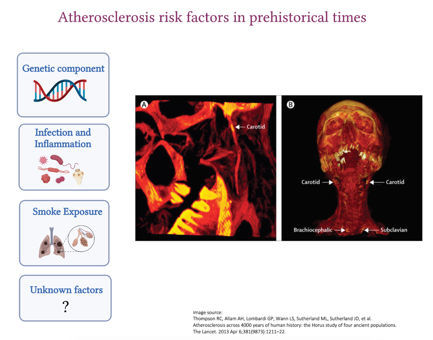
However, atherosclerosis also existed in the ancient civilizations of Egypt, Peru, the American Southwest, and the Aleutian Islands who were all pre-agricultural hunter-gatherers. These observations were pioneered by Czermak in 1852 and taken further by the Horus study published by The Lancet in 2013 where vessel calcification was detected in 34% of the 137 mummies examined. The location of atherosclerosis was very similar to what we see nowadays. The aorta, as well as the femoral, tibial and carotid arteries, were affected, and in older mummies, atherosclerosis was detected in more than one vascular bed. The mummies who were 43 years old at the age of death were more likely to have atherosclerosis compared to those who were 32, highlighting the importance of advanced age in the development of the disease, which we also see nowadays. William Murphy and his colleagues also found carotid calcific atherosclerosis in the Otzi iceman from 3300 BCE.

Since ancient people had a very different lifestyle compared to modern humans, what were the reasons that lead to the development of atherosclerosis back then?

Genetic contribution

Humans have an innate predisposition to atherosclerosis. We now know how important this genetic link is to the disease as the discovery of novel genes involved in atherosclerosis is allowing the development of novel therapies (PCSK9 for example).

Gain of function mutations in lipid-related genes (LDLR, APOB, PCSK9 etc.) cause increased life-long exposure to LDL-C which could not be treated in ancient times. Not to mention the additive contribution of polymorphisms in different genes to atherosclerosis development which we have only started to understand recently (polygenic risk scores).

Inflammation

We now know the major contribution of inflammation to atherosclerosis and how chronic inflammation, which we see in diseases such as rheumatoid arthritis or systemic lupus erythematosus, increases the risk of developing atherosclerosis.

Ancient people had a high exposure to infections, such as tuberculosis and syphilis, against which they had no antimicrobial or vaccines. Chronic inflammation as a result of recurrent untreated infections may have contributed to atherosclerosis development.

Close proximity of these ancient populations to contaminated waters rich in microbes and parasites (such as Schistosoma species, Trichinella spiralis, Taenia species (tapeworm), Plasmodium falciparum (malaria) could have also increased the risk. Systemic inflammation caused by chronic infections in ancient populations could very well have accelerated the development of other inflammatory diseases, such as atherosclerosis. Throughout the years, constant exposure to infections could have led to the selection of genes that provide a strong and effective inflammatory response to Homo sapiens which is only showing to be deleterious nowadays as heightened inflammatory reactions accelerate atherosclerosis and other diseases associated with aging.

Smoke inhalation

Indoor smoke is a risk factor for coronary heart disease and cancer. Ancient people used firewood for the majority of their activities (heating, cooking, and lighting) and their home structures, which were mostly subterranean, had little access to ventilation. Soot deposits were identified in the inner surface of the ribs from burials in southern Turkey suggestive of anthracosis. It was also shown that tobacco was also commonly used in ancient civilizations (especially the Peruvians) which, we now know, contributes to increased inflammation and accelerate atherosclerosis development.

Ancient vs modern atherosclerotic plaque

It remains unclear how the features of the atherosclerotic plaque evolved over the years. Calcifications, which are the ends-stage of atherosclerosis, is what is observed in the remains of the ancient plaques seen in some mummies who were probably important people in their communities. However, we have no clue about the composition of these plaques back then since most of the cellular components would have degraded.

Ancient people who developed atherosclerosis might have not lived long enough to die as a result of their plaque rupturing because their life ended in a tragic fight, a deadly infection or they were poisoned by their enemies. This makes it harder for us to understand if the exposure of ancient people to atherosclerotic risk factors was important enough for them to die from this disease or whether this disease evolved to become more deadly during our modern times. Did the features of what we now call ‘vulnerable plaque’ (high lipid content, thin fibrous cap and presence of intraplaque hemorrhage) exist back then or did the plaque display more stable features?

It may be that since infections, wars and famines are less common in our modern world, people now live long enough to die from the consequences of atherosclerosis which has certainly evolved with our modern lifestyles. However, regardless of the era, atherosclerosis has always been lurking in the shadows throughout the evolution of Homo sapiens, displaying a different face as the years pass by.

References

  1. Thompson RC, Allam AH, Lombardi GP, Wann LS, Sutherland ML, Sutherland JD, et al. Atherosclerosis across 4000 years of human history: the Horus study of four ancient populations. The Lancet. 2013 Apr 6;381(9873):1211–22.
  2. Walker EG. Evidence for prehistoric cardiovascular disease of syphilitic origin on the Northern Plains. Am J Phys Anthropol. 1983;60(4):499–503.
  3. Thomas GS, Wann LS, Allam AH, Thompson RC, Michalik DE, Sutherland ML, et al. Why Did Ancient People Have Atherosclerosis?: From Autopsies to Computed Tomography to Potential Causes. Glob Heart. 2014 Jun 1;9(2):229–37.
  4. Murphy WA, Nedden D zur, Gostner P, Knapp R, Recheis W, Seidler H. The Iceman: Discovery and Imaging. Radiology. 2003 Mar;226(3):614–29.
  5. Clarke EM, Thompson RC, Allam AH, Wann LS, Lombardi GP, Sutherland ML, et al. Is atherosclerosis fundamental to human aging? Lessons from ancient mummies. J Cardiol. 2014 May 1;63(5):329–34.
  6. Marchant J. Mummies reveal that clogged arteries plagued the ancient world. Nature [Internet]. 2013 Mar 11 [cited 2022 Mar 20]; Available from: https://www.nature.com/articles/nature.2013.12568

“The views, opinions, and positions expressed within this blog are those of the author(s) alone and do not represent those of the American Heart Association. The accuracy, completeness, and validity of any statements made within this article are not guaranteed. We accept no liability for any errors, omissions, or representations. The copyright of this content belongs to the author and any liability with regards to infringement of intellectual property rights remains with them. The Early Career Voice blog is not intended to provide medical advice or treatment. Only your healthcare provider can provide that. The American Heart Association recommends that you consult your healthcare provider regarding your health matters. If you think you are having a heart attack, stroke, or another emergency, please call 911 immediately.”

hidden

Turning Back The CLOCK: A Look Into The History of Circadian Oscillations

As an early career researcher, I feel that I lack some critical background knowledge of foundational studies and scientists in research. Most of my blog posts will focus on recent papers or topics, and I’ll dig into the history of that topic to find out how we got to where we are.

Many fantastic papers have been published on the circadian clock lately. Drs. David Montaigne and Bart Staels recently published a Viewpoint on the topic of the circadian clock and cardiovascular disease in Circulation Research.1 My basic understanding of the circadian clock is that transcription factors, CLOCK and BMAL1, are critical proteins. Light is sensed at the retina and the optic nerve transmits a signal to the suprachiasmatic nuclei then throughout the body. Since light (historically) came from the sun, these proteins and the clock functions generally oscillate with day and night. Knocking out or mutating clock proteins can alter metabolism, immunity, and a slew of other systems.

A 2013 paper published in Circulation by Xiaoyue Pan and colleagues showed increased atherosclerosis in mice where the CLOCK protein was mutated, thus disrupting the circadian rhythm.2 The mechanisms appeared to be elevated ApoB48 particles and increased macrophage cholesterol scavenging. In 2009, a Circulation paper by Ciprian Anea and colleagues showed increased collagen deposition and fibrotic remodeling after arterial ligation in the carotid artery of BMAL1 knockout mice.3 Finally, a 2018 Cell Metabolism paper by Carla Winter showed that myeloid cells behave in a circadian fashion.4 These are impressive recent papers, but how did we get to this point? I wanted to gain a better understanding of where the science of biological clocks began.

My first stop was the “circadian clock” page on Wikipedia, but it was lacking any historical context.5 Next, I found a review on Pubmed from 1993 by a scientist named Colin Pittendrigh in the Annual Review of Physiology entitled, Temporal organization: reflections of a Darwinian clock-watcher.6 This review was more of an autobiography than review, but it was a joy to read. I would suggest reading the entire review, but I’m going to summarize some it here. After reading a few paragraphs it became evident that Colin Pittendrigh might be a prominent figure in the science of biological clocks. I did a quick search (the Wikipedia page for “Colin Pittendrigh”) and found this in the second sentence, “…father of the biological clock…”, which came from a Stanford press release upon Pittendrigh’s death.7,8

What I found in Pittendrigh’s 1993 “reflections” was that he was an extraordinary scientist, and an interesting man. Pittendrigh begins by recounting how he became interested in biology. Living in the north of England, he kicked a soccer ball through the window of the town hall and needed to replace it. Needing money, he entered a contest for the best wild flower collection from the local Boy Scouts and won. This sparked his biological interest. During high school, Pittendrigh discovered Charles Darwin’s works which greatly influenced him. He says that his Darwinian beliefs later survived exposure to Lamarckian convictions of a college professor. During wartime in the 1940s, he was in Trinidad and was instructed to focus on breeding vegetables for the campaign. Later, he studied Malaria by focusing on habits of mosquitoes, and eventually he studied drosophila behavior. During these experiences, he observed periodicity in the behaviors of these organisms.6

With influences from colleagues, Pittendrigh reasoned that the biological clock must be endogenous.6 During his time at Princeton and Stanford, Pittendrigh published many papers including five in the journal, Science. Almost all of these were published on the circadian oscillations of plants and animals; however, he had other interests, as well. Notably, a 1965 Science paper titled, Proposed Biological Exploration of Mars between 1969 and 1973.9 Pittendrigh’s career in science was monumental, although he didn’t discover the proteins involved in biological clocks. The CLOCK protein was discovered in 1993 and BMAL1 was discovered in 1997, the year after Pittendrigh’s death.10,11 These discoveries undoubtedly wouldn’t have been made without the pioneering work of Colin Pittendrigh.

Dr. Pittendrigh’s life and discoveries are remarkable and in his writing, he constantly references others that were instrumental to his ideas and discoveries. With mounting pressure to have first or last author publications in our time (forgive the pun), it is easy to overlook others who make scientific progress possible. And although Colin Pittendrigh did not study cardiovascular diseases, his findings are having an impact on cardiovascular medicine now. This highlights the interdisciplinary nature of science and how ideas from diverse fields impacts others. It is always beneficial to read papers, attend seminars, and speak with colleagues from diverse fields to broaden our own experimental approaches and ideas.


References:

  1. David Montaigne & Bart Staels. Time to Check the Clock in Cardiovascular Research and Medicine. Circulation Research. 2018; 123:648–650
  2. Xiaoyue Pan, Xian-Cheng Jiang, and M. Mahmood Hussain. Circulation. 2013 Oct 15; 128(16): 1758–1769.
  3. Anea CB, Zhang M, Stepp DW, Simkins GB, Reed G, Fulton DJ, Rudic RD. Circulation. 2009 Mar 24;119(11):1510-7
  4. Winter C, Silvestre-Roig C, Ortega-Gomez A, Lemnitzer P, Poelman H, Schumski A, Winter J, Drechsler M, de Jong R, Immler R, Sperandio M, Hristov M, Zeller T, Nicolaes GAF, Weber C, Viola JR, Hidalgo A, Scheiermann C, Soehnlein O. Cell Metab. 2018 Jul 3;28(1):175-182.e5
  5. https://en.wikipedia.org/wiki/Circadian_clock
  6. S. Pittendrigh. Temporal organization: reflections of a Darwinian clock-watcher. Annu. Rev. Physiol. 55, 17 16-54. 1993.
  7. https://en.wikipedia.org/wiki/Colin_Pittendrigh
  8. Stanford University News Service. https://news.stanford.edu/pr/96/960325pittendrig.html 1996.
  9. Pittendrigh CS. Proposed Biological Exploration of Mars between 1969 and 1973. Science. 1965 Apr 30;148(3670):667.
  10. King DP, Zhao Y, Sangoram AM, Wilsbacher LD, Tanaka M, Antoch MP, Steeves TD, Vitaterna MH, Kornhauser JM, Lowrey PL, Turek FW, Takahashi JS. Positional cloning of the mouse circadian clock gene. Cell. 1997 May 16;89(4):641-53.
  11. Hogenesch JB, Chan WK, Jackiw VH, Brown RC, Gu YZ, Pray-Grant M, Perdew GH, Bradfield CA (March 1997). “Characterization of a subset of the basic-helix-loop-helix-PAS superfamily that interacts with components of the dioxin signaling pathway”. The Journal of Biological Chemistry. 272 (13): 8581–93.


hidden

Joint Hypertension 2018 Scientific Sessions – You Should Have Been There

hypertension 2018

Just as promised, the Joint Hypertension 2018 Scientific Sessions (Hypertension18) was indeed among the most impactful meetings one could have attended. Council on Hypertension Scientific Sessions Planning Committee Vice Chair Dr. Karen Griffin, FAHA was accurate in her statement that it would be “the premier scientific meeting.” There were experts from all parts of the world covering more cardiovascular topics that I think my fingers could not keep up with in note taking, and each session was more informative than the next with up-to-date information on hypertension.

During the President’s Welcome Address, Dr. Ivor Benjamin, FAHA foreshadowed what was to be expected during the meeting. He gave general overviews of the hypertension guidelines, what the changes mean to clinicians and researchers, as well as the role AHA will play in helping drive those changes forward. His welcome was a great introduction to the ‘Recent Advances in Hypertension’ Session chaired by Drs. Joey Granger from the University of Mississippi Medical Center and John Bisognano from University of Rochester Medical Center. This session covered the new guidelines, implementation, and basic research advances of clinical hypertension moving forward by Drs Basile, Egan, Oparil, and Ellison. The whirlwind of information was just the icebreaker! During the refreshment break and exhibits, I met a number of “Rockstars” including clinicians and researchers from University of Alabama Birmingham, Drs. David and Jennifer Pollock and AHA Early Career blogger Tanja Dudenbostel. Additionally, this was the only time I spent visiting with vendors. Among them, Hulu explained the importance of calibrating automatic blood pressure machines. Historically blood pressure was taken with a manual sphygmomanometer and a technician listening for ausculatory sounds via a stethoscope, but now it is all automated. Generally one machine is used for all patients. This technology forces us to question the accuracy of the readings of the machines. Are they calibrated? Should the BP be taken radially or at the wrist? Should the machine be changed throughout the day? There was Aegis representatives sharing information about products to assist medical professionals determine patient compliance to therapy and toxicology testing equipment. During these conversations, it was surprising to discover some of the rationales behind why people would opt to not take medicine as prescribed.

With my research being focused on oxidative stress-induced vascular injury and since I have become increasingly more interested in health and wellness, I took particular interest in the session focused on “Lifestyle Modifications and Impact on BP” chaired by the Associate Editor of Hypertension, David Harrison, MD, FACC, FAHA, “Recent Advances Obesity and Cardiovascular Disease” chaired by the consulting Editor of Hypertension Suzann Oparil, MD, FAHA, and “Obesity, Diabetes, and Metabolic Syndrome” chaired by Drs. Kamal Rahmouni and Carmen De Miguel. During these sessions, it was not surprising that regular exercise reduced vascular stiffness, but what was noteworthy was that weight training contributes to atherosclerosis. Additionally, the sympathetic nervous system seems to be important in glomerular filtration. Dr. Elizabeth Lambert delivered an intriguing talk about how diet and exercise can significantly decrease metabolic syndrome in middle aged obese individuals, which is consistent with a recent study (Hypertension18 Meeting Report P388) that suggests lifestyle changes can reduce hypertension in both men and women. Further, the study suggests that following the DASH diet, exercising, and weight management over a course of 16 weeks were contributing factors in reducing BP in test subjects. We all know anti-hypertensives work in reducing BP. Lifestyle changes should be the first line of defense in evading hypertension and getting it under control at the onset, according to the American Heart Association/American College of Cardiology  Hypertension Guidelines. We have all heard that we have to get out there and get moving. Choosing the right exercise is just as important as exercising, according to Dr. Tanaka.

I recently wrote a blog discussing metabolic syndrome and therein indicated there is not a direct correlation between obesity and diet. During this conference, Dr. John Hall lectured on the recent advances in CVD and obesity. He suggested that epigenetic transmission of obesity in humans (and others) is associated with increased adiposity and insulin resistance, depletion of nuclear protein, influence chromatin conformation, and altered germ cell methylation and gamete micro RNA.

The new concurrent session Clinical Practice Clinical Science and Primary Care tracks did not go unnoticed. Although I did not get to attend many of these sessions, I did pass them to see that they were well attended. I did attend some of the lunch meetings and they were very insightful. Please refer to my Twitter to see my detailed notes. As mentioned in my pre-conference blog, with all the sessions that were available one should not have had an issue meeting the goals outlined in the program by coordinators (infra vide). Several sessions that met the interest of all researchers/clinicians, early career, and everyone in between. Not a person that attended Hypertension18 could say they could not find a learning opportunity at the Joint Hypertension 2018 Scientific Sessions! Even if one was merely a passerby, there was a session relevant to them. For example, I was on my way to get coffee when I encountered Drs. Yagna Jarajapu from North Dakota State University and Daniel Batlle from University of Chicago discussing research concerning STZ diabetic Foxn1 mice that were ischemic for several days. Subsequently, Eric Metterhausen shared his mission of services (MOS, for you military people) with me as we conversed about field medicine with the United States Public Health Services (USPHS). I did know our US Armed Forces had research officers and divisions of research, but the amount of detail that Major Metterhausen described was a beast that I had not known. Conversations such as these lead to increased mentoring relationship, as well as potential collaborations in research and grant proposals. We all go to conferences to learn, to purchase new research equipment, and to present our data, but we also should not forget to network and build relationships.

Conference Learning Objectives:

  • Discuss changes to the AHA/ACC guidelines for the management of hypertension and their clinical implications.
  • Describe opportunities to improve blood pressure measurement in the clinical setting to provide more accurate results.
  • Identify immune and inflammatory mechanisms that contribute to the development of hypertension and hypertension-related end-organ damage and discuss the research and clinical implications.
  • Educate participants about medical approaches for the management of comorbid obesity in patients with hypertension.


  • Describe participants on the impact of value-based reimbursement on hypertension management and identify opportunities to improve its management.


See you all in Chicago at Scientific Sessions 2018!!!

  • Leave a comment and follow me on Twitter @AnberithaT and @AHAMeetings if you have questions or are interested in something else specifically.


Anberitha Matthews, PhD is a Postdoctoral Fellow at the University of Tennessee Health Science Center in Memphis TN. She is living a dream by researching vascular injury as it pertains to oxidative stress, volunteers with the Mississippi State University Alumni Association, serves as Chapter President and does consulting work with regard to scientific editing.

hidden

How the Immune System Favors Females in Pulmonary Artery Hypertension? Another Regulatory T Cell Story.

While it is commonly thought that cardiovascular disease is a man’s disease, CVD is the number one killer of women with the same number of deaths per year as cancer, diabetes and respiratory disease combined (according to 2015 statistical data from AHA). In addition, women exhibit different and more silent symptoms of heart attacks. There is a lot of interest in the difference between how males and females respond to CVD. A lot of emphasis is put on hormonal differences, but the immune system also seems to play an important role in this disparity. Females have a more robust immune system and therefore respond faster to infections providing more protection than in males. However, a more responsive immune system also means a more reactive immune system that can result in increased incidence of autoimmune diseases, such as rheumatoid arthritis and lupus.

Part of the difference in the immune system response in females can be attributed to the fact that multiple immune-related genes are expressed on the X chromosome. Since females have two alleles of the X chromosomes and males have only one, it is evident that females express more genes that regulate immune system functions. One of these genes is Foxp3, the key transcription factor for regulatory T cells, an adaptive immune cell which I have discussed before in a previous post. Regulatory T cells play an important protective role in CVD, especially in atherosclerosis and hypertension.

Pulmonary artery hypertension (PAH) is a fatal cardio-pulmonary disorder where the pulmonary arterioles narrow leading to a right ventricular fibrosis, heart failure and death. Regulatory T cells play an important role in this disease as animal models that lack regulatory T cells are more susceptible to PAH. Adding regulatory T cells back prevents the development of PAH showing the protective power of these cells. A recent study published in the journal Circulation Research, shows that in the absence of regulatory T cells, females rats are more prone to PAH than male animals due to a lower levels of PGI2, a pulmonary vasodilator, and the lack of the enzyme COX-2 that regulated PGI2. The researchers conducting the study show that by transferring regulatory T cells into these rats, these immune cells were sufficient to restore the levels of COX-2 and PGI2, as well as other immune inhibitory molecules PDL1 and IL-10. The authors suggest that regulatory T cells have both a direct and indirect effects on the arteries. The direct effects are exerted on the endothelial cells directly via COX-2 and PGI2, and the indirect effect is through the release of inhibitory molecules such as IL-10 and TGF, both of which would result in immune suppression and preventing inflammation. The results from this report suggested that females are more reliant on regulatory T cells for protection against PAH.

These new findings highlight the subtlety of immune regulation between females and males and further proves that in addition to hormonal differences, immune regulation disparities between genders that can alter the outcome of cardiovascular diseases. By understanding more about gender differences in CVD and the immune system, and figuring out ways to manipulate these subtle differences, scientists hope to achieve a more personalized and effective therapies to women versus men to combat CVD.


Dalia Gaddis Headshot

Dalia Gaddis is a postdoctoral fellow at the La Jolla Institute for Allergy and Immunology. She has a Ph.D. in microbiology and immunology. She is currently working on understanding the interactions between the immune system and atherosclerosis development


hidden

Form over Abundance: The interesting case of how RNA isoforms in immune cells affect atherosclerosis.

The immune system plays a vital role in heart disease. In my last blog, I wrote about the role of complements in hypertension and lightly mentioned that it affected regulatory T cells. In this post, I would like to shine the spotlight on regulatory T cells, their function and their role in atherosclerosis, drawing attention to a recent publication that describes how the different forms of the RNA material in these cells correlates to cardiac incidents.

Regulatory T cells, or commonly known as Treg cells, belong to the T cell family, a part of the adaptive immune system. Treg cells play an important role in reducing inflammation and keeping the immune system in check. They are involved in multiple inflammatory diseases and their presence is crucial for controlling unnecessary immune activation that can lead to disease. Treg cells are protective in multiple heart conditions. Treg cells play a big role in controlling atherosclerosis progression and reducing the inflammation that occurs during plaque build up. Experiments, where researchers got rid of Treg cells, exacerbate atherosclerosis. Meanwhile transferring Treg cells into animals, reduce the severity of atherosclerosis. In addition to reducing inflammation, Treg cells also play a role in tissue repair as well as modulating lipoprotein metabolism.  Reduction in Treg cells have been shown to correlate with increased risk for myocardial infarction and their numbers are either reduced or they switch to other inflammatory T cells in atherosclerotic lesions.

Treg cells function is dependent on their master transcription factor and regulator, Foxp3. Foxp3 is also the main molecule that defines these cells from other T cells. While only one form of transcribed Foxp3 mRNA is found in mouse cells, in humans, Foxp3 mRNA exists in more than one isoform. The two most dominant isoforms are the full length Foxp3 mRNA (foxp3fl) and a truncated form lacking exon 2 (foxp3Δ2). In a recent research article published in Circulation Research, a group of researchers from Karolinska Insitute in Sweden, examined the role of these two isoforms on Treg cells function and whether their presence correlated with disease incidents. The researchers first found that the activation of Treg cells resulted in more expression of the truncated isoform of foxp3. When they examined patients with atherosclerosis, the researchers found that patients who had unstable plaques; those defined as having one or more cardiac incidents; had lower expression of the truncated isoform of Foxp3 despite the fact that the total mRNA levels of Foxp3 was the same. This suggested that the Treg cells that express this truncated form are more protective against atherosclerosis.

This new research is very interesting and leads to many questions in the field. Does the same hold true with other cardiac disease like myocardial infarction? Can manipulating the Treg cells by expressing only the truncated isoform of Foxp3 reduce cardiac incidents? The researchers found that the truncated isoform of Foxp3 induced a specific glycoprotein that tethered TGFβ, an inhibitory cytokine, to the membrane of Treg cells. If this tethering were achieved differently, would it lead to the same results? There are still a lot more questions to be answered but the current research definitely points to the idea that it is all about form over abundance when it comes to the expression of Foxp3 in Treg cells.

Dalia Gaddis Headshot

Dalia Gaddis is a postdoctoral fellow at the La Jolla Institute for Allergy and Immunology. She has a Ph.D. in microbiology and immunology. She is currently working on understanding the interactions between the immune system and atherosclerosis development.