hidden

Join the race against the clock: Controlling for age in cardiovascular disease

The race against aging has already started. People who want part of this race see the limitless opportunities humans will have if aging is taken out of the equation. Humans will be able to live longer with their loved ones, go back to University when they are 70 years old to study this long-dreamed profession they always wanted to try or take the time to go through every single item on their bucket list, no matter how long the list is. Others are worried about what awaits them at the finish line of the race and what sacrifices will have to be made along the way. They argue that aging is the core of our existence and the reason why we make the choices we make every day. If humans don’t age, will they still find a reason to live and would this type of life be worth living? Regardless of people’s scattered opinions, the remaining question to be answered in our race against the clock is: Can we age disease-free?

As humans began to live longer dying less of problems such as hunger, wars and infections, they were faced with a new type of problem: chronic diseases. As we age and get exposed to different environmental and lifestyle factors, a set of biological and functional changes in our bodies lead to the development of chronic diseases such as cardiovascular diseases (CVD), diabetes, cancer, dementia, arthritis, and the list goes on. Notorious for being the ‘number 1 killer in the world’, preventing CVD has been one of the top priorities in our fight against aging. Age is the best predictor of CVD death, and despite years of research and large amounts of funding spent on biomarker discovery, there are currently no better predictors of CVD death than the age of a person and these are some of the reasons why.

Large blood vessels tend to become stiffer over time as they accumulate more collagen (due to an increase in TGF-b activity leading to collagen synthesis from smooth muscle cells) and lose their elastin content (because of higher metalloproteases and cathepsin activity). This leads to a chronic increase in systolic blood pressure which is worsened by the rise of catecholamines levels usually seen during aging. Both phenomena contribute to left ventricular dysfunction and hypertrophy due to the increase in myocardial oxygen demand. Calcification is another hallmark of aging that also contributes to vessel stiffness and induces stenosis. As we age, skeletal calcium is released and tends to accumulate in the vascular structures.

Apart from leading to vessel stiffness, aging causes the vascular endothelial cell (EC) barrier to become dysfunctional. ECs play a crucial role in maintaining vessel integrity and homeostasis by balancing vasodilatory and vasoconstricting functions and by aligning the vessels with an anti-thrombotic surface. Disruption of this protective barrier over time is characterized by ECs undergoing oxidative stress, reduced nitric oxide (a potent vasodilator) production, increased expression of adhesion molecules (ICAM and VCAM) and secretion of inflammatory chemokines (CXCL8) and cytokines (IL-1b and IL-6). The initiating event of atherosclerosis development starts with endothelial dysfunction which gives way for monocyte infiltration and subsequent foam cell formation contributing to plaque development.

At the molecular level, changes affecting the genome and epigenome are a fundamental feature of aging. With age, the clonal diversity of hematopoietic stem cells decreases resulting in the predominance of one clone. In recent years, clonal hematopoiesis of indeterminate potential (CHIP), which occurs as a result of mutations in transcriptional regulators (DNMT3A, TET2 and ASXL1), was found as a novel CVD risk factor, thereby linking genetic mutations in hematopoietic stem cells, aging and CVD. The number of endothelial progenitor cells also decreases over time which reduces angiogenesis capacity and capillary density leading to microvascular disease (such as peripheral artery disease).

The shortening of chromosome telomers is another molecular change related to aging. As cells replicate, telomeres get shorter until cellular senescence is triggered. Cellular senescence is a cellular protective mechanism that activates NK cells to remove cells with defective genetic material via apoptosis. It has been shown that patients with reduced leukocyte telomere length have increased risk of atherosclerosis. An atherosclerotic plaque, rich in inflammatory cells and trans-differentiated smooth muscle cells, is a dense hypoxic environment characterized by the presence of reactive oxygen species which also induce DNA damage and senescence.

Current therapies for atherosclerosis target some of the pathways of aging highlighted above. While statins are known to reduce plaque lipid content and inflammation, in parallel, they tend to increase calcification leading to vascular stiffness. On the other hand, anti-hypertensive treatments offer benefits beyond reducing CVD mortality, but also decreasing dementia. Recently, novel therapies targeting aging in CVD have focused on stem cell therapy. However, clinical trials using cell therapy to improve left ventricular dysfunction or to reduce cardiovascular events have shown no or modest benefit. This may be because autologous cell therapy of stem cells that already have an ‘aging’ phenotype is not ideal, and these cells may require ex vivo reprograming to improve treatment efficiency.


Since many age-related diseases have similar underlying molecular mechanisms driving them, the future for treating chronic diseases will rely on targeting the mechanisms of aging rather than treating the disease itself. Some of the best ways to slow down aging is by being active, controlling blood glucose levels, opting for diets rich in antioxidants and fibers and introducing low calorie intake periods during the day. However, this usually requires a substantial effort and serious lifestyle changes on our behalf. But, since research on anti-aging therapies and senolytic drugs is booming, it might be possible to slow down aging by taking one or two pills a day without ever needing to change the routines that we are so comfortable with.

References

  1. Paneni F, Diaz Cañestro C, Libby P, Lüscher TF, Camici GG. The Aging Cardiovascular System: Understanding It at the Cellular and Clinical Levels. J Am Coll Cardiol. 2017 Apr 18;69(15):1952–67.
  2. Quyyumi AA, Vasquez A, Kereiakes DJ, Klapholz M, Schaer GL, Abdel-Latif A, et al. PreSERVE-AMI. Circ Res. 2017 Jan 20;120(2):324–31.
  3. Brouilette SW, Moore JS, McMahon AD, Thompson JR, Ford I, Shepherd J, et al. Telomere length, risk of coronary heart disease, and statin treatment in the West of Scotland Primary Prevention Study: a nested case-control study. The Lancet. 2007 Jan 13;369(9556):107–14.
  4. Koopman JJE, Kuipers RS. From arterial ageing to cardiovascular disease. The Lancet. 2017 Apr 29;389(10080):1676–8.
  5. Jaiswal S, Libby P. Clonal haematopoiesis: connecting ageing and inflammation in cardiovascular disease. Nat Rev Cardiol. 2020 Mar;17(3):137–44.
  6. Antonangeli F, Zingoni A, Soriani A, Santoni A. Senescent cells: Living or dying is a matter of NK cells. J Leukoc Biol. 2019 Jun;105(6):1275–83.
  7. What is the Age of My Heart? – Calculate Your Own Heart Age • MyHeart [Internet]. MyHeart. 2015 [cited 2022 May 16]. Available from: https://myheart.net/articles/what-is-the-age-of-my-heart-calculate-your-own-heart-age/

“The views, opinions, and positions expressed within this blog are those of the author(s) alone and do not represent those of the American Heart Association. The accuracy, completeness, and validity of any statements made within this article are not guaranteed. We accept no liability for any errors, omissions, or representations. The copyright of this content belongs to the author and any liability with regards to infringement of intellectual property rights remains with them. The Early Career Voice blog is not intended to provide medical advice or treatment. Only your healthcare provider can provide that. The American Heart Association recommends that you consult your healthcare provider regarding your health matters. If you think you are having a heart attack, stroke, or another emergency, please call 911 immediately.”
hidden

What to look forward to at Vascular Discovery 2022

The primary goal of the Vascular Discovery: From Genes to Medicine Scientific Sessions is to provide a forum for the timely exchange of information about new and emerging scientific research in lipids and lipoproteins, arteriosclerosis, thrombosis, vascular biology, genomics, precision medicine, peripheral vascular disease, and vascular surgery.

This meeting is planned to kick-off on Thursday the 12th of May at 7:00 am with an early career round table discussion session moderated by Robert C. Bauer, PhD and Cynthia St. Hilaire PhD. It will be the perfect setting to discuss ideas, including starting your own lab, mentoring, project management and work-life integration with friendly faces around a hot cup of coffee.

After the conference opening, Vascular Discovery 2022 will officially launch with plenary session 1, which includes several concurrent sessions, each covering novel concepts of atherosclerosis. The new discoveries related to inflammation and atherosclerosis will be presented during a packed oral abstract presentations session including the role of autophagy in foam cells as well as the link between plasma cholesterol and the chromatin landscape of bone marrow monocytes. In parallel, another session will be running for those interested in listening to the new research related to the cellular biology of the vessel wall. These topics were launched by a presentation by Jiliang Zhou, MD, PhD about the discovery of long non-coding RNA CARMN and smooth muscle cells. Some of the exciting topics which will be covered during this session are related to the interplay between the endothelium and the inner vasculature. An exciting talk to look forward to is by Paul Cheng, MD, PhD from Stanford University, about the human arterial cell atlas. Discussions on the interplay between COVID-19 and thrombosis will also take place in a concurrent session.

After lunch, the day continues with presentations by the 2022 ATVB Journal Early Career Investigator Award recipients and followed by plenary session 2 with subjects covering lipid metabolism, vascular cells and thrombosis, or hot topics presented by the AHA Career Development Awardees. The day will end with a poster session and reception, which will be a great networking opportunity!

The second day of Vascular Discovery 2022 is also scheduled to start with a special session for Early Career Researchers specifically focused on perfecting your CV and motivation letter. Plenary session three will begin with the stimulating topic of the shapeshifters in the vascular disease, mostly focused on endothelial and smooth muscle cell plasticity, before dividing into three concurrent sessions which will handle various topics from novel therapeutic targets in atherosclerosis to the vascular effects of antithrombotic therapy as well as health disparities in peripheral vascular disease. Plenary session four will include the Page Junior Faculty Research Award Competition featuring exciting topics with a major interest in smooth muscle cells differentiation before splitting into concurrent sessions covering metabolic disorders, platelet production, signaling, and function, or polygenic risk scores for risk stratification. The Hot Off the Bench Oral Abstract Presentations will be a great way to meet the authors of the hottest research currently being done on vascular diseases.

The last day of Vascular Discovery 2022 will be highlighted by the plenary session 6 with a list of invited lecture series covering genomic aging in cardiovascular disease and cerebrovascular disease.

After meeting virtually for the past two years, Vascular Discovery 2022 will be an in-person meeting this year, reuniting us again to discuss our passion for science, form new collaborations, share experiences and finally see each other again after a long break.

Registration for the Vascular Discovery Conference is now open!

“The views, opinions, and positions expressed within this blog are those of the author(s) alone and do not represent those of the American Heart Association. The accuracy, completeness, and validity of any statements made within this article are not guaranteed. We accept no liability for any errors, omissions, or representations. The copyright of this content belongs to the author and any liability with regards to infringement of intellectual property rights remains with them. The Early Career Voice blog is not intended to provide medical advice or treatment. Only your healthcare provider can provide that. The American Heart Association recommends that you consult your healthcare provider regarding your health matters. If you think you are having a heart attack, stroke, or another emergency, please call 911 immediately.”

hidden

Atherosclerosis in Prehistorical Times

Since some of the risk factors for atherosclerosis such as eating fast food, lacking physical activity, and developing diabetes appeared with the modernization of our societies, it is natural to think that atherosclerosis is a disease of the modern world.

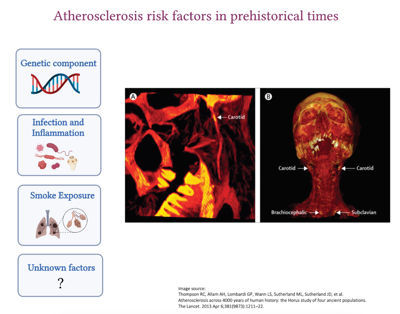
However, atherosclerosis also existed in the ancient civilizations of Egypt, Peru, the American Southwest, and the Aleutian Islands who were all pre-agricultural hunter-gatherers. These observations were pioneered by Czermak in 1852 and taken further by the Horus study published by The Lancet in 2013 where vessel calcification was detected in 34% of the 137 mummies examined. The location of atherosclerosis was very similar to what we see nowadays. The aorta, as well as the femoral, tibial and carotid arteries, were affected, and in older mummies, atherosclerosis was detected in more than one vascular bed. The mummies who were 43 years old at the age of death were more likely to have atherosclerosis compared to those who were 32, highlighting the importance of advanced age in the development of the disease, which we also see nowadays. William Murphy and his colleagues also found carotid calcific atherosclerosis in the Otzi iceman from 3300 BCE.

Since ancient people had a very different lifestyle compared to modern humans, what were the reasons that lead to the development of atherosclerosis back then?

Genetic contribution

Humans have an innate predisposition to atherosclerosis. We now know how important this genetic link is to the disease as the discovery of novel genes involved in atherosclerosis is allowing the development of novel therapies (PCSK9 for example).

Gain of function mutations in lipid-related genes (LDLR, APOB, PCSK9 etc.) cause increased life-long exposure to LDL-C which could not be treated in ancient times. Not to mention the additive contribution of polymorphisms in different genes to atherosclerosis development which we have only started to understand recently (polygenic risk scores).

Inflammation

We now know the major contribution of inflammation to atherosclerosis and how chronic inflammation, which we see in diseases such as rheumatoid arthritis or systemic lupus erythematosus, increases the risk of developing atherosclerosis.

Ancient people had a high exposure to infections, such as tuberculosis and syphilis, against which they had no antimicrobial or vaccines. Chronic inflammation as a result of recurrent untreated infections may have contributed to atherosclerosis development.

Close proximity of these ancient populations to contaminated waters rich in microbes and parasites (such as Schistosoma species, Trichinella spiralis, Taenia species (tapeworm), Plasmodium falciparum (malaria) could have also increased the risk. Systemic inflammation caused by chronic infections in ancient populations could very well have accelerated the development of other inflammatory diseases, such as atherosclerosis. Throughout the years, constant exposure to infections could have led to the selection of genes that provide a strong and effective inflammatory response to Homo sapiens which is only showing to be deleterious nowadays as heightened inflammatory reactions accelerate atherosclerosis and other diseases associated with aging.

Smoke inhalation

Indoor smoke is a risk factor for coronary heart disease and cancer. Ancient people used firewood for the majority of their activities (heating, cooking, and lighting) and their home structures, which were mostly subterranean, had little access to ventilation. Soot deposits were identified in the inner surface of the ribs from burials in southern Turkey suggestive of anthracosis. It was also shown that tobacco was also commonly used in ancient civilizations (especially the Peruvians) which, we now know, contributes to increased inflammation and accelerate atherosclerosis development.

Ancient vs modern atherosclerotic plaque

It remains unclear how the features of the atherosclerotic plaque evolved over the years. Calcifications, which are the ends-stage of atherosclerosis, is what is observed in the remains of the ancient plaques seen in some mummies who were probably important people in their communities. However, we have no clue about the composition of these plaques back then since most of the cellular components would have degraded.

Ancient people who developed atherosclerosis might have not lived long enough to die as a result of their plaque rupturing because their life ended in a tragic fight, a deadly infection or they were poisoned by their enemies. This makes it harder for us to understand if the exposure of ancient people to atherosclerotic risk factors was important enough for them to die from this disease or whether this disease evolved to become more deadly during our modern times. Did the features of what we now call ‘vulnerable plaque’ (high lipid content, thin fibrous cap and presence of intraplaque hemorrhage) exist back then or did the plaque display more stable features?

It may be that since infections, wars and famines are less common in our modern world, people now live long enough to die from the consequences of atherosclerosis which has certainly evolved with our modern lifestyles. However, regardless of the era, atherosclerosis has always been lurking in the shadows throughout the evolution of Homo sapiens, displaying a different face as the years pass by.

References

  1. Thompson RC, Allam AH, Lombardi GP, Wann LS, Sutherland ML, Sutherland JD, et al. Atherosclerosis across 4000 years of human history: the Horus study of four ancient populations. The Lancet. 2013 Apr 6;381(9873):1211–22.
  2. Walker EG. Evidence for prehistoric cardiovascular disease of syphilitic origin on the Northern Plains. Am J Phys Anthropol. 1983;60(4):499–503.
  3. Thomas GS, Wann LS, Allam AH, Thompson RC, Michalik DE, Sutherland ML, et al. Why Did Ancient People Have Atherosclerosis?: From Autopsies to Computed Tomography to Potential Causes. Glob Heart. 2014 Jun 1;9(2):229–37.
  4. Murphy WA, Nedden D zur, Gostner P, Knapp R, Recheis W, Seidler H. The Iceman: Discovery and Imaging. Radiology. 2003 Mar;226(3):614–29.
  5. Clarke EM, Thompson RC, Allam AH, Wann LS, Lombardi GP, Sutherland ML, et al. Is atherosclerosis fundamental to human aging? Lessons from ancient mummies. J Cardiol. 2014 May 1;63(5):329–34.
  6. Marchant J. Mummies reveal that clogged arteries plagued the ancient world. Nature [Internet]. 2013 Mar 11 [cited 2022 Mar 20]; Available from: https://www.nature.com/articles/nature.2013.12568

“The views, opinions, and positions expressed within this blog are those of the author(s) alone and do not represent those of the American Heart Association. The accuracy, completeness, and validity of any statements made within this article are not guaranteed. We accept no liability for any errors, omissions, or representations. The copyright of this content belongs to the author and any liability with regards to infringement of intellectual property rights remains with them. The Early Career Voice blog is not intended to provide medical advice or treatment. Only your healthcare provider can provide that. The American Heart Association recommends that you consult your healthcare provider regarding your health matters. If you think you are having a heart attack, stroke, or another emergency, please call 911 immediately.”

hidden

The Researcher’s Ultimate Toolkit: The PPI network Passion, Perseverance, and Interaction.

I had the pleasure of having a one-on-one virtual meet-up with Mabruka Alfaidi MD, PhD who won the ATVB Investigator in Training Award Competition during last year’s Vascular Discovery 2021 meeting based on her fascinating work on endothelial cells and IL-1b signaling pathway as well as her active involvement with the research community. We discussed her career path and her future projects which we couldn’t do without also going over the many hurdles that come our way as researchers. I decided to summarize the main themes that we tackled in a researcher’s toolkit which encompasses key ingredients to sustain a career in research: The PPI network.

Passion: Follow your passion, but it’s ok to be unsure

Mabruka Alfaidi is a postdoc at Louisiana State University and is currently an instructor seeking her research independence. For Mabruka, her passion for research started when she was a clinical cardiologist and when she realized that one needed to do more to save patients’ lives. Her PhD at the University of Sheffield in the UK opened her eyes to the field of IL1-b signaling in endothelial cells which further developed into her research passion and the basis on which she would like to build her future research career. Research without passion is unsustainable. Passion is the fuel which will motivate us to wake up in the morning (or in the night) and check the latest research, be inspired by the newest methods and design novel hypotheses. However, it is ok to be unsure sometimes when trying to figure out our next step; do we want to stay in academia, or should we venture into setting up this start-up that we always dreamed of? Nothing is really set in stone because research, just like our passion for it, is a dynamic process and it evolves.

Perseverance: It’s ok to fail

A career in basic research is impossible without facing failures and hardships. For Mabruka who started her research path with a medical background, failure, which is assimilated to losing a person’s life in medical practice, was not an option. However, life as a basic researcher is surrounded by failures. We have all struggled with experiments not working, manuscripts getting rejected, grant applications not receiving any interest and the list goes on. So, becoming a basic researcher coming from a medical background is definitely an adjustment. In those moments where doubt creeps into our heads and our confidence and self-esteem is at its lowest, it is important to be surrounded by the right people and inspiring mentors to give us this moral boost that we need to persevere and remind us that it is ok to fail.

Interaction: Network and share your experience, you are not alone

It is common for researchers to feel isolated in their own niche of research and drown in it. However, sharing one’s experience with the research community is important to learn from others and be presented with new opportunities. Mabruka’s experience with the AHA community helped  in her career development as the organization provides funding opportunities for early career researchers as well as those seeking independence and is an important platform where basic and clinical researchers can communicate and find common ground. However, Mabruka’s involvement with AHA and other research communities is because she feels that it is important for a researcher to always ‘give back’ to the community as a way to acknowledge the help and contribution they received and carry on the flame.

“The views, opinions, and positions expressed within this blog are those of the author(s) alone and do not represent those of the American Heart Association. The accuracy, completeness, and validity of any statements made within this article are not guaranteed. We accept no liability for any errors, omissions, or representations. The copyright of this content belongs to the author and any liability with regards to infringement of intellectual property rights remains with them. The Early Career Voice blog is not intended to provide medical advice or treatment. Only your healthcare provider can provide that. The American Heart Association recommends that you consult your healthcare provider regarding your health matters. If you think you are having a heart attack, stroke, or another emergency, please call 911 immediately.”

hidden

Space traveling and CVD risk: RACE to MACE

Picture Reference: https://twitter.com/glitchandgrace/status/711782004497326081 @glitchandgrace

Lately, the space race has been the talk of the town and while it might seem that we are on the verge between science-fiction and reality, there is a long way to go before humans can overcome the health hazards associated with space travel and adapt to their new environment. Humans will likely be exposed to many health hazards emerging from the “space exposome” during their journey into space. Many of these health hazards can affect the cardiovascular (CV) system.

Astronauts are required to undergo an extensive training program including engineering and paramedic courses, intensive health check and various assessments to see how they can cope under stressful conditions. From the start, there is a specific selection process to identify the most ‘fit’ of people who can qualify for space travel. However, even the most ‘fit’ of people can’t escape the health hazards that are awaiting us when we leave mother Earth.

One of the most prominent health hazards associated with long distance space travel affecting the CV system is space radiation. Space radiation usually consists of solar radiation and, the more dangerous, cosmic radiation. Among the components of cosmic radiation, the HZE ions are the most hazardous to the human body because they are highly penetrating and can even generate secondary particles when they interact with shielding materials such as spacecraft or spacesuit. Space radiation damages DNA either directly by energy absorption leading to clustered DNA damage, mutations, chromosome exchanges, carcinogenesis and apoptotic cell death, or indirectly via the production of reactive oxygen species (ROS) from the radiolysis of water molecules.

Exposure to various types of radiation can lead to radiation-induced cardiovascular disease (RICVD) which is a known complication in patients undergoing radiation therapy to treat thoracic cancers and in Japanese atomic bomb survivors. RICVD following space radiation exposure can develop acutely in the form of pericarditis or chronically leading to myocardial remodeling and fibrosis, accelerated development of atherosclerosis, cardiomyopathies, valve abnormalities, arrhythmias and conduction disorders. RICVD can also develop 10-15 years following exposure.

The number of astronauts that have surpassed the low Earth Orbit, where the space radiation amount becomes significantly higher, were only from the Apollo mission which limits the amount of data available. Therefore, several studies to investigate RICVD have been done in animal models using different types of radiations to mimic, to a certain degree, RICVD in humans. These studies have shown that 56Fe ions, the most prominent heavy ion found in cosmic radiation, lead to cardiac hypertrophy and myocardial remodeling. In addition, people that have been exposed to higher levels of radiation compared to the general population had increased risk of myocardial infarction due to atherosclerosis. Mouse models further showed that the effects of irradiation are rather local than systemic where atherosclerosis developed in the areas which were specifically subject to irradiation using 56Fe ions. Plaques in these mice had a thickened intima (carotid), indicating a damage to the arterial wall, and a large necrotic core (aortic root) which increases the risk of instability and thrombogenic complications. The biological processes that have been suggested to induce RICVD include endothelial dysfunction leading to a pro-inflammatory and a pro-fibrogenic environment, apoptotic cell death of various cardiovascular cell types and alterations in DNA methylation.

To date, extensive research is going into improving the shielding methods to protect astronauts from the effects of space radiation. The administration of anti-oxidants such as N-acetyl cysteine (NAC), ascorbic acid (vitamin C), vitamin B, coenzyme Q10 and vitamin E can complement the vitamin deficiencies that humans are subject to during space travel and also remove the generated ROS limiting DNA damage and protecting from space radiation exposure. Some drugs such as angiotensin converting enzyme (ACE) inhibitors and statins have also shown to reduce radiation-induced cardiopulmonary complications and radiation induced atherosclerosis in animal models respectively but need further exploration. Of note, altered DNA methylation can serve as an early biomarker for space radiation exposure and might lead to personalized treatment based on the level of altered DNA methylation after exposure. Space radiation can also increase the risk of cancer and diseases of the central nervous system such as impaired motor function, neurobehavioral changes, Alzheimer’s disease or accelerated aging which can complicate an existing CVD.

Apart from space radiation, other health hazards are also associated with space traveling. Prolonged exposure to microgravity induces bone and muscle atrophy as well as cardiovascular deconditioning. This requires humans to exercise constantly to remain healthy which could be beneficial for CV outcomes. Being confined in a closed environment, disrupted circadian rhythms and the stress for being away from mother Earth also add another layer of psychological and mental challenges to be overcome when venturing into space.


References:

  1. Meerman M, Bracco Gartner TCL, Buikema JW, Wu SM, Siddiqi S, Bouten CVC, et al. Myocardial Disease and Long-Distance Space Travel: Solving the Radiation Problem. Front Cardiovasc Med. 2021;8:27.
  2. Patel ZS, Brunstetter TJ, Tarver WJ, Whitmire AM, Zwart SR, Smith SM, et al. Red risks for a journey to the red planet: The highest priority human health risks for a mission to Mars. Npj Microgravity. 2020 Nov 5;6(1):1–13.
  3. Patel S. The effects of microgravity and space radiation on cardiovascular health: From low-Earth orbit and beyond. IJC Heart Vasc. 2020 Oct 1;30:100595.
  4. Delp MD, Charvat JM, Limoli CL, Globus RK, Ghosh P. Apollo Lunar Astronauts Show Higher Cardiovascular Disease Mortality: Possible Deep Space Radiation Effects on the Vascular Endothelium. Sci Rep. 2016 Jul 28;6(1):29901.
  5. Glitch And Grace Art. Love #glitchandgrace #watercolour #watercolor #space #anatomical-heart #lovewins https://t.co/RIugIHw13W [Internet]. @glitchandgrace. 2016 [cited 2022 Jan 11]. Available from: https://twitter.com/glitchandgrace/status/711782004497326081
hidden

Cut from the same clot? – High-risk primary prevention vs secondary prevention in CVD

Atherosclerotic cardiovascular disease (CVD) prevention has been traditionally divided into primary or pre-event prevention and secondary or re-event prevention. The AHA 2021 joint session with the American Society of Preventive Cardiology entitled “Blurred Lines-Overlap in High-Risk Primary Prevention vs Secondary Prevention” challenged this traditional concept. The session tackled the case of a high-risk patient where using stricter treatment approaches, usually applied in secondary prevention, might be more relevant for her primary prevention. The invited speakers gave an overview of the different treatment options currently available for high-risk primary prevention and highlighted the knowledge gaps in the field.

It is well established that most cardiovascular events (CVE), such as myocardial infarction (MI) and stroke, occur in patients with prior no symptoms. When it comes to predicting CVE, it has been shown that the plaque burden (calculated by Coronary Artery Calcium (CAC) score for example) is a better predictor than the severity of stenosis. This is because the type and certain features of an atherosclerotic plaque can render it more vulnerable regardless of the stenosis degree.


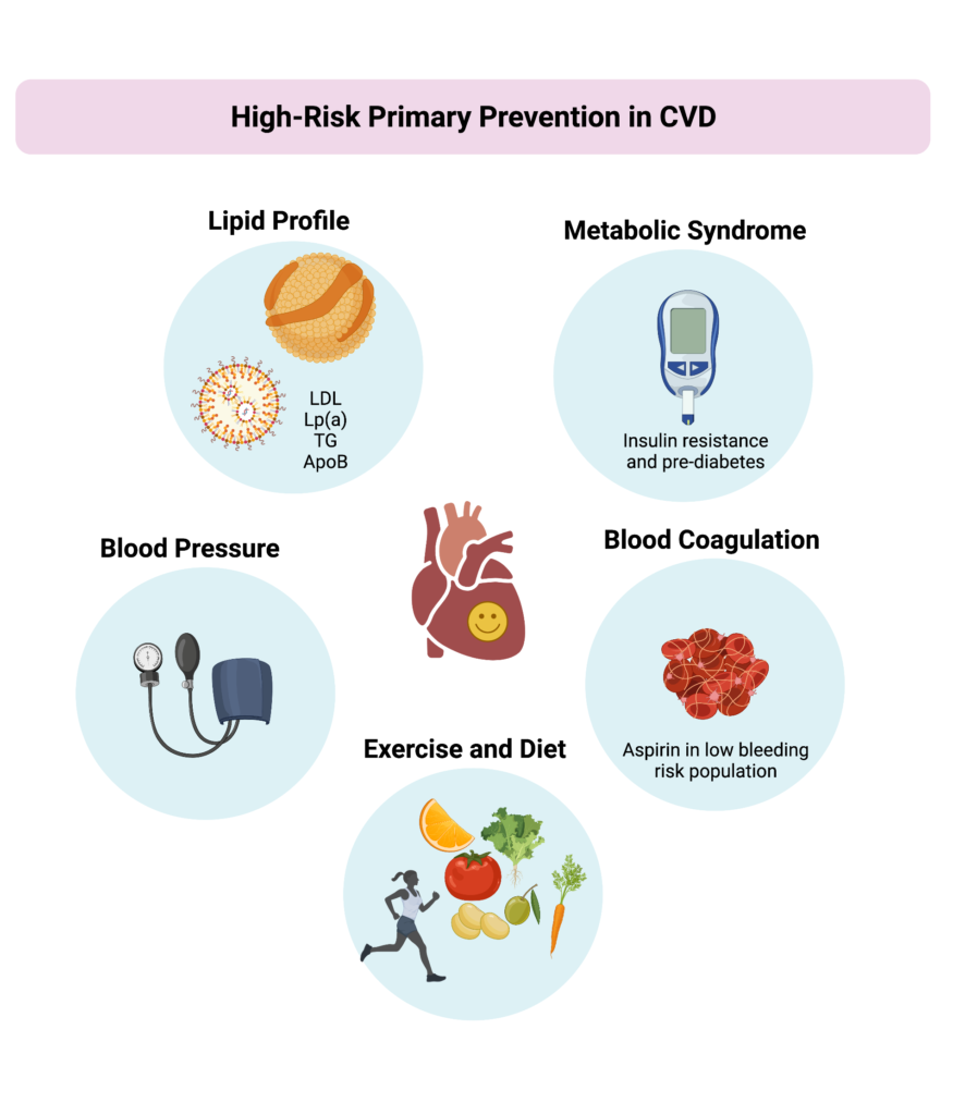
The lines between primary and secondary prevention become blurry when studies such as the one recently conducted by Peng and colleagues demonstrated that primary prevention individuals with very high CAC score (~900) had a similar rate of CVE compared to stable treated high risk secondary prevention patients such as those in the FOURRIER trial. These results show that high-risk primary prevention population might benefit from intense management. Additional risk factors can be taken into account when optimizing the treatment while keeping in mind that not all high-risk primary prevention patients are the same despite presenting with a high CAC score. Before selecting a treatment strategy for a high-risk patient in primary prevention, it is important to evaluate the severity of the patient’s risk factors and act on them accordingly.

Reducing LDL-C levels, ideally below 70 mg/dl, is a good starting point. This can be done using intensive statin therapy or adding ezetimibe or a PCSK9 inhibitor for patients with additional risk factors who do not achieve LDL-C targets. It is of note that PCSK9 inhibitors can reduce Lp(a), triglycerides and ApoB levels which are known to increase CVD risk. The ongoing VESALIUS trial is evaluating the effect of using the PCSK9 inhibitor evolocumab on CVE in primary prevention high-risk patients with no history of myocardial infarction or stroke. For patients with high triglyceride levels, the results of the REDUCE-IT trial demonstrated a reduction of CVE (CV deaths, MI and stroke) when using icosapent ethyl, an EPA derivative. In this trial, 1% of patients had an increase in atrial fibrillation and atrial flutter which can be managed considering the advantageous reduction in stroke risk. However, the REDUCE-IT trial was conducted in a prevalent secondary prevention population and the benefit in high-risk primary prevention needs to be further investigated.

Metabolic syndrome is another risk factor than can be monitored in high-risk primary prevention as it increases the risk of CVE and type 2 diabetes. SGTL2 inhibitors have been shown to reduce CVE in patients with heart failure and reduced ejection fraction offering a benefit of using this anti-diabetic drug class in a non-diabetic population. About a third of the patients enrolled in the CANVAS trial were high-risk primary prevention and had a reduction of CVE while using SGLT2 inhibitors. The REWIND trial on the other hand, which enrolled about 69% of high-risk primary prevention patients, showed that GLP1-RA, another class of anti-diabetics, decreased the occurrence of CVE. However, SGLT2 and GLP1-RA are not yet FDA approved for high-risk primary prevention and the current alternative is managing lifestyle. Patients with metabolic syndrome can be advised to improve the quality (Mediterranean and DASH diets) and quantity (1600 to 3000 calories) of their diet and encouraged to include a fasting period of 14 hours per day to improve their blood pressure and atherogenic lipids profile.

In addition to controlling lipids, blood pressure and pre-diabetes, aspirin can also be considered for high-risk patients in primary prevention but only in those with low bleeding risk as shown by the recent data from the Dallas Heart Study (DHS).

References

  1. Van Venrooij FV, Stolk RP, Banga JD, Erkelens DW, Grobbee DE. Primary and secondary prevention in cardiovascular disease: an old-fashioned concept? J Intern Med. 2002;251(4):301–6.
  2. Mortensen MB, Dzaye O, Steffensen FH, B øtker HE, Jensen JM, R  ønnow SNP, et al. Impact of Plaque Burden Versus Stenosis on Ischemic Events in Patients With Coronary Atherosclerosis. J Am Coll Cardiol. 2020 Dec 15;76(24):2803–13.
  3. Peng AW, Dardari ZA, Blumenthal RS, Dzaye O, Obisesan OH, Iftekhar Uddin S m., et al. Very High Coronary Artery Calcium (≥1000) and Association With Cardiovascular Disease Events, Non–Cardiovascular Disease Outcomes, and Mortality. Circulation. 2021 Apr 20;143(16):1571–83.
  4. Arbab-Zadeh A, Fuster V. From Detecting the Vulnerable Plaque to Managing the Vulnerable Patient: JACC State-of-the-Art Review. J Am Coll Cardiol. 2019 Sep 24;74(12):1582–93.
  5. Amgen. A Double-blind, Randomized, Placebo-controlled, Multicenter Study to Evaluate the Impact of Evolocumab on Major Cardiovascular Events in Patients at High Cardiovascular Risk Without Prior Myocardial Infarction or Stroke [Internet]. clinicaltrials.gov; 2021 Dec [cited 2021 Dec 10]. Report No.: NCT03872401. Available from: https://clinicaltrials.gov/ct2/show/NCT03872401
  6. McMurray JJV, Solomon SD, Inzucchi SE, Køber L, Kosiborod MN, Martinez FA, et al. Dapagliflozin in Patients with Heart Failure and Reduced Ejection Fraction. N Engl J Med. 2019 Nov 21;381(21):1995–2008.
  7. Wilkinson MJ, Manoogian ENC, Zadourian A, Lo H, Fakhouri S, Shoghi A, et al. Ten-Hour Time-Restricted Eating Reduces Weight, Blood Pressure, and Atherogenic Lipids in Patients with Metabolic Syndrome. Cell Metab. 2020 Jan 7;31(1):92-104.e5.
  8. Bhatt DL, Steg PG, Miller M, Brinton EA, Jacobson TA, Ketchum SB, et al. Cardiovascular Risk Reduction with Icosapent Ethyl for Hypertriglyceridemia. N Engl J Med. 2019 Jan 3;380(1):11–22.
  9. Ajufo E, Ayers CR, Vigen R, Joshi PH, Rohatgi A, de Lemos JA, et al. Value of Coronary Artery Calcium Scanning in Association With the Net Benefit of Aspirin in Primary Prevention of Atherosclerotic Cardiovascular Disease. JAMA Cardiol. 2021 Feb 1;6(2):179–87.

“The views, opinions and positions expressed within this blog are those of the author(s) alone and do not represent those of the American Heart Association. The accuracy, completeness and validity of any statements made within this article are not guaranteed. We accept no liability for any errors, omissions or representations. The copyright of this content belongs to the author and any liability with regards to infringement of intellectual property rights remains with them. The Early Career Voice blog is not intended to provide medical advice or treatment. Only your healthcare provider can provide that. The American Heart Association recommends that you consult your healthcare provider regarding your personal health matters. If you think you are having a heart attack, stroke or another emergency, please call 911 immediately.”

hidden

Forever Young at Heart: Aging and Cardiovascular Risk

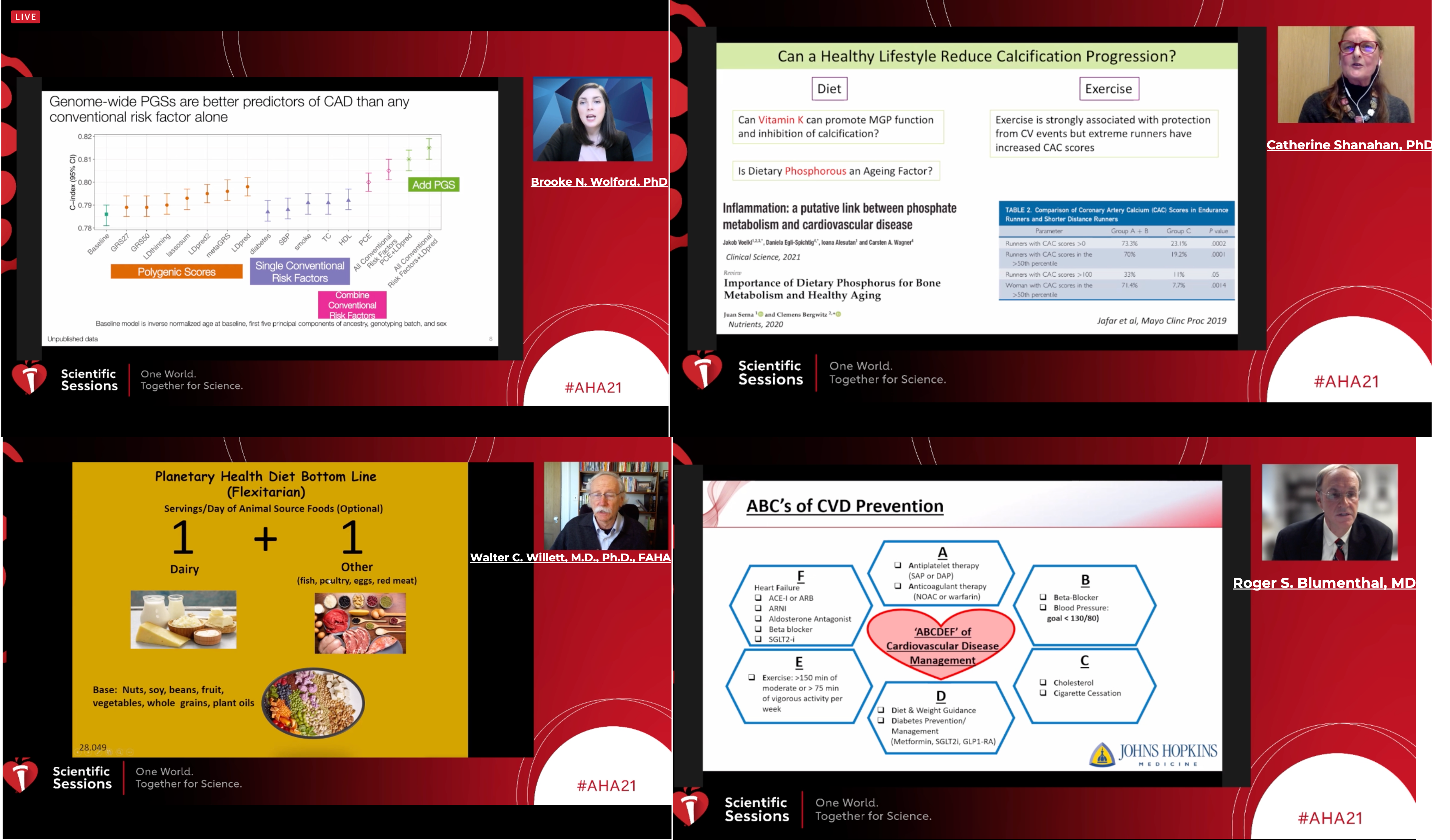
One of the hot topics from this year’s AHA21 Scientific Sessions main event was how to promote healthy vascular aging. Age is the strongest predictor for cardiovascular (CV) risk and risk factors such as genetic predisposition and hyperlipidemia increase CV risk. The general recommendations to reduce CV risk were elegantly presented by Dr Roger S. Blumenthal, MD from Johns Hopkins Ciccarone Center during the “Novel Strategies to Promote Healthy Vascular Aging” AHA21 session. These recommendations mainly focused on promoting a healthy lifestyle via an ABCDE toolbox.

It is important to first Assess someone’s CV risk which can be done using the coronary artery calcium score for example. A novel approach to improve current methods of CV risk assessment above and beyond the current methods is via the implementation of a polygenic risk scores (PGS) which was discussed by Dr Brooke N. Wolford, PhD from the University of Michigan. PGS rely on GWAS data to estimate an individual’s predisposition to a disease and have shown to reclassify about 10% of individuals that were initially misclassified from a ‘low risk’ or ‘intermediate risk’ category into the ‘high risk’ category. The limitation of using PGS however is that it requires genetic testing which might not be currently accessible to all patients. However, the advances in genetic screening technologies might render this possible in the near future.

Blood pressure control is crucial to prevent cardiovascular disease since hypertension increases the risk of stroke, heart attack and heart failure. Controlling blood pressure can be achieved by following a healthy Diet. In light of the growing awareness about climate change, people have been wondering how they can have a diet that is both healthy and sustainable. This contemporary topic was presented by Walter C. Willett, M.D., Ph.D., FAHA from Harvard T.H. Chan School of Public Health and introduced the concept of planetary health diet. This ‘flexitarian’ diet favors plant-based food while allowing one dairy serving a day and other animal products (such as fish, poultry, eggs and meat) to a maximum of two servings a week.  Red meat consumption, which is known to have deleterious effects on planet sustainability, increases LDL-Cholesterol levels in comparison to high quality plant protein diet (legumes, soy, nuts). This means that replacing red meat proteins with plant based proteins can be healthy for us and for the planet.

Exercise in known to reduce all-cause mortality and cardiovascular disease in all ages and even a small amount of aerobic exercise is enough to reduce CV risk. Dr Anthony J. Donato, PhD, MS from the University of Utah presented the benefits of exercise on endothelial dilatation which is a parameter that usually decreases with age and is associated with CV risk. Endothelial cells lose their ability to secrete nitric oxide with time which reduces their vasodilatory properties. Exercise is known to promote a healthier endothelium by increasing endothelial dilatation and reducing endothelial oxidative stress.

A topic that has gained a lot of attention recently in the field of cardiovascular disease and ageing is senescence. Senescence, or biological aging, is induced by DNA damage and leads to metabolically active cells that do not replicate. Vascular senescence increases inflammation and oxidative stress within the vasculature which leads to endothelial dysfunction and smooth muscle cell trans-differentiation. This exciting topic was covered by Dr Catherine Shanahan, PhD from King’s College London where she showed that senescence can accelerate smooth muscle cells ectopic calcification leading to early aging vessels. Senescence is emerging as a key player in chronic diseases and an attractive mechanism that can be targeted to increase longevity.


References:


  1. Choi SW, Mak TS-H, O’Reilly PF. Tutorial: a guide to performing polygenic risk score analyses. Nat Protoc. 2020 Sep;15(9):2759–72.
  2. Willett W, Rockström J, Loken B, Springmann M, Lang T, Vermeulen S, et al. Food in the Anthropocene: the EAT–Lancet Commission on healthy diets from sustainable food systems. The Lancet. 2019 Feb 2;393(10170):447–92.
  3. Guasch-Ferré M, Satija A, Blondin SA, Janiszewski M, Emlen E, O’Connor LE, et al. Meta-Analysis of Randomized Controlled Trials of Red Meat Consumption in Comparison With Various Comparison Diets on Cardiovascular Risk Factors. Circulation. 2019 Apr 9;139(15):1828–45.
  4. Donato AJ, Morgan RG, Walker AE, Lesniewski LA. Cellular and molecular biology of aging endothelial cells. J Mol Cell Cardiol. 2015 Dec;89(Pt B):122–35.
  5. Pierce GL, Donato AJ, LaRocca TJ, Eskurza I, Silver AE, Seals DR. Habitually exercising older men do not demonstrate age-associated vascular endothelial oxidative stress. Aging Cell. 2011 Dec;10(6):1032–7.
  6. Sanchis P, Ho CY, Liu Y, Beltran LE, Ahmad S, Jacob AP, et al. Arterial “inflammaging” drives vascular calcification in children on dialysis. Kidney Int. 2019 Apr 1;95(4):958–72.

“The views, opinions and positions expressed within this blog are those of the author(s) alone and do not represent those of the American Heart Association. The accuracy, completeness and validity of any statements made within this article are not guaranteed. We accept no liability for any errors, omissions or representations. The copyright of this content belongs to the author and any liability with regards to infringement of intellectual property rights remains with them. The Early Career Voice blog is not intended to provide medical advice or treatment. Only your healthcare provider can provide that. The American Heart Association recommends that you consult your healthcare provider regarding your personal health matters. If you think you are having a heart attack, stroke or another emergency, please call 911 immediately.”