hidden

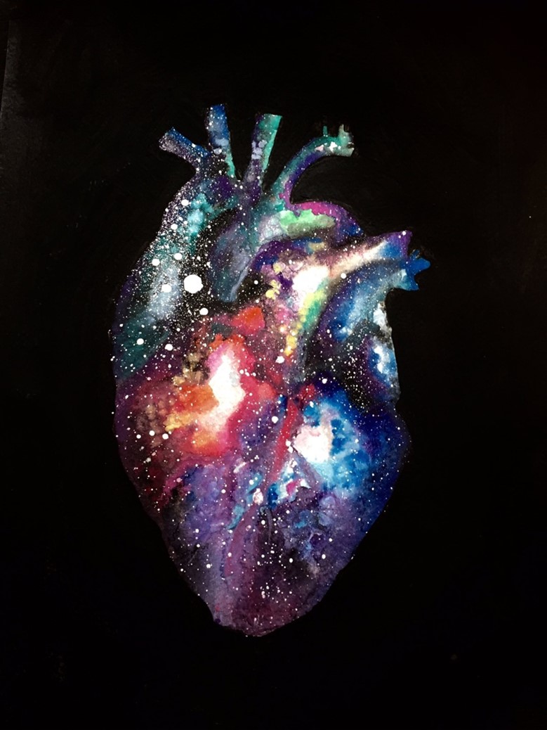
Join the race against the clock: Controlling for age in cardiovascular disease

The race against aging has already started. People who want part of this race see the limitless opportunities humans will have if aging is taken out of the equation. Humans will be able to live longer with their loved ones, go back to University when they are 70 years old to study this long-dreamed profession they always wanted to try or take the time to go through every single item on their bucket list, no matter how long the list is. Others are worried about what awaits them at the finish line of the race and what sacrifices will have to be made along the way. They argue that aging is the core of our existence and the reason why we make the choices we make every day. If humans don’t age, will they still find a reason to live and would this type of life be worth living? Regardless of people’s scattered opinions, the remaining question to be answered in our race against the clock is: Can we age disease-free?

As humans began to live longer dying less of problems such as hunger, wars and infections, they were faced with a new type of problem: chronic diseases. As we age and get exposed to different environmental and lifestyle factors, a set of biological and functional changes in our bodies lead to the development of chronic diseases such as cardiovascular diseases (CVD), diabetes, cancer, dementia, arthritis, and the list goes on. Notorious for being the ‘number 1 killer in the world’, preventing CVD has been one of the top priorities in our fight against aging. Age is the best predictor of CVD death, and despite years of research and large amounts of funding spent on biomarker discovery, there are currently no better predictors of CVD death than the age of a person and these are some of the reasons why.

Large blood vessels tend to become stiffer over time as they accumulate more collagen (due to an increase in TGF-b activity leading to collagen synthesis from smooth muscle cells) and lose their elastin content (because of higher metalloproteases and cathepsin activity). This leads to a chronic increase in systolic blood pressure which is worsened by the rise of catecholamines levels usually seen during aging. Both phenomena contribute to left ventricular dysfunction and hypertrophy due to the increase in myocardial oxygen demand. Calcification is another hallmark of aging that also contributes to vessel stiffness and induces stenosis. As we age, skeletal calcium is released and tends to accumulate in the vascular structures.

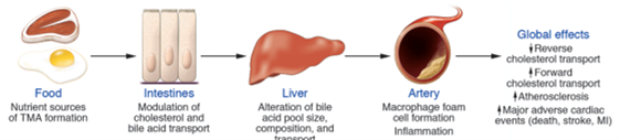
Apart from leading to vessel stiffness, aging causes the vascular endothelial cell (EC) barrier to become dysfunctional. ECs play a crucial role in maintaining vessel integrity and homeostasis by balancing vasodilatory and vasoconstricting functions and by aligning the vessels with an anti-thrombotic surface. Disruption of this protective barrier over time is characterized by ECs undergoing oxidative stress, reduced nitric oxide (a potent vasodilator) production, increased expression of adhesion molecules (ICAM and VCAM) and secretion of inflammatory chemokines (CXCL8) and cytokines (IL-1b and IL-6). The initiating event of atherosclerosis development starts with endothelial dysfunction which gives way for monocyte infiltration and subsequent foam cell formation contributing to plaque development.

At the molecular level, changes affecting the genome and epigenome are a fundamental feature of aging. With age, the clonal diversity of hematopoietic stem cells decreases resulting in the predominance of one clone. In recent years, clonal hematopoiesis of indeterminate potential (CHIP), which occurs as a result of mutations in transcriptional regulators (DNMT3A, TET2 and ASXL1), was found as a novel CVD risk factor, thereby linking genetic mutations in hematopoietic stem cells, aging and CVD. The number of endothelial progenitor cells also decreases over time which reduces angiogenesis capacity and capillary density leading to microvascular disease (such as peripheral artery disease).

The shortening of chromosome telomers is another molecular change related to aging. As cells replicate, telomeres get shorter until cellular senescence is triggered. Cellular senescence is a cellular protective mechanism that activates NK cells to remove cells with defective genetic material via apoptosis. It has been shown that patients with reduced leukocyte telomere length have increased risk of atherosclerosis. An atherosclerotic plaque, rich in inflammatory cells and trans-differentiated smooth muscle cells, is a dense hypoxic environment characterized by the presence of reactive oxygen species which also induce DNA damage and senescence.

Current therapies for atherosclerosis target some of the pathways of aging highlighted above. While statins are known to reduce plaque lipid content and inflammation, in parallel, they tend to increase calcification leading to vascular stiffness. On the other hand, anti-hypertensive treatments offer benefits beyond reducing CVD mortality, but also decreasing dementia. Recently, novel therapies targeting aging in CVD have focused on stem cell therapy. However, clinical trials using cell therapy to improve left ventricular dysfunction or to reduce cardiovascular events have shown no or modest benefit. This may be because autologous cell therapy of stem cells that already have an ‘aging’ phenotype is not ideal, and these cells may require ex vivo reprograming to improve treatment efficiency.


Since many age-related diseases have similar underlying molecular mechanisms driving them, the future for treating chronic diseases will rely on targeting the mechanisms of aging rather than treating the disease itself. Some of the best ways to slow down aging is by being active, controlling blood glucose levels, opting for diets rich in antioxidants and fibers and introducing low calorie intake periods during the day. However, this usually requires a substantial effort and serious lifestyle changes on our behalf. But, since research on anti-aging therapies and senolytic drugs is booming, it might be possible to slow down aging by taking one or two pills a day without ever needing to change the routines that we are so comfortable with.

References

  1. Paneni F, Diaz Cañestro C, Libby P, Lüscher TF, Camici GG. The Aging Cardiovascular System: Understanding It at the Cellular and Clinical Levels. J Am Coll Cardiol. 2017 Apr 18;69(15):1952–67.
  2. Quyyumi AA, Vasquez A, Kereiakes DJ, Klapholz M, Schaer GL, Abdel-Latif A, et al. PreSERVE-AMI. Circ Res. 2017 Jan 20;120(2):324–31.
  3. Brouilette SW, Moore JS, McMahon AD, Thompson JR, Ford I, Shepherd J, et al. Telomere length, risk of coronary heart disease, and statin treatment in the West of Scotland Primary Prevention Study: a nested case-control study. The Lancet. 2007 Jan 13;369(9556):107–14.
  4. Koopman JJE, Kuipers RS. From arterial ageing to cardiovascular disease. The Lancet. 2017 Apr 29;389(10080):1676–8.
  5. Jaiswal S, Libby P. Clonal haematopoiesis: connecting ageing and inflammation in cardiovascular disease. Nat Rev Cardiol. 2020 Mar;17(3):137–44.
  6. Antonangeli F, Zingoni A, Soriani A, Santoni A. Senescent cells: Living or dying is a matter of NK cells. J Leukoc Biol. 2019 Jun;105(6):1275–83.
  7. What is the Age of My Heart? – Calculate Your Own Heart Age • MyHeart [Internet]. MyHeart. 2015 [cited 2022 May 16]. Available from: https://myheart.net/articles/what-is-the-age-of-my-heart-calculate-your-own-heart-age/

“The views, opinions, and positions expressed within this blog are those of the author(s) alone and do not represent those of the American Heart Association. The accuracy, completeness, and validity of any statements made within this article are not guaranteed. We accept no liability for any errors, omissions, or representations. The copyright of this content belongs to the author and any liability with regards to infringement of intellectual property rights remains with them. The Early Career Voice blog is not intended to provide medical advice or treatment. Only your healthcare provider can provide that. The American Heart Association recommends that you consult your healthcare provider regarding your health matters. If you think you are having a heart attack, stroke, or another emergency, please call 911 immediately.”

hidden

Climate Change and Cardiovascular Diseases

Climate change is partly due to the increased atmospheric concentration of greenhouse gases emitted by burning fossil fuels like oil, natural gas, and methane produced by ruminant agricultural animals. The earth’s temperature has augmented by 0.85°C in the last century, and the rate of global warming has increased to 0.18°C/decade in the last three decades. The altering temperature, in particular increasing heat, is one of the critical features of climate change and can significantly affect cardiac health. In addition, extreme weather events, rising sea levels, and lack of food and water are expected outcomes of climate change1.

Cardiovascular Diseases (CVDs) are the leading cause of death globally, and climate change can worsen CVD incidence further. Heatwaves are expected to be more frequent and prolonged due to ongoing climate change. The intense heat can cause mortality and morbidity due to heatstroke, which is defined as hyperthermia associated with a systemic inflammatory response resulting in multiple organ failure and predominant encephalopathy. Heat stress is associated with acute cardiac events where heated blood circulates in peripheral circulation, and heat tolerance is impaired due to insufficient cardiac output to meet the body’s needs for sufficient heat loss1.

There is a U-shaped relationship between temperature and all-cause mortality where mortality increases with the shift from ‘optimum temperature’ at both cold and hot ends. In the Netherlands, the lowest mortality rate was observed with an average temperature of 16.5°C, and CVD mortalities accounted for 57% of cold-related death2. In 1976, daily deaths from coronary thrombosis increased two-fold during the London heatwave3. Additionally, daily mortality due to congestive heart failure is strongly associated with maximum daily temperature in Montreal, with an exponential increase starting at 25°C4. Similarly, during comparatively hotter summer, a U-shaped relationship between outer temperature and coronary artery disease deaths is reported in Taiwan5.

Lifestyle modifications on a large population scale are required to reduce the emission of greenhouse gases, thereby mitigating the extent of climate change. Reducing the use of motor vehicles for short-distance commuting can help reduce the emissions of greenhouse gases related to transportation and air pollution that can have adverse effects on health. Further, reducing the consumption of ruminant meat such as sheep and cows and increasing the use of renewable energy, including solar radiation or wind power, can help mitigate climate change, air pollution, and the risks of heart diseases1.

Another essential remedy to diminish climate change can be ‘active transport or self-transport that encompasses more physical activity and involves walking, cycling, and use of public transport as a mode of transportation. This will not only reduce the emanation of greenhouse gases but increased physical activity can confer several cardiac health benefits1. An extra kilometer walk is associated with a 4.8% reduction in risk of obesity which is a significant risk factor of CVD, whereas an extra hour spent in car/day is lined with a 6% increase in the likelihood of obesity6. Additionally, a study done in Finland reported a significant reduction in CVD risk and all-cause mortality in women who walked or cycled 15 minutes or more for work7.

In conclusion, we need to modify our lifestyle and make healthier choices to protect our hearts and earth. If you are further interested in the topic, you can get a detailed insight in the review article published in the Cardiology journal1 titled “The effects of climate change on cardiac health”.

 REFERENCE

  1. De Blois J, Kjellstrom T, Agewall S, Ezekowitz JA, Armstrong PW, Atar D. The Effects of Climate Change on Cardiac Health. Cardiology. 2015;131(4):209-17. doi:10.1159/000398787
  2. Kunst AE, Looman CW, Mackenbach JP. Outdoor air temperature and mortality in The Netherlands: a time-series analysis. Am J Epidemiol. Feb 01 1993;137(3):331-41. doi:10.1093/oxfordjournals.aje.a116680
  3. Keatinge WR, Coleshaw SR, Easton JC, Cotter F, Mattock MB, Chelliah R. Increased platelet and red cell counts, blood viscosity, and plasma cholesterol levels during heat stress, and mortality from coronary and cerebral thrombosis. Am J Med. Nov 1986;81(5):795-800. doi:10.1016/0002-9343(86)90348-7
  4. Kolb S, Radon K, Valois MF, Héguy L, Goldberg MS. The short-term influence of weather on daily mortality in congestive heart failure. Arch Environ Occup Health. 2007;62(4):169-76. doi:10.3200/AEOH.62.4.169-176
  5. Pan WH, Li LA, Tsai MJ. Temperature extremes and mortality from coronary heart disease and cerebral infarction in elderly Chinese. Lancet. Feb 11 1995;345(8946):353-5. doi:10.1016/s0140-6736(95)90341-0
  6. Frank LD, Andresen MA, Schmid TL. Obesity relationships with community design, physical activity, and time spent in cars. Am J Prev Med. Aug 2004;27(2):87-96. doi:10.1016/j.amepre.2004.04.011
  7. Barengo NC, Hu G, Lakka TA, Pekkarinen H, Nissinen A, Tuomilehto J. Low physical activity as a predictor for total and cardiovascular disease mortality in middle-aged men and women in Finland. Eur Heart J. Dec 2004;25(24):2204-11. doi:10.1016/j.ehj.2004.10.009

“The views, opinions, and positions expressed within this blog are those of the author(s) alone and do not represent those of the American Heart Association. The accuracy, completeness, and validity of any statements made within this article are not guaranteed. We accept no liability for any errors, omissions, or representations. The copyright of this content belongs to the author and any liability with regards to infringement of intellectual property rights remains with them. The Early Career Voice blog is not intended to provide medical advice or treatment. Only your healthcare provider can provide that. The American Heart Association recommends that you consult your healthcare provider regarding your health matters. If you think you are having a heart attack, stroke, or another emergency, please call 911 immediately.”

hidden

Mental stress may lead to poor cardiovascular outcomes

The presence of mental-stress-induced myocardial ischemia is associated with an increased risk of cardiovascular death and nonfatal myocardial infarction, as per an interesting study published in JAMA Network by researchers from Emory University.1

Several studies have revealed the correlation between acute mental stress and the onset of myocardial ischemia seen on myocardial perfusion imaging, strengthening the concept of mental stress and coronary heart disease (CHD). These studies parallelly enrolled patients with stable CHD in the Mental Stress Ischemia Prognosis Study (MIPS) and Myocardial Infarction and Mental Stress Study 2(MIMS2). All participants underwent clinical and psychological assessment at baseline, standardized mental stress test, and myocardial perfusion imaging at rest, with mental stress and exercise or pharmacological stress test.

638 and 313 participants were enrolled in MIPS and MIMS2 study, respectively. Mental stress-induced ischemia was seen in 15%(MIPS) and 17%(MIMS2) participants. Over a medial follow up of 5 years, the pooled results of both studies revealed a higher event rate of cardiovascular death or myocardial infarction (6.9 per 100 patient-years) in positive mental stress-induced ischemia compared to those without ischemia (2.6 per 100 patient-years) (HR: 2.5, 95% CI: 1.8-3.5).1 There was an even higher rate in patients with conventional and mental-stress-induced ischemia (8.1 events per 100 patient-years, HR:3.8, 95% CI: 2.6-5.6).  Interestingly, participants with conventional stress ischemia did not have an increased risk of cardiovascular events. The study also revealed statistically significant higher heart failure incidence in patients with mental stress-induced ischemia. 1

The Brain-Heart axis has been an active area of research over the last decade2; the current study further strengthens this correlation. This study differentiates between the ischemia incidence and outcomes based on mental and conventional stress, which has not been reported in prior studies. It is noteworthy that patients with mental stress-induced ischemia have a higher incidence than conventional ischemia. ere have been other studies in the past which have investigated the impact of acute mental stress leading to decline in cardiac function known as Takotsubo cardiomyopathy, or commonly known as broken-heart syndrome.3 Interestingly, a few smaller studies have shown that an ecstatic happiness state can also lead to Takotsubo cardiomyopathy.3 Nevertheless, the acute decline in these scenarios are mostly transient, but, as per the current study effect of mental stress induced ischemia my lead to prolonged adverse outcomes.

Further studies evaluating the screening for mental stress-induced ischemia and potential early interventions can pave the pathway for reducing CHD, thereby strengthening the concept of the Brain-Heart axis.

https://twitter.com/DrDavidKatz/status/1459574297601658883

https://twitter.com/andrebez/status/1479425795949056004

References.

  1. Vaccarino V, Almuwaqqat Z, Kim JH, et al. Association of Mental Stress–Induced Myocardial Ischemia With Cardiovascular Events in Patients With Coronary Heart Disease. JAMA. 2021;326(18):1818-1828.
  2. Tahsili-Fahadan P, Geocadin RG. Heart–Brain Axis. Circulation Research. 2017;120(3):559-572.
  3. Lyon AR, Citro R, Schneider B, et al. Pathophysiology of Takotsubo Syndrome: JACC State-of-the-Art Review. J Am Coll Cardiol. 2021;77(7):902-921.


hidden

Space traveling and CVD risk: RACE to MACE

Picture Reference: https://twitter.com/glitchandgrace/status/711782004497326081 @glitchandgrace

Lately, the space race has been the talk of the town and while it might seem that we are on the verge between science-fiction and reality, there is a long way to go before humans can overcome the health hazards associated with space travel and adapt to their new environment. Humans will likely be exposed to many health hazards emerging from the “space exposome” during their journey into space. Many of these health hazards can affect the cardiovascular (CV) system.

Astronauts are required to undergo an extensive training program including engineering and paramedic courses, intensive health check and various assessments to see how they can cope under stressful conditions. From the start, there is a specific selection process to identify the most ‘fit’ of people who can qualify for space travel. However, even the most ‘fit’ of people can’t escape the health hazards that are awaiting us when we leave mother Earth.

One of the most prominent health hazards associated with long distance space travel affecting the CV system is space radiation. Space radiation usually consists of solar radiation and, the more dangerous, cosmic radiation. Among the components of cosmic radiation, the HZE ions are the most hazardous to the human body because they are highly penetrating and can even generate secondary particles when they interact with shielding materials such as spacecraft or spacesuit. Space radiation damages DNA either directly by energy absorption leading to clustered DNA damage, mutations, chromosome exchanges, carcinogenesis and apoptotic cell death, or indirectly via the production of reactive oxygen species (ROS) from the radiolysis of water molecules.

Exposure to various types of radiation can lead to radiation-induced cardiovascular disease (RICVD) which is a known complication in patients undergoing radiation therapy to treat thoracic cancers and in Japanese atomic bomb survivors. RICVD following space radiation exposure can develop acutely in the form of pericarditis or chronically leading to myocardial remodeling and fibrosis, accelerated development of atherosclerosis, cardiomyopathies, valve abnormalities, arrhythmias and conduction disorders. RICVD can also develop 10-15 years following exposure.

The number of astronauts that have surpassed the low Earth Orbit, where the space radiation amount becomes significantly higher, were only from the Apollo mission which limits the amount of data available. Therefore, several studies to investigate RICVD have been done in animal models using different types of radiations to mimic, to a certain degree, RICVD in humans. These studies have shown that 56Fe ions, the most prominent heavy ion found in cosmic radiation, lead to cardiac hypertrophy and myocardial remodeling. In addition, people that have been exposed to higher levels of radiation compared to the general population had increased risk of myocardial infarction due to atherosclerosis. Mouse models further showed that the effects of irradiation are rather local than systemic where atherosclerosis developed in the areas which were specifically subject to irradiation using 56Fe ions. Plaques in these mice had a thickened intima (carotid), indicating a damage to the arterial wall, and a large necrotic core (aortic root) which increases the risk of instability and thrombogenic complications. The biological processes that have been suggested to induce RICVD include endothelial dysfunction leading to a pro-inflammatory and a pro-fibrogenic environment, apoptotic cell death of various cardiovascular cell types and alterations in DNA methylation.

To date, extensive research is going into improving the shielding methods to protect astronauts from the effects of space radiation. The administration of anti-oxidants such as N-acetyl cysteine (NAC), ascorbic acid (vitamin C), vitamin B, coenzyme Q10 and vitamin E can complement the vitamin deficiencies that humans are subject to during space travel and also remove the generated ROS limiting DNA damage and protecting from space radiation exposure. Some drugs such as angiotensin converting enzyme (ACE) inhibitors and statins have also shown to reduce radiation-induced cardiopulmonary complications and radiation induced atherosclerosis in animal models respectively but need further exploration. Of note, altered DNA methylation can serve as an early biomarker for space radiation exposure and might lead to personalized treatment based on the level of altered DNA methylation after exposure. Space radiation can also increase the risk of cancer and diseases of the central nervous system such as impaired motor function, neurobehavioral changes, Alzheimer’s disease or accelerated aging which can complicate an existing CVD.

Apart from space radiation, other health hazards are also associated with space traveling. Prolonged exposure to microgravity induces bone and muscle atrophy as well as cardiovascular deconditioning. This requires humans to exercise constantly to remain healthy which could be beneficial for CV outcomes. Being confined in a closed environment, disrupted circadian rhythms and the stress for being away from mother Earth also add another layer of psychological and mental challenges to be overcome when venturing into space.


References:

  1. Meerman M, Bracco Gartner TCL, Buikema JW, Wu SM, Siddiqi S, Bouten CVC, et al. Myocardial Disease and Long-Distance Space Travel: Solving the Radiation Problem. Front Cardiovasc Med. 2021;8:27.
  2. Patel ZS, Brunstetter TJ, Tarver WJ, Whitmire AM, Zwart SR, Smith SM, et al. Red risks for a journey to the red planet: The highest priority human health risks for a mission to Mars. Npj Microgravity. 2020 Nov 5;6(1):1–13.
  3. Patel S. The effects of microgravity and space radiation on cardiovascular health: From low-Earth orbit and beyond. IJC Heart Vasc. 2020 Oct 1;30:100595.
  4. Delp MD, Charvat JM, Limoli CL, Globus RK, Ghosh P. Apollo Lunar Astronauts Show Higher Cardiovascular Disease Mortality: Possible Deep Space Radiation Effects on the Vascular Endothelium. Sci Rep. 2016 Jul 28;6(1):29901.
  5. Glitch And Grace Art. Love #glitchandgrace #watercolour #watercolor #space #anatomical-heart #lovewins https://t.co/RIugIHw13W [Internet]. @glitchandgrace. 2016 [cited 2022 Jan 11]. Available from: https://twitter.com/glitchandgrace/status/711782004497326081
hidden

Reducing Disparities in Access to Cardiovascular Disease Prevention with the Polypill

This year’s AHA 2020 Scientific Session is taking place using combined modalities, including live, simulive, and on-demand sessions. Despite the change from the traditional in-person modality to the virtual approach, listening to the opening session and findings from emerging science reminded me of the mission of the American Heart Association to be a relentless force for a world of longer, healthier lives. This year’s scientific sessions also align with a wide range of events we all have experienced this year, ranging from the COVID-19 pandemic, to racial/ethnic, gender, and income disparities leading to health inequity in our society. These further present a call to action in order to address these very same societal issues that are likely to impact on health equity for the most vulnerable groups.

The cardiovascular polypill, or combined aspirin, cholesterol, and blood-pressure-lowering agents into a single pill has been proposed for nearly a decade as a complementary option in the prevention of cardiovascular diseases in intermediate- and high-risk patient populations.1 Yet there have been previous limitations in understanding its efficacy and relative safety in developing countries.2  The findings of the International Polycap Study (TIPS)-3 presented by Dr. Salim Yusuf during the late-breaking science session bring a ray of hope to the global disparities in cardiovascular disease prevention.3 The study resulted in a 30% reduction in cardiovascular risk with a combined regimen composed of Aspirin and a polypill (atenolol, ramipril, hydrochlorothiazide, and a statin).3  Based on the TIPS-3 study, the polypill approach presents a safe and cost-effective strategy with the potential for satisfactory medication adherence.

While these findings are promising for developing countries, the polypill may present a viable solution to underserved, low-income minority groups in developed countries.4  Another important takeaway from this study was the inclusion of women, who represented 53% of the sample.  Their inclusion in global studies such as this one also highlights the move into health equity and awareness of women’s health globally at a time when cardiovascular disease continue to present women’s greatest health threat. Traditionally, the enrollment of women in clinical trials has been limited. This has resulted in a limited understanding of risk factors and benefits from treatment regimens for cardiovascular disease-specific to women.5

As we observe the benefits related to polypill, it is also important to keep in mind that it may not align with the medical trend in developed countries for precision medicine, leading to individualized, targeted therapy.6  With cardiovascular disease remaining the leading cause of mortality and morbidity in developed and developing countries, and low-income, ethnic minorities affected by it, the question remains on long-term, best preventive strategies in the reduction of cardiovascular risk factors for all. It will also be important to measure long-term outcomes related to polypill strategies in future studies.


References:

  1. Lafeber M, Spiering W, Singh K, Guggilla RK, Patil V, Webster R; SPACE collaboration. The cardiovascular polypill in high-risk patients. Eur J Prev Cardiol. 2012 Dec;19(6):1234-42. doi: 10.1177/1741826711428066. Epub 2011 Oct 21. PMID: 22019908.
  2. Nguyen C, Cheng-Lai A. The polypill: a potential global solution to cardiovascular disease. Cardiol Rev. 2013 Jan-Feb;21(1):49-54. doi: 10.1097/CRD.0b013e3182755429. PMID: 23018668.
  3. Joseph P, Pais P, Dans AL, Bosch J, Xavier D, Lopez-Jaramillo P, Yusoff K, Santoso A, Talukder S, Gamra H, Yeates K, Lopez PC, Tyrwhitt J, Gao P, Teo K, Yusuf S; TIPS-3 Investigators. The International Polycap Study-3 (TIPS-3): Design, baseline characteristics and challenges in conduct. Am Heart J. 2018 Dec;206:72-79. doi: 10.1016/j.ahj.2018.07.012. Epub 2018 Aug 2. PMID: 30342297; PMCID: PMC6299262.
  4. Muñoz D, Uzoije P, Reynolds C, Miller R, Walkley D, Pappalardo S, Tousey P, Munro H, Gonzales H, Song W, White C, Blot WJ, Wang TJ. Polypill for Cardiovascular Disease Prevention in an Underserved Population. N Engl J Med. 2019 Sep 19;381(12):1114-1123. doi: 10.1056/NEJMoa1815359. PMID: 31532959; PMCID: PMC6938029.
  5. Saeed A, Kampangkaew J, Nambi V. Prevention of Cardiovascular Disease in Women. Methodist Debakey Cardiovasc J. 2017 Oct-Dec;13(4):185-192. doi: 10.14797/mdcj-13-4-185. PMID: 29744010; PMCID: PMC5935277.
  6. Psaty BM, Dekkers OM, Cooper RS. Comparison of 2 Treatment Models: Precision Medicine and Preventive Medicine. JAMA. 2018 Aug 28;320(8):751-752. doi: 10.1001/jama.2018.8377. PMID: 30054607.


“The views, opinions and positions expressed within this blog are those of the author(s) alone and do not represent those of the American Heart Association. The accuracy, completeness and validity of any statements made within this article are not guaranteed. We accept no liability for any errors, omissions or representations. The copyright of this content belongs to the author and any liability with regards to infringement of intellectual property rights remains with them. The Early Career Voice blog is not intended to provide medical advice or treatment. Only your healthcare provider can provide that. The American Heart Association recommends that you consult your healthcare provider regarding your personal health matters. If you think you are having a heart attack, stroke or another emergency, please call 911 immediately.”

hidden

Modifiable Factors Influence Non-modifiable Factors for Cardiovascular Health?

The scientific community continues its full force swing at reducing cardiovascular disease risk. In the Scientific Session titled “Microbiome in Cardiovascular Disease,” the complexity of accounting for human variation was the theme. The important difference and interactions between non-modifiable (genetics) and modifiable (diet, exercise, smoking, etc..) factors were presented. Dr. Katherine Tucker opened up the session by highlighting work from Thanassoulis et. al., 2012, which identified 13 single nucleotide polymorphisms (SNPs) to generate a genetic risk score (GRS) to predict cardiovascular events and coronary artery calcium (CAC). Single nucleotide polymorphisms are the most common type of genetic variation among people and are used to help quantify the variation in individuals (1). The CAC score comes from a test that quantifies the amount of calcium accumulation in the walls of the coronary arteries. A lower score represents a greater risk and a lower score relates to a lower risk of heart disease. Dr. Tucker went on to explain the genetic risk is influenced by individual environmental factors (i.e. smoking, exercise, and diet) (2). Recent data from the CARDIA study supports this in reporting that, “low-carbohydrate diets at a younger age is associated with an increased risk of subsequent CAC progression, particularly when animal protein or fat are chosen to replace carbohydrates. (3).”

Figure 1 source: https://doi.org/10.1093/nutrit/nux001

The changes in macronutrient content in a diet is related to what happens in the gut. Within the gut, there are trillions of bacteria that make up a microbiome. An individual microbiome modulates the immune system and metabolic processes. The microbiome influence on human health is so pronounced in that it actively reprograms the genome in response to the environment, changing the bacteria phyla ratios that lead to down-stream effects that could influence cardiovascular health (Figure 1) (2). Dietary fiber and prebiotic consumption are two components that modulate the composition of the gut microbiome (Figure 2) (4). Also, there is some great news for you Kombucha fans out there! Fermented foods have some benefits for the gut.

Figure 2. Source: https://doi.org/10.1093/nutrit/nux062

Bhat and Kapila 2017 further highlight diet in a review stating “The composition of the gut microbiota has a tremendous influence on host metabolism.” Perhaps specific dietary interventions can reduce the risk of cardiovascular disease with the focus on obtaining an optimal microbiota composition. Zhang et. al., 2020, showed how detrimental diets with contain highly processed foods can be the bacteria in our gut (Figure 3) (5).

Figure 3. Source: https://doi.org/10.1093/ajcn/nqaa276

To further highlight how much people differ from one another, Dr. Tang from the Cleveland Clinic explained only 37% of the gut is actually shared between twins. In addition, there are significant diurnal variations in response to meals consumed among people. The work presented the relationship between microbiota and trimethylamine (TMA)/trimethylamine–N-oxide(TMAO) generation. Elevated TMAO levels predict major adverse cardiac events like death, myocardial infarction (MI), and stroke (Figure 4) (6). Dr. Tang explained that risk is highest with people who displayed the highest baseline levels of two TMAO precursors choline or L-carnitine, while some may show no risk with higher levels. Dr. Tang emphasized the variation again among individuals.

Figure 4. Source: Tang, W. W., & Hazen, S. L. (2014)

We are only scratching the surface with the modifiable risk factors for heart disease. Specifically, the gut shows an area rich for investigation. The gut microbiota contributes to human physiology and diseases and it is something to be excited about for biomedical researchers.


References:

  1. Thanassoulis G, Peloso GM, Pencina MJ, Hoffmann U, Fox CS, Cupples LA, Levy D, D’Agostino RB, Hwang SJ, O’Donnell CJ. A genetic risk score is associated with incident cardiovascular disease and coronary artery calcium: the Framingham Heart Study. Circ Cardiovasc Genet. 2012 Feb 1;5(1):113-21. doi: 10.1161/CIRCGENETICS.111.961342. Epub 2012 Jan 10.
  2. Mohd Iqbal Bhat, Rajeev Kapila, Dietary metabolites derived from gut microbiota: critical modulators of epigenetic changes in mammals, Nutrition Reviews, Volume 75, Issue 5, May 2017, Pages 374–389, https://doi.org/10.1093/nutrit/nux001
  3. Gao, J. W., Hao, Q. Y., Zhang, H. F., Li, X. Z., Yuan, Z. M., Guo, Y., … & Liu, P. M. (2020). Low-Carbohydrate Diet Score and Coronary Artery Calcium Progression: Results From the CARDIA Study. Arteriosclerosis, Thrombosis, and Vascular Biology, ATVBAHA-120.
  4. Genelle R Healey, Rinki Murphy, Louise Brough, Christine A Butts, Jane Coad, Interindividual variability in gut microbiota and host response to dietary interventions, Nutrition Reviews, Volume 75, Issue 12, December 2017, Pages 1059–1080, https://doi.org/10.1093/nutrit/nux062
  5. Zefeng Zhang, Sandra L Jackson, Euridice Martinez, Cathleen Gillespie, Quanhe Yang, Association between ultraprocessed food intake and cardiovascular health in US adults: a cross-sectional analysis of the NHANES 2011–2016, The American Journal of Clinical Nutrition, https://doi.org/10.1093/ajcn/nqaa276
  6. Tang, W. W., & Hazen, S. L. (2014). The contributory role of gut microbiota in cardiovascular disease. The Journal of clinical investigation, 124(10), 4204-4211.


“The views, opinions and positions expressed within this blog are those of the author(s) alone and do not represent those of the American Heart Association. The accuracy, completeness and validity of any statements made within this article are not guaranteed. We accept no liability for any errors, omissions or representations. The copyright of this content belongs to the author and any liability with regards to infringement of intellectual property rights remains with them. The Early Career Voice blog is not intended to provide medical advice or treatment. Only your healthcare provider can provide that. The American Heart Association recommends that you consult your healthcare provider regarding your personal health matters. If you think you are having a heart attack, stroke or another emergency, please call 911 immediately.”

hidden

Bending the Curve for CV Disease- Precision or PolyPill?

Source: https://www.phri.ca/

Drs. Yusuf and Pais from the Population Health Research Institute in Ontario, Canada presented data from the International Polycap Study (TIPS)-3 study[1] as part of the Late-Breaking Science Session: Bending the Curve for CV Disease-Precision or PolyPill? at the AHA20 Scientific Sessions. The aim of this study was to try to simplify primary prevention via a ‘polypill’ (Polycap) for not only cardiovascular disease (CVD) but also conditions with similar risk profiles, such as breast cancer and osteoporosis. The polypill contains 3 blood pressure medications (hydrochlorothiazide (25mg), atenolol (100 mg), ramipril (10mg)) and a statin (simvastatin (40 mg). They are searching for a ‘magic bullet’ if you will, to reduce these chronic diseases with a high burden in the U.S and around the world. Precision medicine can be effective but is costly. The use of a polypill can help to reduce the curve of disease burden or at least shift it towards reducing the number of high cardiovascular risk people worldwide.

Source: Joseph et al. The International Polycap Study-3 (TIPS-3): Design, baseline characteristics and challenges in conduct. Am Heart J. 2018 206:72-79

This study enrolled 5,713 middle aged participants from 10 different countries (Including India, Tanzania, and Tunisia). With a 2x2x2 factorial design, randomized controlled trial investigators aimed to assess the effectiveness of PolyCap the ‘Polypill’.  Participants were eligible for the study if they did not have prior heart disease or stroke. Participants were excluded if they had any contraindications to the study medications, low and symptomatic  hypotension, history of malignancy, and inability to attend follow-up. There were three treatment arms. The participants were randomized to the polypill vs placebo. In addition, participants were also randomized to receive aspirin (75 mg) and vitamin D (60,000 IU monthly) each vs. placebo. The primary outcome was major cardiovascular disease (CVD) (CV death, non-fatal stroke, non-fatal MI), plus heart failure, resuscitated and cardiac arrest, or revascularization with evidence of ischemia in participants taking Polycap versus placebo. For the aspirin arm, the primary outcome was composite CV events ( CV death, MI or stroke) and cancer. For vitamin D arm, the primary outcome was risk of fractures in participants taking Vitamin D. The data presented at AHA2020 Scientific Sessions was for the Polypill with and without aspirin alone vs. placebo. This was an intention to treat analysis. Investigators also conducted a sensitivity analysis for those who were not able to adhere to medications and identified outcomes at 30 days in the active and placebo groups.

Source: Joseph et al. The International Polycap Study-3 (TIPS-3): Design, baseline characteristics and challenges in conduct. Am Heart J. 2018 206:72-79

After a follow-up time up to 5 years, the investigators enrolled a cohort of 53% women with intermediate CVD risk based on the IH (INTERHEART) risk score (1.5 % per year risk of CVD). For participants taking the Polypill vs. placebo, there was a significant mean reduction in systolic blood pressure by approximately 5 mm Hg and LDL-C by approximately 19 mg/dL. There was a 21% reduction in the primary outcome; however, overall mortality was not significantly different. The greatest reduction was seen with revascularization with a 60% reduction compared to the placebo. There was a reduction in cancer outcomes as well, but not significantly; this is likely related to low events. The bleeding risk profile was low. With the combination of aspirin and the Polypill, there was a 31 % risk reduction compared to placebo, aspirin alone, and the Polypill alone ( compared to 14% with aspirin vs. placebo alone)  in the composite primary outcome but no overall mortality benefit. This was mainly driven by a reduction in stroke. CVD death and cancer were significantly reduced by 30% compared to placebo. There was also a reduction with systolic blood pressure and LDL-C as seen with the Polypill alone. Aspirin alone did not show any difference with major/minor bleeding or GI bleeding likely related to having a run-in period and a lower dose of asa (75 mg). In both cases, the heart failure rate was higher in both groups but this was not significant with a wide confidence interval with low event. It is important to note that lifestyle modification teaching was also instituted and the reduction in outcomes is therefore contributed to both the medication and education.  One main issue was adherence to the medications (just two pills) up to 43%! This was in part due to COVID19 by the end of the study.  Per the sensitivity analysis, the outcomes of those with some adherence (<30 days) were still significantly lower than the placebo. Taking something for even a short period of time is better than nothing.

The authors highlight the significant limitation of non-adherence which can create a selection bias in the data. However,  if only half eligible people adhere to this regimen, 3-5 million CVD events can be avoided each year globally. They note that the challenge of adherence lies in social determinants of health, which have a great impact on CVD outcomes. More needs to be done to understand cost-effective ways to ‘bend the CVD curve’ by identifying effective implementation programs (including telehealth) to distribute this combination of medications.

References:

Joseph et al. The International Polycap Study-3 (TIPS-3): Design, baseline characteristics and challenges in conduct. Am Heart J. 2018 206:72-79


hidden

The Role of Intestinal Microbiota and Cardiovascular Disease

In recent years, the role of intestinal microbiome and host health has gained wide interest due to many findings suggesting gut microbiota may play a role in the development and maintenance of cardiovascular disease (CVD) and metabolic disorders, such as hypertension, obesity, diabetes mellitus, and metabolic syndromes. Hypertension, one of the important risk factors for CVD, plays a significant role in intestinal dysbiosis. Dysbiosis is a change in the gut microbiota that imbalance the ratio of Firmicutes (F) to Bacteroidetes (B) (F/B) and is considered as a biomarker for gut dysbiosis. When environmental factors, dietary habits, medications such as antibiotics, intestinal infections or other factors alter the species and balance of the intestinal microorganism ecosystem in the adult gut, dysbiosis can take place, causing inflammation and metabolic disorders, thus promoting the development of CVD.

The recently discovered contribution of intestinal microbiota and its contribution to the development of CVDs and its risk factors has significantly increased attention to the important connection between the heart and the gut. The intestinal microbiota function as a filter of our largest environmental exposure, that is what we eat. What we eat provides nutrients for intestinal microbial metabolism. Several intestinal microbial metabolites are biologically active and could possibly affect the host phenotype and health outcomes.

The gut microbiota resembles a large virtual endocrine organ that is capable of responding and reacting to circulating signaling molecules within the host. Intestinal microbiota- host interaction occurs through many pathways, including trimethylamine-N-oxide (TMAO) and short chain fatty acids (SCFA). This interaction has been shown to affect the host phenotypes relevant to cardiovascular disease, ranging from inflammation, obesity, and insulin resistance, to more direct process like atherosclerosis and susceptibility to hypercoagulability. Furthermore, multiple animal and human clinical studies revealed striking association between either gut microbiota composition, or their derived metabolites, and both the presence and incident development of CVD.

The healthy gut microbiome

 The composition of the healthy gut microbiome can vary significantly across individuals. However, this composition is relatively stable over time. The gut microbiome is primarily composed of species within the Bacteroidetes, Firmicutes, Actinobacteria, Proteobacteria and Cerrucomicrobia phylae. The precise composition of species varies among individuals due to variety of genetic and environmental factors, including diet and medications used such as antibiotics. It is not surprising that microbial metabolite profiles are strongly associated with enterotypes. For instance, Bacteroidetes bacteria predominantly metabolize proteins, whereas, Prevotella species are saccharolytic bacteria that primarily metabolize carbohydrates. The gut microbiota has evolved to play a symbiotic role in extracting calories from indigestible macromolecules. Indigestible carbs and proteins are fermented by the colonic bacteria to form short chain fatty acid (SCFA) such as acetate, propionate and butyrate which play a significant role in weight loss and provide various health benefits. Microorganisms that are incapable of catabolism of indigestible macromolecules, use the SCFA produced by other microbiota as fuel in a process called cross-feeding. In addition to its role as an important energy source for both host and microbiota, the SCFA is important in regulation of the inflammatory response. Furthermore, commensal gut bacteria are necessary for dampening the immune response to non-pathogenic bacteria, hence they protect the host from the harms of sterile inflammation and they are also responsible for establishing an intact gut epithelial barrier, thus preserving the digestive and absorptive functions of the intestine and protecting from the invasion of pathogens and toxic metabolites into the circulation.

 The Intestinal Microbiota and CVD:

 The unique link between the gut microbiota and cardiovascular diseases has been described recently with the discovery of the link of Trimethylamine-N-Oxide (TMAO) to atherosclerosis. TMAO is produced by the breakdown of Phosphatidylcholine and other trimethylamine containing compounds by the intestinal bacteria. In a recent human experiment that consisted of ingestion of two hard boiled eggs (high in phosphatidylcholine) and deuterium [d9]-labeled phosphatidylcholine before and after suppression of intestinal microbiota with oral broad-spectrum antibiotics, it was found that circulating TMAO and its d9 isotopologue (both molecules are derived from the metabolism of phosphatidylcholine) was remarkably elevated after the phosphatidylcholine challenge.  However, plasma levels of TMAO were markedly suppressed after the administration of antibiotics and then reappeared after withdrawal of antibiotics. In a large independent clinical cohort (n=4,007), patients in the highest quartile of plasma TMAO levels had a 2.5-folds higher risk of major adverse cardiovascular event than patients in the lowest quartile. Furthermore, higher fasting plasma levels were found to correlate with the risk of incident major adverse cardiovascular events independent of the classic cardiovascular risk factors. In mouse models, studies confirmed that dietary supplementation of Choline or TMAO increased TMAO levels, macrophage foam cell formation and inflammation, and atherosclerosis development. Moreover, TMAO has also been shown to enhance platelet hyperactivity and thrombosis risk. In a human cohort study, there was a dose-dependent association between plasma TMAO levels and platelets aggregation. This association explains the increased risk of cardiovascular events with high TMAO levels.

Ahmadmehrabi S, Tang WHW. Gut microbiome and its role in cardiovascular diseases. Curr Opin Cardiol. 2017;32(6):761-766. doi:10.1097/HCO.0000000000000445

Dysbiosis has been linked to increased CVD risk. A lower ratio of Bacteroidetes to Firmicutes has been associated with significantly increased risk to hypertension, diabetes mellitus, obesity and atherosclerosis. When there is decreased intestinal microbiota diversity (decreased Bacteroidetes to Firmicutes ratio) there will be an increase in the plasma TMAO levels and reduced SCFA level which is important for increasing insulin sensitivity, secretion of the satiety hormone GLP1, lower BMI and increase HDL levels. Higher levels of TMAO and lower levels of SCFA has been associated with increased risk for Type 2 Diabetes (T2DM) and metabolic syndrome. Hypertension has also shown to be associated with gut dysbiosis; however, the exact mechanism is still unknown.


Therapeutic Interventions:

The recent discovery of the TMAO and SCFA pathways and evidence for links between gut dysbiosis and several risk factors for cardiovascular disease now provides new opportunities for therapeutic interventions. Now knowing that alteration in the gut microbiota community is associated with much pathology, therapeutic interventions aimed at restoring microbial composition balance present an auspicious therapeutic approach. A fiber rich diet has been reported to increase SCFA- producing microbiota and lower blood pressure in patients with end-stage-renal disease. Dietary supplements of prebiotics, which are typically food indigestible molecules have a favorable impact on intestinal microbiota composition and can be beneficial. Similarly, probiotics, which are compilation of live bacteria administered to promote gut microbiome health have shown to have beneficial effects on the gut microbial environment and to be associated with cardioprotective effects. While prebiotics and probiotics focus on eliciting the growth of healthy gut bacteria, antibiotics treatment is focused on reducing the harmful bacteria content. However, the lack of specificity of broad-spectrum antibiotics makes them a less favorable approach.

With the recent discovery of the unique pathways between the gut microbiome and the heart and their association with CVD, we are presented with a new and a promising opportunity for CVD treatment and prevention. The most up-to-date discoveries and use of a structural analog of choline,3,3-dimethyl-1-butanol (DMB), was shown to inhibit TMA production and reduce circulating plasma TMAO levels and to reduce macrophage foam cell formation and risk of atherosclerosis, more importantly, this small-molecule inhibitor shown not be lethal to the gut microbiota ecosystem.

References:

  1. Ahmadmehrabi S, Tang WHW. Gut microbiome and its role in cardiovascular diseases. Curr Opin Cardiol. 2017;32(6):761-766. doi:10.1097/HCO.0000000000000445
  2. Yang T, Richards EM, Pepine CJ, Raizada MK. The gut microbiota and the brain-gut-kidney axis in hypertension and chronic kidney disease. Nat Rev Nephrol. 2018;14(7):442-456. doi:10.1038/s41581-018-0018-2
  3. Jin M, Qian Z, Yin J, Xu W, Zhou X. The role of intestinal microbiota in cardiovascular disease. J Cell Mol Med. 2019;23(4):2343-2350. doi:10.1111/jcmm.14195
  4. Tang WHW, Bäckhed F, Landmesser U, Hazen SL. Intestinal Microbiota in Cardiovascular Health and Disease: JACC State-of-the-Art Review. J Am Coll Cardiol. 2019;73(16):2089-2105. doi:10.1016/j.jacc.2019.03.024
  5. Yoshida N, Yamashita T, Hirata KI. Gut Microbiome and Cardiovascular Diseases. Diseases. 2018;6(3):56. Published 2018 Jun 29. doi:10.3390/diseases6030056

“The views, opinions and positions expressed within this blog are those of the author(s) alone and do not represent those of the American Heart Association. The accuracy, completeness and validity of any statements made within this article are not guaranteed. We accept no liability for any errors, omissions or representations. The copyright of this content belongs to the author and any liability with regards to infringement of intellectual property rights remains with them. The Early Career Voice blog is not intended to provide medical advice or treatment. Only your healthcare provider can provide that. The American Heart Association recommends that you consult your healthcare provider regarding your personal health matters. If you think you are having a heart attack, stroke or another emergency, please call 911 immediately.”

hidden

Cardiovascular diseases in women: the heart of the matter

It was 4 am one winter night on call when I got paged:

“Youngish diabetic female, mid-thirties, chest pain for a few hours. Unremarkable ECG. Let me send troponins and see. Doesn’t seem cardiac.”

“Doesn’t seem cardiac”

Dismissed, just like that, because she was young, and because she was a woman.

A proper listen to her symptoms revealed that this could indeed, be cardiac. She was admitted, her troponins were raised, a coronary angiography done a few hours later showed an occluded principal obtuse marginal branch which was stented. She was symptom-free the same day.

Fortunately for her, a definitive culprit lesion in her coronaries could be identified, that was amenable to stenting and thus treated. For the majority of women with non-obstructive coronaries, presenting with myocardial infarction with non-obstructive coronary arteries (MINOCA)1 or ischemia with no obstructive coronary arteries (INOCA), investigations would very likely have stopped right there, with that normal coronary angiography. Dismissed.

CVD in women

Cardiovascular disease (CVD) is the number one cause of mortality among women across the globe.2 Despite improved treatment algorithms and the enormous strides made in cardiovascular care, women continue to have worse clinical outcomes than men, partly owing to them being underdiagnosed, understudied and undertreated.

One size does not fit all: A spectrum of differences

The inherent biological differences between men and women, in addition to the socio-cultural attributes of gender, mean that women have very different characteristics of ischemia in terms of symptoms, triggers, and aetiologies.3

Symptoms: While chest pain is the predominant presenting symptom in both men and women in acute coronary syndrome (ACS), historically, women have been known to present with more “atypical” symptoms such as neck pain, fatigue, dyspnea or nausea, often triggered by emotional stress but even this time-honored notion has been challenged by a recent study that found that typical symptoms were more common among women and have greater predictive value in women than in men with myocardial infarction.4

Co-morbidities: Women with ACS are known to be older, with a clustering of risk factors and greater prevalence of co-morbidities.3  Particularly, diabetes, smoking and a family history of ischaemic heart disease have been shown to have a stronger impact on event rates among women.3 Younger women with ACS have been found to have a worse pre-event health status (both physical and mental) in comparison to men.5

The age paradox: Premenopausal women are thought to be relatively protected against CVD compared to similar-aged men, owing to favorable effects of estrogen on cardiovascular function and metabolism. Intriguingly though, recent studies report an increase in hospitalization rates of ACS among young women, despite a decline among younger men. The mechanisms behind these differences remain a fairly understudied area.

Delayed presentation: Women are also known to present later, frequently attributing their symptoms to a non-cardiac-related condition such as acid reflux, stress, or anxiety.2,3 This inaccurate symptom attribution, in addition to a lack of awareness of risk, and barriers to self-care in general, lead to a delay in seeking treatment, contributing to poorer outcomes.

Different etiologies: By virtue of an obstructed coronary artery, my patient got lucky in terms of prompt diagnosis and treatment. In about 10% of all patients, and in about a third of women, such a culprit coronary lesion cannot be identified on angiography.2,3 Furthermore, microvascular angina affects close to a half of patients with non-obstructive coronary arteries.7 This coronary microvascular dysfunction (CMD) is defined as the presence of symptoms and objective evidence of ischemia in absence of obstructive coronary artery disease, with blood flow reserve and/or inducible microvascular spasmAngina with no obstructive coronary arteries is twice as prevalent in women as in men, 7 and might also contribute to the pathogenesis of heart failure with preserved ejection fraction (HFpEF), which is also more commonly observed in women.9

Women are still under-studied in clinical trials

In the face of such a formidable gender disparity in CVD, women continue to be under-represented in some areas of cardiovascular clinical trials, particularly in ischaemic heart disease and heart failure drug trials, the most common cardiovascular conditions affecting women. In fact, a number of pivotal cardiovascular drug trials of 2019 had less than a quarter of women enroll.12-15 Interestingly, the PARAGON-HF trial, where 51.7% of patients were women, found a heterogeneity in treatment response: women with HFpEF responded better to valsartan-sacubitril, with a 28% reduction (rate ratio 0.73) in the primary endpoint.

In a compelling 2018 editorial, doctors Pilote and Raparelli explore the practical reasons for under-enrollment of women in cardiovascular drug trials, notably male-patterned inclusion criteria and gender-related barriers to screening and participation in trials, such as caretaking roles and low socioeconomic status. While proposing interventions to mitigate this issue (childcare and such support for women during time spent as a research participant, inclusion criteria that consider sex differences in pathophysiology, prespecified subgroup analyses, etc.), they warn that such under-representation of women could lead to sex-biased outcome measurements and missed opportunities to transfer results in clinical practice.

The issue, in essence, is not just about researching CVD in women: even within this large cohort, differences in symptoms, presentation and outcomes, heterogeneity related to age, ethnicity and geographic locations exist. Why younger women with ACS tend to have unfavorable prognoses is an as-yet unanswered question, with huge scope for research, as is microvascular dysfunction, known to be more prevalent among women.

What can be done?

With February being national heart month, and the American Heart Association’s #GoRedForWomen campaign soaring at its highest, it seems like a good time to reflect on what can (and should) be done for women with CVD. Because there is plenty left to do.

Raise awareness: It’s vital that both women and men are aware that heart disease is as big a killer in women as in men. The AHA’s signature women’s initiative Go Red for Women (https://www.goredforwomen.org/) and the sub-initiatives of Wear Red Day are great platforms dedicated to increase women’s heart health awareness. The Women’s Heart Alliance (https://www.womensheartalliance.org/) is another organization working to promote gender equity in research, prevention, awareness and treatment.

Enroll more women in clinical trials: it’s important to identify barriers accounting for the low inclusion of women in clinical trials, and actively intervene to overcome them.

Women’s Heart Health Clinic: a number of programs have successfully initiated women’s heart health clinics, exclusively catering to the diagnosis and treatment of this often-underestimated patient group.

Get more women involved: at every level, be it as clinical trialists, advocates, physicians, nurses or other health-care providers.

As physicians, perhaps the best thing we can do for our female patients is to pay more attention. Don’t dismiss a symptom, because nothing should “not seem cardiac” until proven otherwise.

So, yes:

Listen to her.

Diagnose her.

Investigate her.

Study her.

Treat her.

And don’t just #GoRedForWomen in February. #GoRedForWomen throughout the year.


References

  1. Pasupathy S, Tavella R, Beltrame JF. Myocardial Infarction With Nonobstructive Coronary Arteries (MINOCA): The Past, Present, and Future Management. Circulation. 2017;135(16):1490-1493.
  2. Mehta LS, Beckie TM, DeVon HA, Grines CL, Krumholz HM, Johnson Mnet al; American Heart Association Cardiovascular Disease in Women and Special Populations Committee of the Council on Clinical Cardiology, Council on Epidemiology and Prevention, Council on Cardiovascular and Stroke Nursing, and Council on Quality of Care and Outcomes Research. Acute Myocardial Infarction in Women: A Scientific Statement From the American Heart Association. Circulation. 2016;133(9):916-47.
  3. Haider A, Bengs S, Luu J, Osto E, Siller-Matula JM, Muka T, et al. Sex and gender in cardiovascular medicine: presentation and outcomes of acute coronary syndrome. European Heart Journal (2019) 0, 1–14.
  4. Ferry AV, Anand A, Strachan FE, Mooney L, Stewart SD, Marshall L, et al. Presenting Symptoms in Men and Women Diagnosed With Myocardial Infarction Using Sex-Specific Criteria. J Am Heart Assoc. 2019 Sep 3;8(17):e012307.
  5. Dreyer RP, Smolderen KG, Strait KM, Beltrame JF, Lichtman JH, Lorenze NP, et al. Gender differences in prevent health status of young patients with acute myocardial infarction: a VIRGO study analysis. Eur Heart J Acute Cardiovasc Care 2016;5:43–54.
  6. Arora S, Stouffer GA, Kucharska-Newton AM, Qamar A, Vaduganathan M, Pandey A, et al. Twenty year trends and sex differences in young adults hospitalized acute myocardial infarction: the ARIC Community Surveillance Study. Circulation. 2019;139:1047–1056.
  7. 037137Jespersen L, Hvelplund A, Abildstrom SZ, Pedersen F, Galatius S, Madsen JK, et al. Stable angina pectoris with no obstructive coronary artery disease is associated with increased risks of major adverse cardiovascular events. Eur Heart J 2012;33:734–744.
  8. Ong P, Camici PG, Beltrame JF, Crea F, Shimokawa H, Sechtem U,et al. International standardization of diagnostic criteria for microvascular angina. Int J Cardiol 2018;250:16–20.
  9. Srivaratharajah K1 Coutinho T, deKemp R, Liu P, Haddad H, Stadnick E, et al. Reduced Myocardial Flow in Heart Failure Patients With Preserved Ejection Fraction. Circ Heart Fail. 2016;9(7).
  10. Scott PE, Unger EF, Jenkins MR, Southworth MR, McDowell TY, Geller RJ, et al. Participation of Women in Clinical Trials Supporting FDA Approval of Cardiovascular Drugs. J Am Coll Cardiol. 2018;71(18):1960-1969.
  11. Pilote L, Raparelli V. Participation of Women in Clinical Trials: Not Yet Time to Rest Our Laurels. J Am Coll Cardiol. 2018;71(18):1970-1972.
  12. Mehran R, Baber U, Sharma SK, Cohen DJ, Angiolillo DJ, Briguori C, et al. Ticagrelor with or without Aspirin in High-Risk Patients after PCI. N Engl J Med. 2019;381(21):2032-2042.
  13. McMurray JJV, Solomon SD, Inzucchi SE, Køber L, Kosiborod MN, Martinez FA, et al; DAPA-HF Trial Committees and Investigators. Dapagliflozin in Patients with Heart Failure and Reduced Ejection Fraction. N Engl J Med. 2019;381(21):1995-2008.
  14. Schüpke S, Neumann FJ, Menichelli M, Mayer K, Bernlochner I, Wöhrle J, et al; ISAR-REACT 5 Trial Investigators. Ticagrelor or Prasugrel in Patients with Acute Coronary Syndromes. N Engl J Med. 2019 ;381(16):1524-1534.
  15. Presented by Dr Judith S. Hochman at the American Heart Association Scientific Sessions (AHA 2019), Philadelphia, PA, November 2019. https://www.ischemiatrial.org/system/files/attachments/ISCHEMIA%20MAIN%2012.03.19%20MASTER.pdf


“The views, opinions and positions expressed within this blog are those of the author(s) alone and do not represent those of the American Heart Association. The accuracy, completeness and validity of any statements made within this article are not guaranteed. We accept no liability for any errors, omissions or representations. The copyright of this content belongs to the author and any liability with regards to infringement of intellectual property rights remains with them. The Early Career Voice blog is not intended to provide medical advice or treatment. Only your healthcare provider can provide that. The American Heart Association recommends that you consult your healthcare provider regarding your personal health matters. If you think you are having a heart attack, stroke or another emergency, please call 911 immediately.”

hidden

Follow Your Heart But Take Your Mind With You: Insights on Vascular Dementia

Cardiovascular diseases such as diabetes and hypertension are established risk factors for mild cognitive impairment (MCI) and vascular dementia (VD). Vascular pathology occurs alongside neurodegenerative disease pathology, and both are associated with interactive effects on the clinical presentation of VD1. Several cardiovascular risk factors of VD could be modified during the preclinical course of the disease during midlife rather than later in life or closer to VD onset2,3.

In this year at ISC19, Angela L. Jefferson reported results supporting that age-related aortic stiffness contributes to transmission of damaging pulsatility and reduction in blood flow. This contributes to blood brain barrier compromise, resulting in reduced cerebral perfusion and subsequent tissue damage4. Brain MRI results suggest that vascular dysregulation may drive neurodegeneration over time, possibly due to neurofibrillary tangle formation or synaptic degradation4.

Looking from another angle, Lawrence J. Fine presented on the interplay between CVD and VD in epidemiological studies. According to data from the American Heart Association, loss of a perfect cardiovascular health during midlife is concurrently associated with steep increase in risk of MCI and vascular dementia. A recent report from the Women Health Initiative Memory Study (WHIMS) assessed MCI and Parkinson’s disease (PD) in women with myocardial infarction (MI). The data suggests modest absolute numbers, but higher rates of MCI and Parkinson’s disease (PD) cases in women with myocardial infarction (MI) (adjusted HR for PD or MCI was 2.23, 95% CI 1.51 to 3.30)5.

Investigators of the SprintMind trial examined the effect of one or more intensive high blood pressure treatment than is currently recommended. SprintMind was a randomized controlled trial that compared intensive treatment (goal SBP < 120 mm hg) to standard treatment (goal SBP < 140 mm Hg). Patients with major CVD as strokes, diabetes and congestive heart failure were excluded. The results suggest that intensive blood pressure control causes no harm on cognition with actual reduction in MCI risk compared to standard treatment6.

Overall, these observations add novel insights on the association between CVD and VD. More data is needed to assess the extent to which CVD contributes to the occurrence of MCI and dementia in more diverse populations and over longer follow-up periods.


REFERENCES

  1. Yaffe, Kristine. “Prevention of cognitive impairment with intensive systolic blood pressure control.” Jama (2019).
  2. Gottesman, Rebecca F., et al. “Associations between midlife vascular risk factors and 25-year incident dementia in the Atherosclerosis Risk in Communities (ARIC) cohort.” Jama neurology 74.10 (2017): 1246-1254.
  3. Gottesman, Rebecca F., et al. “Association between midlife vascular risk factors and estimated brain amyloid deposition.” Jama 317.14 (2017): 1443-1450.
  4. Jefferson, Angela L., et al. “Higher Aortic Stiffness Is Related to Lower Cerebral Blood Flow and Preserved Cerebrovascular Reactivity in Older Adults.” Circulation 138.18 (2018): 1951-1962.
  5. Haring, Bernhard, et al. “Cardiovascular Disease and Cognitive Decline in Postmenopausal Women: Results from the Women’s Health Initiative Memory Study.” Journal of the American Heart Association 2.6 (2013): e000369.
  6. Williamson, Jeff D. “A randomized trial of intensive versus standard systolic blood pressure control and the risk of mild cognitive impairment and dementia: results from SPRINT MIND.” Alzheimer’s & Dementia: The Journal of the Alzheimer’s Association 14.7 (2018): P1665-P1666.