hidden

Who Am I? Ruminations of a Cardiology Fellow

I walked out of the hospital after my last inpatient shift of my first year of Cardiology fellowship and let out a sigh. It felt like I was releasing a breath I’d been holding in for years. Four years ago, as I started intern year, I told myself that I would dedicate the first years of my career to becoming first the best internist, and then the best cardiologist that I could be. This decision was borne out of a desire to fully invest in my training, but also to some extent out of imposter syndrome. I worried (as many new interns do) about not being smart enough or good enough, so my natural reaction was to work hard to become a better physician.

The ensuing four years, during which I immersed myself in clinical medicine, have been transformative. My experiences as a budding internist and cardiologist reaffirmed time and time again that I chose the right path for me. Though I cannot say that my initial insecurities totally went away, somewhere along the way, I learned to accept and set aside feelings of inadequacy so that I could do my job and take care of my patients. My patients have been my guiding light and have taught me everything I know. Moreover, as a first year Cardiology fellow, I spent countless hours learning the amazing intricacies of cardiovascular pathophysiology. While I have much left to learn in the remainder of my fellowship, I feel much more adept at managing typical cardiovascular problems.

However, these incredible experiences have also come at a personal cost. I have missed weddings, birthdays, funerals of loved ones. I have relinquished hobbies that I used to cherish so that I could prioritize self care, sleep or precious moments with friends and family. Some days I can’t help but wonder if I lost a little bit of myself in devoting myself so wholly in caring for others. Have I sanded away the aspects of my personality that made me unique in service of my training? Who am I, beyond my job as a physician?

Now, don’t get me wrong – I love my job. There’s nothing I’d rather be than a cardiologist. I love being in the echo lab or the cath lab. The cardiac intensive care unit is my happy place. Being in the hospital feels like being at home. But my job also can’t be all that I am.

In my favorite AHA Early Career Voice blog post, Dr. Nasrien Ibrahim wrote about how important it is to “bring your whole self to work.” I loved the piece so much that it inspired me to apply to be a blogger for the AHA Early Career Blogger program myself. I loved the concept that we should bring all parts of ourselves with us in our daily life. “The authentic you,” as Dr. Ibrahim called it. Now, reading that piece back two years later, I ask myself – what parts of myself do I bring with me? What parts have I left behind? What other parts of myself do I need to cultivate and nurture again?

When I move on to my second year of Cardiology fellowship, I hope to have more time to answer these questions and rediscover who I am outside of my day job. It is a given that I will continue to improve my skills as a cardiologist. But I also hope to continue current hobbies, reignite old passions, and maybe even discover some new ones. I hope to write more. I hope to travel more. I hope to spend more time with my loved ones. Only then can I bring “my whole self” with me every day.

To every rising Cardiology fellow reading this, I say: The ride will be wild, but enjoy it as much as you can. Never forget why you decided to become a cardiologist in the first place, especially when you feel overwhelmed by all the demands being placed on your time or the seemingly endless consults you are seeing. Give yourself a chance to do the things that make you happy when you are not at work, but also give yourself some grace and recognize that it can also be ok to do nothing if that is what you need to recharge your battery. When in doubt, look to your patient and ask yourself what you would want from your cardiologist if you were in their shoes. And when you have the time, space and mental bandwidth, ask yourself: who am I and how do I ensure that I preserve myself in this journey?

“The views, opinions, and positions expressed within this blog are those of the author(s) alone and do not represent those of the American Heart Association. The accuracy, completeness, and validity of any statements made within this article are not guaranteed. We accept no liability for any errors, omissions, or representations. The copyright of this content belongs to the author and any liability with regards to infringement of intellectual property rights remains with them. The Early Career Voice blog is not intended to provide medical advice or treatment. Only your healthcare provider can provide that. The American Heart Association recommends that you consult your healthcare provider regarding your health matters. If you think you are having a heart attack, stroke, or another emergency, please call 911 immediately.”

hidden

How Poster Presentations Are Vital to the Conference

The poster presentation provides an excellent platform to communicate research findings and promote networking visually. Poster presentation helps make clever use of time and space for scientific discussion among conference attendees, thus are beneficially for both presenter and the conference organizer1,2. The outbreak of COVID-19 dramatically changed our lives, especially traveling and gatherings. With most scientific conferences online or hybrid, we now have an option of “e-posters”. Instead of printing your poster, you can submit a PDF version and insert a 2 to 5 minutes long video discussing your poster. In my opinion, e-posters are more beneficial as they allow you to share your work from the comfort of your home. Besides, speaking from personal experience, you can present your work at more conferences and across the borders without worrying about funding.

What are the benefits of poster/e-poster presentation, and why should you consider doing it?1

  1. Posters offer the opportunity to network and communicate with other attendees from your field, leading to collaborations and job offers. My own postdoctoral offer results from a conversation during a poster session.
  2. Due to time constraints and limited slot availability, not every trainee receives a chance for oral presentation. However, poster session allows a vast majority of trainees to share their work. Besides, question & answer session for oral presentation is typically 5 to 10 minutes and does not allow in-depth conversations. Conversely, poster sessions usually last a couple of hours, thus providing a platform for more extended discussion without time constraints.
  3. You can discuss specific aspects of your research receive detailed critiques, and impartial discussion/review by peers that can help you improve your work, ideas, or potential manuscripts. It can even provide you a new direction for your project.
  4. Poster presentations are an excellent platform to improve your presentation skills. One advantage is that those poster sessions allow you to explain your research differently. You can present as short as 3 minutes but can go up to 15-20 minutes. You also learn to communicate your work visually and verbally to a broader audience.
  5. In my opinion, abstracts and poster presentations are one of the ways to show your potential employees that you are active in the scientific community and participate in the meeting. Besides, almost all the conferences have poster awards thus, you have an opportunity to keep a feather in your cap or, should I say, resume.

What are some of the things to keep in mind for poster/e-poster presentation? 

Remember, the poster’s purpose is to visually communicate the latest research in a quick, efficient, and effective manner2. One should design your poster considering the readers and venue, especially doing an e-poster. Most people will view e-poster from their laptop screen; therefore, font size needs to be adjusted so that they don’t have trouble reading. Besides, a person might stop on your poster for less than a minute before deciding whether to engage in in-depth conversation or not. Therefore, I cannot emphasize enough the inclusion of graphical abstract, and your overall poster should synchronize as an illustrated abstract of your work. For more tips for designing a poster, please read a detailed article by Dr. Adam M. Persky2.

Two personal tips, something I learned over the year, are that:

  1. Don’t hesitate to invite people to your poster, especially when doing an e-poster. I have had valuable input to my projects from every person I invited to my e-poster. It helped me build my network too.
  2. Prepare your talk based on your audience. Suppose you attend a general regional conference, where people from all fields of science like engineering and biology are coming. In that case, you should be ready with a layman summary of your work so that people from other fields can understand your work. However, if you are going to conferences focused on your area of research, then you can include detailed results.

What are Vascular Discovery 2022 abstract submission categories?

  • Aortic Aneurysm Disease
  • Apolipoproteins and Lipid Metabolism
  • Blood Coagulation, Platelet Function and Antithrombotic Therapy
  • Cardiovascular Precision Medicine
  • COVID-19
  • Functional Genomics and Epigenetics in Vascular Disease
  • Immune Mechanisms of Atherosclerosis
  • Immunity and Inflammation in Vascular Biology
  • Metabolic Disorders and Atherosclerosis
  • Molecular and Cellular Mechanisms of Atherosclerosis
  • Molecular, Developmental and Cellular Biology of Vessel Wall
  • Peripheral Artery Disease, Carotid Artery Disease and Stroke
  • Population Science and Genetics in Vascular Discovery
  • Sex Differences in Cardiovascular Disease
  • Therapeutic Targets in Atherosclerosis
  • Venous Thromboembolism and Lymphatic Disease

What are the Vascular Discovery 2022 poster award categories?

  • Kenneth M. Brinkhous Young Investigator Prize in Thrombosis and the Irvine H. Page Young Investigator Research Award: Finalists will receive a chance to present oral abstracts during the meeting, and winners will be announced during council dinner. The Brinkhaus Prize recognizes outstanding endeavors in thrombosis, whereas the Page Award encourages investigators to continue careers in arteriosclerosis and vascular biology. Both awards are aimed to recognize new investigators.
  • ATVB Emerging Scientist Award for Women is sponsored by the ATVB Women’s Leadership Committee and recognizes excellent research conducted by women in the field of arteriosclerosis, thrombosis, and vascular biology. The finalists will present their abstracts during the poster session, give a 2-minute rapid-fire presentation during the Mentor of Women Award Luncheon, and the winner will be announced during the Council Dinner.
  • ATVB Investigators in Training Award recognizes pre-doctoral and postdoctoral in arteriosclerosis, thrombosis, and vascular biology who are members of the ATVB Council. The finalists will present their poster abstracts, and the winner will be announced at the Council Dinner.
  • ATVB Diversity Outreach Travel Grants encourages minority, early career investigators and students, to participate in the Vascular Discovery: From Genes to Medicine Scientific Sessions
  • 2 awards sponsored by the PVD Council (these are not competitions, and the winners will be recognized during the Council Dinner):
    • The Alan T. Hirsch, MD Mid-Career Award in Vascular Medicine supports mid-career level investigators studying the peripheral vascular disease. This award encourages participation in the PVD Council and AHA activities by providing travel funds to attend the Vascular Discovery: From Genes to Medicine Scientific Sessions, present research in oral or poster format, and engage in discussion with senior investigators.
    • The Robert W. Hobson, II, MD Early Career Investigator Award recognizes excellence in basic or clinical science in early career investigators in vascular and endovascular medicine, vascular surgery, or vascular biology.

Registration for the Vascular Discovery Conference is now open. Advance pricing opens on 4/1.  Register now for the best price!

REFERENCE

  1. Lab O. 7 reasons why you should present posters at a conference. Accessed March 20, 2022. https://people.clas.ufl.edu/oppenhe/2014/06/09/7-reasons-why-you-should-present-posters-at-a-conference/
  2. Persky AM. Scientific Posters: A Plea from a Conference Attendee. Am J Pharm Educ. Dec 25 2016;80(10):162. doi:10.5688/ajpe8010162

“The views, opinions, and positions expressed within this blog are those of the author(s) alone and do not represent those of the American Heart Association. The accuracy, completeness, and validity of any statements made within this article are not guaranteed. We accept no liability for any errors, omissions, or representations. The copyright of this content belongs to the author and any liability with regards to infringement of intellectual property rights remains with them. The Early Career Voice blog is not intended to provide medical advice or treatment. Only your healthcare provider can provide that. The American Heart Association recommends that you consult your healthcare provider regarding your health matters. If you think you are having a heart attack, stroke, or another emergency, please call 911 immediately.”

hidden

Blood Pressure and Hypertension Control Matter for Young Adults

Many young adults (18-39 year-olds) view themselves as physically healthy and may wonder why their doctor is concerned about their blood pressure. However, being young does not prevent you from developing elevated or high blood pressure. Uncontrolled blood pressure in young adults is a significant public health concern. In the U.S., 1 in 5 young men and 1 in 6 young women have hypertension. Hypertension control also varies by age group, with only 39% of U.S. young adults with hypertension having achieved control (blood pressure < 140/90 mmHg) compared with 58% of middle-aged adults (40-59 year-olds) and 54% of older adults (≥60 year-olds). Importantly, given that young adults with hypertension have more prolonged exposure to high blood pressure, they ultimately have a higher lifetime risk for cardiovascular disease. Early monitoring, diagnosis, and managed treatment are important to reduce the risk of serious medical conditions associated with uncontrolled hypertension.

Here’s a quick primer on blood pressure values and meanings and the effect of elevated blood pressure on cardiac structure and functioning:

Blood pressure is the force that blood applies to the walls of arteries as it’s pumped throughout the body.

Generally, your arteries can withstand some pressure, but there are limits to what the arteries can handle. For this reason, blood pressure is measured and monitored, and the values are categorized based on how the level of pressure affects our health. The four blood pressure categories are:

  • Normal: systolic less than 120, and diastolic less than 80
  • Elevated: 120 – 129, and less than 80
  • Hypertension (stage 1): 130 – 139, or 80 – 89
  • Hypertension (stage 2): 140 or higher, or 90 or higher
  • Hypertensive crisis: higher than 180, and/or higher than 120

Only normal blood pressure is considered healthy, while elevated or high blood pressure is associated with damaging the heart and arteries by forcing the heart to pump harder. When the heart works harder to pump blood, this can cause the heart muscles to thicken (altering the structure of the heart) and make it harder for the heart to fill with and pump blood (altering the functioning of the heart). The body’s arteries will also begin to narrow and harden, limiting the normal flow of blood.

Fortunately, high blood pressure is treatable and preventable. But uncontrolled hypertension affects nearly half of adults in the U.S., with many people unaware they even have the condition. The CDC recommends that knowing key facts about hypertension, getting your blood pressure checked regularly, and taking action to control your blood pressure if it is high is key to lowering your risk.

Source: “6 Facts About High Blood Pressure.” Venngage. https://venngage.net/pl/bVswgLzcpM

Since hypertension does not cause noticeable symptoms, it mustn’t be ignored. Over time, high blood pressure quietly damages the circulatory system and increases one’s risk of developing adverse health conditions – thus, hypertension is known as a silent killer. Additionally, high blood pressure is associated with poorer outcomes with COVID.

Steps to lower your blood pressure are often considered manageable and include common lifestyle modifications:

  • Smoking cessation
  • Maintaining a healthy weight
  • Consuming low levels of salt
  • Getting plenty of exercise
  • Limiting alcohol
  • Eating healthy

However, the patient experience among young adults with hypertension suggests significant barriers to receiving adequate blood pressure control management exist for this population. In a multi-center qualitative study, Johnson et al. (2016) identified unique emergent themes among young adults with hypertension that differed from prior hypertension qualitative studies in older age groups. Young adults voiced that the chronic disease diagnosis and the recommended lifestyle modifications made them feel older than their biological age. The participants also mentioned ongoing adverse psychological effects associated with their diagnosis and feeling a sense of self-blame and shame. This may be a critical point of intervention for healthcare teams to understand and address the negative emotional and mental health effects that a hypertension diagnosis has on young adults. Other emergent themes identified in the focus groups included the cost-benefit analysis performed by young adults when determining the necessity of recommended blood pressure treatment plan (e.g., lifestyle modifications, medication) and concern about experiencing negative social stigma based on their behavior choices reflecting new lifestyle modifications. Finally, most participants reported discarding hypertension education materials after leaving the clinic, citing that the materials were not tailored to young adults and their lifestyles.

These themes identified important barriers to young adult patients’ education on hypertension awareness and risks and opportunities for hypertension treatment non-adherence related to both medication and lifestyle modifications. Young adults with hypertension represent a unique population that could benefit from targeted interventions to improve hypertension control and cardiovascular disease prevention.

References:

  1. Centers for Disease Control and Prevention. Hypertension Cascade: Hypertension Prevalence, Treatment and Control Estimates Among US Adults Aged 18 Years and Older Applying the Criteria From the American College of Cardiology and American Heart Association’s 2017 Hypertension Guideline—NHANES 2013–2016. Atlanta, GA: US Department of Health and Human Services; 2019.
  2. Virani SS, Alonso A, Benjamin EJ, Bittencourt MS, Callaway CW, Carson AP, et al. Heart disease and stroke statistics-2020 update: a report from the American Heart Association. Circulation 2020;141:e139-596.
  3. Wall HK, Hannan JA, Wright JS. Patients with undiagnosed hypertension: Hiding in plain sight. JAMA2014;312(19):1973–1974.
  4. Parcha V, Patel N, Kalra R, Arora G, Arora P. Prevalence, Awareness, Treatment, and Poor Control of Hypertension Among Young American Adults: Race-Stratified Analysis of the National Health and Nutrition Examination Survey. Mayo Clin Proc. 2020 Jul;95(7):1390-1403. doi: 10.1016/j.mayocp.2020.01.041. PMID: 32622447.
  5. Johnson HM, Warner RC, LaMantia JN, Bowers BJ. “I have to live like I’m old.” Young adults’ perspectives on managing hypertension: a multi-center qualitative study. BMC Family Practice. 2016 Dec;17(1):1-9.
  6. https://www.houstonmethodist.org/blog/articles/2020/jan/why-your-blood-pressure-matters-even-in-your-20s-and-30s/
  7. https://www.cdc.gov/bloodpressure/5_surprising_facts.htm
  8. “6 Facts About High Blood Pressure.” Venngage. https://venngage.net/pl/bVswgLzcpM

“The views, opinions, and positions expressed within this blog are those of the author(s) alone and do not represent those of the American Heart Association. The accuracy, completeness, and validity of any statements made within this article are not guaranteed. We accept no liability for any errors, omissions, or representations. The copyright of this content belongs to the author and any liability with regards to infringement of intellectual property rights remains with them. The Early Career Voice blog is not intended to provide medical advice or treatment. Only your healthcare provider can provide that. The American Heart Association recommends that you consult your healthcare provider regarding your health matters. If you think you are having a heart attack, stroke, or another emergency, please call 911 immediately.”

hidden

Atherosclerosis in Prehistorical Times

Since some of the risk factors for atherosclerosis such as eating fast food, lacking physical activity, and developing diabetes appeared with the modernization of our societies, it is natural to think that atherosclerosis is a disease of the modern world.

However, atherosclerosis also existed in the ancient civilizations of Egypt, Peru, the American Southwest, and the Aleutian Islands who were all pre-agricultural hunter-gatherers. These observations were pioneered by Czermak in 1852 and taken further by the Horus study published by The Lancet in 2013 where vessel calcification was detected in 34% of the 137 mummies examined. The location of atherosclerosis was very similar to what we see nowadays. The aorta, as well as the femoral, tibial and carotid arteries, were affected, and in older mummies, atherosclerosis was detected in more than one vascular bed. The mummies who were 43 years old at the age of death were more likely to have atherosclerosis compared to those who were 32, highlighting the importance of advanced age in the development of the disease, which we also see nowadays. William Murphy and his colleagues also found carotid calcific atherosclerosis in the Otzi iceman from 3300 BCE.

Since ancient people had a very different lifestyle compared to modern humans, what were the reasons that lead to the development of atherosclerosis back then?

Genetic contribution

Humans have an innate predisposition to atherosclerosis. We now know how important this genetic link is to the disease as the discovery of novel genes involved in atherosclerosis is allowing the development of novel therapies (PCSK9 for example).

Gain of function mutations in lipid-related genes (LDLR, APOB, PCSK9 etc.) cause increased life-long exposure to LDL-C which could not be treated in ancient times. Not to mention the additive contribution of polymorphisms in different genes to atherosclerosis development which we have only started to understand recently (polygenic risk scores).

Inflammation

We now know the major contribution of inflammation to atherosclerosis and how chronic inflammation, which we see in diseases such as rheumatoid arthritis or systemic lupus erythematosus, increases the risk of developing atherosclerosis.

Ancient people had a high exposure to infections, such as tuberculosis and syphilis, against which they had no antimicrobial or vaccines. Chronic inflammation as a result of recurrent untreated infections may have contributed to atherosclerosis development.

Close proximity of these ancient populations to contaminated waters rich in microbes and parasites (such as Schistosoma species, Trichinella spiralis, Taenia species (tapeworm), Plasmodium falciparum (malaria) could have also increased the risk. Systemic inflammation caused by chronic infections in ancient populations could very well have accelerated the development of other inflammatory diseases, such as atherosclerosis. Throughout the years, constant exposure to infections could have led to the selection of genes that provide a strong and effective inflammatory response to Homo sapiens which is only showing to be deleterious nowadays as heightened inflammatory reactions accelerate atherosclerosis and other diseases associated with aging.

Smoke inhalation

Indoor smoke is a risk factor for coronary heart disease and cancer. Ancient people used firewood for the majority of their activities (heating, cooking, and lighting) and their home structures, which were mostly subterranean, had little access to ventilation. Soot deposits were identified in the inner surface of the ribs from burials in southern Turkey suggestive of anthracosis. It was also shown that tobacco was also commonly used in ancient civilizations (especially the Peruvians) which, we now know, contributes to increased inflammation and accelerate atherosclerosis development.

Ancient vs modern atherosclerotic plaque

It remains unclear how the features of the atherosclerotic plaque evolved over the years. Calcifications, which are the ends-stage of atherosclerosis, is what is observed in the remains of the ancient plaques seen in some mummies who were probably important people in their communities. However, we have no clue about the composition of these plaques back then since most of the cellular components would have degraded.

Ancient people who developed atherosclerosis might have not lived long enough to die as a result of their plaque rupturing because their life ended in a tragic fight, a deadly infection or they were poisoned by their enemies. This makes it harder for us to understand if the exposure of ancient people to atherosclerotic risk factors was important enough for them to die from this disease or whether this disease evolved to become more deadly during our modern times. Did the features of what we now call ‘vulnerable plaque’ (high lipid content, thin fibrous cap and presence of intraplaque hemorrhage) exist back then or did the plaque display more stable features?

It may be that since infections, wars and famines are less common in our modern world, people now live long enough to die from the consequences of atherosclerosis which has certainly evolved with our modern lifestyles. However, regardless of the era, atherosclerosis has always been lurking in the shadows throughout the evolution of Homo sapiens, displaying a different face as the years pass by.

References

  1. Thompson RC, Allam AH, Lombardi GP, Wann LS, Sutherland ML, Sutherland JD, et al. Atherosclerosis across 4000 years of human history: the Horus study of four ancient populations. The Lancet. 2013 Apr 6;381(9873):1211–22.
  2. Walker EG. Evidence for prehistoric cardiovascular disease of syphilitic origin on the Northern Plains. Am J Phys Anthropol. 1983;60(4):499–503.
  3. Thomas GS, Wann LS, Allam AH, Thompson RC, Michalik DE, Sutherland ML, et al. Why Did Ancient People Have Atherosclerosis?: From Autopsies to Computed Tomography to Potential Causes. Glob Heart. 2014 Jun 1;9(2):229–37.
  4. Murphy WA, Nedden D zur, Gostner P, Knapp R, Recheis W, Seidler H. The Iceman: Discovery and Imaging. Radiology. 2003 Mar;226(3):614–29.
  5. Clarke EM, Thompson RC, Allam AH, Wann LS, Lombardi GP, Sutherland ML, et al. Is atherosclerosis fundamental to human aging? Lessons from ancient mummies. J Cardiol. 2014 May 1;63(5):329–34.
  6. Marchant J. Mummies reveal that clogged arteries plagued the ancient world. Nature [Internet]. 2013 Mar 11 [cited 2022 Mar 20]; Available from: https://www.nature.com/articles/nature.2013.12568

“The views, opinions, and positions expressed within this blog are those of the author(s) alone and do not represent those of the American Heart Association. The accuracy, completeness, and validity of any statements made within this article are not guaranteed. We accept no liability for any errors, omissions, or representations. The copyright of this content belongs to the author and any liability with regards to infringement of intellectual property rights remains with them. The Early Career Voice blog is not intended to provide medical advice or treatment. Only your healthcare provider can provide that. The American Heart Association recommends that you consult your healthcare provider regarding your health matters. If you think you are having a heart attack, stroke, or another emergency, please call 911 immediately.”

hidden

Gender Disparity in the Guideline Authorship, More Work Needs to Be Done on the International Level

Women have been widely underrepresented in cardiology over the past decades. Lately, over the last decade, the American College of Cardiology/American Heart Association (ACC/AHA) has made active efforts to bridge this gap. Other international societies such as the European Society of Cardiology (ESC) and the Canadian Cardiovascular Society (CCS) have also made similar efforts. However, the fruition of these efforts remains questionable. Although there is closure parity in the number of men and women entering medical school, the percentage of women continues to decrease as they advance in their career from medical school to residency and further to fellowship. This phenomenon has been called a “leaky pipeline,” which continues to drop down further going into academics and progressing to leadership.1

As per recent original research published in the Journal of American Heart Association, there is persistent disparity in including women in the guideline authorships from the ACC/AHA, ESC, and CCS guidelines from 2006-2020.2 The authors extracted all published guidelines from 2006-2020, reporting 80 ACC/AHA (1288 authors, 28% women), 64 CCS (988 authors, 26% women), 59 ESC (1157 authors, 16% women) guidelines suggesting vast underrepresentation of women in the leadership. There is a positive trend towards inclusion of women in the ACC/AHA guidelines, from11 (12.6%) in 2006 to 63 (42.6%) in 2020 (average annual percentage change, 6.6% [2.3% to 11.1%];P=0.005).2 There was a similar increase in the inclusion of women in the ESC guidelines as well, from 1 (7.1%) in 2006 to 23 (25.8%) in 2020 (average annual percentage change, 6.6% [0.2% to 13.5%]; P=0.04). Interestingly, the inclusion of women in CCS guidelines remained similar over the years.

In recent years, there has been a comparatively higher inclusion of women in ACC/AHA than CCS and ESC. This could be reflective of earlier efforts initiated by ACC/AHA back in 1995 by setting up nationwide and statewide women in cardiology chapters to promote women in cardiology. The study reported a higher inclusion of women in the guideline writing group when a woman was a chair or at least one of the chairs was women in the ACC(48% versus 30% versus 21%; P<0.0001) and ESC (43% versus 34% versus 14%; P<0.0001) guidelines; however, a similar trend was not seen in the CCS guidelines. These results are intriguing, as guideline writing committees are chosen independently by the task force group without direct input from the chairs. These results suggest inherent bias in the selection of writing group members.2

The authors also report women authors’ inclusion in general cardiology and subspecialties, reporting a higher inclusion of women in pediatric cardiology and heart failure followed by general cardiology and lowest in interventional and electrophysiology guidelines. The lower inclusion of women in the intervention and electrophysiology guidelines is likely secondary to fewer women in these fields; this has been likely attributed to the procedure-oriented areas and women shying away from these fields due to potential radiation exposure. Currently, professional societies like Women as One SCAI have put special efforts to promote women in the procedure-oriented fields and decrease overall radiation exposure.3-6

Another interesting aspect of this study was the repetition of the unique authors (the same authors being included in multiple guidelines) revealed 31.9% of women authors were repeat authors, which was similar to 32.9% of men authors. However, the highest frequency of inclusion of repeated men authors was higher than women. The authors propose limiting the number of times an author can be included on guidelines as a potential way to encourage more women in cardiology in the leadership.

It is important to achieve parity in the guideline authorship group as this group should reflect the population we serve. Prior studies have also supported that having a diverse physician group or patient treated by physicians of similar racial and ethnic backgrounds has better clinical outcomes. Thus, concerted efforts to plug the leaky pipeline at every step can help achieve gender parity in cardiology and promote leadership among women in cardiology.1

Prominent researcher and senior author Dr. Martha Gulati says: “This work was led by fellow-in-training Dr. Devesh Rai. He was particularly interested in the need for the inclusion of women in cardiology. I was honored to serve as the senior author and mentor of Dr. Rai and am grateful that the upcoming generation of cardiologists, regardless of whether male or female, are interested in seeing a change in our cardiology community in terms of diversity, equity, and inclusion.

Our work demonstrated that there is a significant disparity in the inclusion of women on all national guideline committees within AHA/ACC, ESC, and the CCS. Additionally, women are less likely to serve as a chair of cardiology guidelines. Further advocacy is required to promote equity, diversity, and inclusion in our cardiology guidelines globally.”

Reference

  1. Arnett DK. Plugging the Leaking Pipeline. Circulation: Cardiovascular Quality and Outcomes. 2015;8:S63-S64.
  2. Rai D, Kumar A, Waheed SH, Pandey R, Guerriero M, Kapoor A, Tahir MW, Zahid S, Hajra A, Balmer‐Swain M, Castelletti S, Maas AHEM, Grapsa J, Mulvagh S, Zieroth S, Kalra A, Michos ED and Gulati M. Gender Differences in International Cardiology Guideline Authorship: A Comparison of the US, Canadian, and European Cardiology Guidelines From 2006 to 2020. Journal of the American Heart Association. 2022;11:e024249.
  3. Cardiology ACo. Welcome to the Women in Cardiology (WIC) Member Section!
  4. Cardiology ESo. EAPCI Women Committee.
  5. Cardiology ESo. Women in Electrophysiology.
  6. Interventions SoCA. Women in Innovations.

“The views, opinions, and positions expressed within this blog are those of the author(s) alone and do not represent those of the American Heart Association. The accuracy, completeness, and validity of any statements made within this article are not guaranteed. We accept no liability for any errors, omissions, or representations. The copyright of this content belongs to the author and any liability with regards to infringement of intellectual property rights remains with them. The Early Career Voice blog is not intended to provide medical advice or treatment. Only your healthcare provider can provide that. The American Heart Association recommends that you consult your healthcare provider regarding your health matters. If you think you are having a heart attack, stroke, or another emergency, please call 911 immediately.”

hidden

Taking the Guesswork out of HFpEF

With an aging population and a higher burden of comorbidities, the proportion of heart failure patients with a preserved ejection fraction, i.e. ejection fraction ≥ 50% is increasing.1 Heart failure with preserved ejection fraction (HFpEF) now accounts for more than half of all heart failure hospitalizations. Despite the increasing prevalence, HFpEF remains a nebulous entity. HFpEF is often alluded to without a complete understanding of the underlying pathophysiology. Diagnosing HFpEF can be challenging as opposed to heart failure with reduced ejection fraction (HFrEF). With a normal ejection fraction, attributing dyspnea to cardiac congestion without performing invasive hemodynamic testing requires good clinical suspicion and judgment. Moreover, euvolemic patients with compensated HFpEF can have elevated filling pressures and dyspnea only with exertion. Non-invasive measurement of cardiac pressures can be inconclusive and invasive cardiopulmonary exercise testing (CPET) is considered the gold standard in this population.2 However its routine use is not feasible as it is an invasive and technically complex procedure with limited availability. An algorithm incorporating clinical and non-invasive parameters can help stratify patients’ probability of having HFpEF. The utility of risk scores, such as the CHA2DS2-VASc, TIMI, and Wells’ scores, is well established in the field of cardiovascular disease. There has been research into developing similar algorithms/ prediction scores for the diagnosis of HFpEF. Here, we discuss 2 proposed scoring systems for HFpEF- H2FPEF and HFA-PEFF.

H2FPEF Score

Reddy and colleagues at Mayo Clinic, Rochester developed the H2FPEF score to help physicians discriminate HFpEF from non-cardiac causes of dyspnea in symptomatic patients without obvious fluid overload.3 The score was developed from a cohort of 414 patients with an ejection fraction ≥ 50%, who underwent invasive hemodynamic exercise testing for definitive evaluation of unexplained dyspnea. Different clinical and echocardiographic markers were evaluated through logistic regression to identify variables associated with HFpEF. Ultimately 6 routinely available variables (BMI > 30 kg/m2, atrial fibrillation, hypertension treated with ≥ 2 medications, pulmonary artery systolic pressure > 35 mmHg, age > 60 years, and E/e’ > 9) were used for the model. Each variable was assigned a point based on the strength of association observed with HFpEF diagnosed via invasive testing (Figure 1). The final score had good discriminatory power (area under the curve = 0.84) for differentiating HFpEF from other causes of dyspnea. As the score increased from 0 to 9, so did the probability of HFpEF. The robustness of the model was validated through sensitivity analyses and a test cohort of 100 patients. The authors proposed a Bayesian approach- using a low score (0-1) to rule out HFpEF, a high score (6-9) to make a diagnosis of HFpEF, and an intermediate score (2-5) to consider additional testing.

The major limitation of this important model is the setting of the study. It was conducted at a single institute serving as a referral center, which may not truly represent the general population. It is reassuring that the score has been validated in small external cohorts.4,5 Moreover, an analysis from the TOPCAT trial population showed that patients with a higher H2FPEF score had an increased risk of adverse outcomes, suggesting a prognostic value of the score.6

Figure 1. H2FPEF Score proposed by Reddy et al. https://doi.org/10.1161/CIRCULATIONAHA.118.034646

HFA-PEFF Score

In 2020, the Heart Failure Association (HFA) and the European Society of Cardiology (ESC) released a consensus recommendation for diagnosing HFpEF, proposing the stepwise HFA-PEFF algorithm.7 This recommendation centers around the use of the HFA-PEFF score. The proposed scoring system uses echocardiographic parameters and natriuretic peptide (BNP and NT-proBNP) levels. The variables are divided into major and minor criteria across 3 domains- functional, morphological, and biomarker (Figure 2). Parameters within a domain are not additive, hence the score can be used even when certain values are not available. Each domain can contribute a maximum of 2 points and the total score ranges from 0 to 6. A total score of 5-6 is considered diagnostic of HFpEF while a score of 0-1 makes HFpEF unlikely. An intermediate score of 2-4 warrants further testing with non-invasive or invasive functional testing.

Notably, the HFA-PEFF score did not utilize demographic and clinical parameters and the power of the score was not assessed. However, an independent study later demonstrated its validity in two separate cohorts.8

Figure 2. HFA-PEFF Score proposed by HFA and ESC. https://doi.org/10.1002/ejhf.1741

Both of the above scoring systems have been received with enthusiasm, given the lack of a clear definition and diagnostic framework for HFpEF. Studies evaluating the 2 scores have also been published. Parcha and colleagues studied the generalizability of the H2FPEF and HFA-PEFF scores in an analysis of participants with unexplained dyspnea from prior HFpEF trials and the Atherosclerosis Risk in Communities (ARIC) study.9 They found that both the scores could rule out HFpEF with a greater than 99% success rate but the H2FPEF score had a higher specificity than the HFA-PEFF score. Amanai and colleagues calculated H2FPEF and HFA-PEFF scores in patients with HFpEF referred for stress echocardiography.10 They found that both scores had similarly high positive and negative predictive values and a correlation with abnormal hemodynamics during exercise. Another study in the ARIC population also found that both high H2PEFF and HFA-PEFF scores were associated with increased risk of heart failure hospitalizations or death, suggesting the prognostic value of both.11

Both H2FPEF and HFA-PEFF are validated and easy-to-use scores using readily available clinical, laboratory, and echocardiographic parameters. The use of these scores in the appropriate patient and context can aid in the timely and accurate diagnosis of HFpEF. Growing recognition and emergence of effective therapies such as SGLT2 inhibitors are important strides for improving outcomes for patients with HFpEF.

References:

  1. Borlaug BA. Evaluation and management of heart failure with preserved ejection fraction. Nat Rev Cardiol. 2020;17(9):559-573. doi:10.1038/s41569-020-0363-2
  2. Sorajja P, Borlaug BA, Dimas VV, et al. SCAI/HFSA clinical expert consensus document on the use of invasive hemodynamics for the diagnosis and management of cardiovascular disease. Catheter Cardiovasc Interv. 2017;89(7):E233-E247. doi:10.1002/ccd.26888
  3. Reddy YNV, Carter RE, Obokata M, Redfield MM, Borlaug BA. A Simple, Evidence-Based Approach to Help Guide Diagnosis of Heart Failure With Preserved Ejection Fraction. Circulation. 2018;138(9):861-870. doi:10.1161/CIRCULATIONAHA.118.034646
  4. Sepehrvand N, Alemayehu W, Dyck GJB, et al. External Validation of the H2F-PEF Model in Diagnosing Patients With Heart Failure and Preserved Ejection Fraction. Circulation. 2019;139(20):2377-2379. doi:10.1161/CIRCULATIONAHA.118.038594
  5. Segar MW, Patel KV, Berry JD, Grodin JL, Pandey A. Generalizability and Implications of the H2FPEF Score in a Cohort of Patients With Heart Failure With Preserved Ejection Fraction. Circulation. 2019;139(15):1851-1853. doi:10.1161/CIRCULATIONAHA.118.039051
  6. Myhre PL, Vaduganathan M, Claggett BL, et al. Application of the H2 FPEF score to a global clinical trial of patients with heart failure with preserved ejection fraction: the TOPCAT trial. Eur J Heart Fail. 2019;21(10):1288-1291. doi:10.1002/ejhf.1542
  7. Pieske B, Tschöpe C, de Boer RA, et al. How to diagnose heart failure with preserved ejection fraction: the HFA-PEFF diagnostic algorithm: a consensus recommendation from the Heart Failure Association (HFA) of the European Society of Cardiology (ESC). Eur J Heart Fail. 2020;22(3):391-412. doi:10.1002/ejhf.1741
  8. Barandiarán Aizpurua A, Sanders-van Wijk S, Brunner-La Rocca HP, et al. Validation of the HFA-PEFF score for the diagnosis of heart failure with preserved ejection fraction. Eur J Heart Fail. 2020;22(3):413-421. doi:10.1002/ejhf.1614
  9. Parcha V, Malla G, Kalra R, et al. Diagnostic and prognostic implications of heart failure with preserved ejection fraction scoring systems. ESC Heart Fail. 2021;8(3):2089-2102. doi:10.1002/ehf2.13288
  10. Amanai S, Harada T, Kagami K, et al. The H2FPEF and HFA-PEFF algorithms for predicting exercise intolerance and abnormal hemodynamics in heart failure with preserved ejection fraction. Sci Rep. 2022;12(1):13. doi:10.1038/s41598-021-03974-6
  11. Selvaraj S, Myhre PL, Vaduganathan M, et al. Application of Diagnostic Algorithms for Heart Failure With Preserved Ejection Fraction to the Community. JACC Heart Fail. 2020;8(8):640-653. doi:10.1016/j.jchf.2020.03.013

“The views, opinions, and positions expressed within this blog are those of the author(s) alone and do not represent those of the American Heart Association. The accuracy, completeness, and validity of any statements made within this article are not guaranteed. We accept no liability for any errors, omissions, or representations. The copyright of this content belongs to the author and any liability with regards to infringement of intellectual property rights remains with them. The Early Career Voice blog is not intended to provide medical advice or treatment. Only your healthcare provider can provide that. The American Heart Association recommends that you consult your healthcare provider regarding your health matters. If you think you are having a heart attack, stroke, or another emergency, please call 911 immediately.”

hidden

Climate Change and Cardiovascular Diseases

Climate change is partly due to the increased atmospheric concentration of greenhouse gases emitted by burning fossil fuels like oil, natural gas, and methane produced by ruminant agricultural animals. The earth’s temperature has augmented by 0.85°C in the last century, and the rate of global warming has increased to 0.18°C/decade in the last three decades. The altering temperature, in particular increasing heat, is one of the critical features of climate change and can significantly affect cardiac health. In addition, extreme weather events, rising sea levels, and lack of food and water are expected outcomes of climate change1.

Cardiovascular Diseases (CVDs) are the leading cause of death globally, and climate change can worsen CVD incidence further. Heatwaves are expected to be more frequent and prolonged due to ongoing climate change. The intense heat can cause mortality and morbidity due to heatstroke, which is defined as hyperthermia associated with a systemic inflammatory response resulting in multiple organ failure and predominant encephalopathy. Heat stress is associated with acute cardiac events where heated blood circulates in peripheral circulation, and heat tolerance is impaired due to insufficient cardiac output to meet the body’s needs for sufficient heat loss1.

There is a U-shaped relationship between temperature and all-cause mortality where mortality increases with the shift from ‘optimum temperature’ at both cold and hot ends. In the Netherlands, the lowest mortality rate was observed with an average temperature of 16.5°C, and CVD mortalities accounted for 57% of cold-related death2. In 1976, daily deaths from coronary thrombosis increased two-fold during the London heatwave3. Additionally, daily mortality due to congestive heart failure is strongly associated with maximum daily temperature in Montreal, with an exponential increase starting at 25°C4. Similarly, during comparatively hotter summer, a U-shaped relationship between outer temperature and coronary artery disease deaths is reported in Taiwan5.

Lifestyle modifications on a large population scale are required to reduce the emission of greenhouse gases, thereby mitigating the extent of climate change. Reducing the use of motor vehicles for short-distance commuting can help reduce the emissions of greenhouse gases related to transportation and air pollution that can have adverse effects on health. Further, reducing the consumption of ruminant meat such as sheep and cows and increasing the use of renewable energy, including solar radiation or wind power, can help mitigate climate change, air pollution, and the risks of heart diseases1.

Another essential remedy to diminish climate change can be ‘active transport or self-transport that encompasses more physical activity and involves walking, cycling, and use of public transport as a mode of transportation. This will not only reduce the emanation of greenhouse gases but increased physical activity can confer several cardiac health benefits1. An extra kilometer walk is associated with a 4.8% reduction in risk of obesity which is a significant risk factor of CVD, whereas an extra hour spent in car/day is lined with a 6% increase in the likelihood of obesity6. Additionally, a study done in Finland reported a significant reduction in CVD risk and all-cause mortality in women who walked or cycled 15 minutes or more for work7.

In conclusion, we need to modify our lifestyle and make healthier choices to protect our hearts and earth. If you are further interested in the topic, you can get a detailed insight in the review article published in the Cardiology journal1 titled “The effects of climate change on cardiac health”.

 REFERENCE

  1. De Blois J, Kjellstrom T, Agewall S, Ezekowitz JA, Armstrong PW, Atar D. The Effects of Climate Change on Cardiac Health. Cardiology. 2015;131(4):209-17. doi:10.1159/000398787
  2. Kunst AE, Looman CW, Mackenbach JP. Outdoor air temperature and mortality in The Netherlands: a time-series analysis. Am J Epidemiol. Feb 01 1993;137(3):331-41. doi:10.1093/oxfordjournals.aje.a116680
  3. Keatinge WR, Coleshaw SR, Easton JC, Cotter F, Mattock MB, Chelliah R. Increased platelet and red cell counts, blood viscosity, and plasma cholesterol levels during heat stress, and mortality from coronary and cerebral thrombosis. Am J Med. Nov 1986;81(5):795-800. doi:10.1016/0002-9343(86)90348-7
  4. Kolb S, Radon K, Valois MF, Héguy L, Goldberg MS. The short-term influence of weather on daily mortality in congestive heart failure. Arch Environ Occup Health. 2007;62(4):169-76. doi:10.3200/AEOH.62.4.169-176
  5. Pan WH, Li LA, Tsai MJ. Temperature extremes and mortality from coronary heart disease and cerebral infarction in elderly Chinese. Lancet. Feb 11 1995;345(8946):353-5. doi:10.1016/s0140-6736(95)90341-0
  6. Frank LD, Andresen MA, Schmid TL. Obesity relationships with community design, physical activity, and time spent in cars. Am J Prev Med. Aug 2004;27(2):87-96. doi:10.1016/j.amepre.2004.04.011
  7. Barengo NC, Hu G, Lakka TA, Pekkarinen H, Nissinen A, Tuomilehto J. Low physical activity as a predictor for total and cardiovascular disease mortality in middle-aged men and women in Finland. Eur Heart J. Dec 2004;25(24):2204-11. doi:10.1016/j.ehj.2004.10.009

“The views, opinions, and positions expressed within this blog are those of the author(s) alone and do not represent those of the American Heart Association. The accuracy, completeness, and validity of any statements made within this article are not guaranteed. We accept no liability for any errors, omissions, or representations. The copyright of this content belongs to the author and any liability with regards to infringement of intellectual property rights remains with them. The Early Career Voice blog is not intended to provide medical advice or treatment. Only your healthcare provider can provide that. The American Heart Association recommends that you consult your healthcare provider regarding your health matters. If you think you are having a heart attack, stroke, or another emergency, please call 911 immediately.”

hidden

Inflammation: a missing target in coronary heart disease treatment

The role of inflammation in coronary artery disease’s pathophysiology dates back to more than 100 years ago. By the end of the 18th century, Virchow described atherosclerosis as “endarteritis deformans” for the first time. Since then, many studies at the cellular level have shown that lipid accumulation in blood vessels cannot justify the development and progression of the atherosclerosis process. Today, it is established that metabolic factors in conjunction with the inflammatory process lead to the initiation and progression of atherosclerosis. Still, the interaction of innate and adaptive immune systems for the development of atherosclerosis is not fully understood.

Despite significant progress in cardiovascular disease therapies, patients with cardiovascular disease are at high risk of adverse clinical outcomes. Current treatments have focused on lowering low-density lipoprotein-cholesterol concentration, inhibiting platelet activation and coagulation cascades, controlling blood pressure and glucose levels. None of these FDA-approved therapies have targeted the inflammatory pathways involved in atherosclerosis.

Clinical studies have emerged in the cardiovascular field to target inflammation in the past five years. Canakinumab, a monoclonal antibody targeting interleukin-1β, was one of the first anti-inflammatory medications shown to lower the risk of adverse cardiovascular events. In 2017, Ridker and colleagues1 showed that canakinumab led to a significantly lower rate of recurrent cardiovascular events than placebo, independent of lipid-level lowering among patients with previous myocardial infarction and a high-sensitivity C-reactive protein level of 2 mg or more per liter. Two years later, in 2019, Ridker and colleagues2 published the efficacy of low-dose methotrexate to prevent atherosclerotic events. Unlike the Canakinumab Anti-Inflammatory Thrombosis Outcome Study (CANTOS), methotrexate-an antimetabolite medication indicated for the treatment of autoimmune diseases and a variety of cancers- not only failed to show any efficacy in lowering adverse cardiovascular events among patients with previous myocardial infarction or multivessel coronary disease but also resulted in elevations in liver enzyme levels, reductions in leukocyte counts, hematocrit levels, and a higher incidence of non–basal-cell skin cancers than placebo. The negative result implies the need for explicitly targeting the inflammatory pathways directly involved in atherosclerosis. In 2020, 2 studies evaluated the efficacy of colchicine in reducing atherosclerotic events. Both studies3, 4 showed that patients with chronic coronary artery disease who received colchicine 0.5mg daily had a lower risk of cardiovascular events compared who received placebo. Lastly, a double-blind, randomized, placebo-controlled phase 2 trial5 evaluated the efficacy of ziltivekimab-a human monoclonal IL-6 inhibitor- among chronic kidney disease patients with elevated high-sensitivity CRP. The study showed that ziltivekimab significantly reduced biomarkers of inflammation relevant to atherosclerosis. The study paves the way for conducting a large-scale cardiovascular outcomes trial to investigate the effect of ziltivekimab at high risk of cardiovascular events.

In today’s practice, monoclonal antibodies targeting interleukins are standard therapies in many medicine subspeciality like oncology (many cancers: lymphoma, leukemia), rheumatology (autoimmune disease: rheumatoid arthritis, gout), gastroenterology (Crohn’s disease), and infectious disease (COVID-19 treatment). In the cardiovascular field, although randomized trials are emerging about the efficacy of monoclonal antibodies targeting inflammatory pathways to reduce the cardiovascular risk in patients with atherosclerotic disease, still further evidence is needed. The role of inflammation in atherosclerosis is well-established, and cardiologists may need to better familiarize themselves with inflammatory pathways involved in atherosclerosis since many anti-inflammatory medications will probably be routinely prescribed in the near future to lower the elevated cardiovascular risk.

References:

  1. Ridker PM, Everett BM, Thuren T, MacFadyen JG, Chang WH, Ballantyne C, Fonseca F, Nicolau J, Koenig W, Anker SD, Kastelein JJP, Cornel JH, Pais P, Pella D, Genest J, Cifkova R, Lorenzatti A, Forster T, Kobalava Z, Vida-Simiti L, Flather M, Shimokawa H, Ogawa H, Dellborg M, Rossi PRF, Troquay RPT, Libby P, Glynn RJ and Group CT. Antiinflammatory Therapy with Canakinumab for Atherosclerotic Disease. N Engl J Med. 2017;377:1119-1131.
  2. Ridker PM, Everett BM, Pradhan A, MacFadyen JG, Solomon DH, Zaharris E, Mam V, Hasan A, Rosenberg Y, Iturriaga E, Gupta M, Tsigoulis M, Verma S, Clearfield M, Libby P, Goldhaber SZ, Seagle R, Ofori C, Saklayen M, Butman S, Singh N, Le May M, Bertrand O, Johnston J, Paynter NP, Glynn RJ and Investigators C. Low-Dose Methotrexate for the Prevention of Atherosclerotic Events. N Engl J Med. 2019;380:752-762.
  3. Tardif JC, Kouz S, Waters DD, Bertrand OF, Diaz R, Maggioni AP, Pinto FJ, Ibrahim R, Gamra H, Kiwan GS, Berry C, Lopez-Sendon J, Ostadal P, Koenig W, Angoulvant D, Gregoire JC, Lavoie MA, Dube MP, Rhainds D, Provencher M, Blondeau L, Orfanos A, L’Allier PL, Guertin MC and Roubille F. Efficacy and Safety of Low-Dose Colchicine after Myocardial Infarction. N Engl J Med. 2019;381:2497-2505.
  4. Nidorf SM, Fiolet ATL, Mosterd A, Eikelboom JW, Schut A, Opstal TSJ, The SHK, Xu XF, Ireland MA, Lenderink T, Latchem D, Hoogslag P, Jerzewski A, Nierop P, Whelan A, Hendriks R, Swart H, Schaap J, Kuijper AFM, van Hessen MWJ, Saklani P, Tan I, Thompson AG, Morton A, Judkins C, Bax WA, Dirksen M, Alings M, Hankey GJ, Budgeon CA, Tijssen JGP, Cornel JH, Thompson PL and LoDoCo2 Trial I. Colchicine in Patients with Chronic Coronary Disease. N Engl J Med. 2020;383:1838-1847.
  5. Ridker PM, Devalaraja M, Baeres FMM, Engelmann MDM, Hovingh GK, Ivkovic M, Lo L, Kling D, Pergola P, Raj D, Libby P, Davidson M and Investigators R. IL-6 inhibition with ziltivekimab in patients at high atherosclerotic risk (RESCUE): a double-blind, randomized, placebo-controlled, phase 2 trial. Lancet. 2021;397:2060-2069.


“The views, opinions, and positions expressed within this blog are those of the author(s) alone and do not represent those of the American Heart Association. The accuracy, completeness, and validity of any statements made within this article are not guaranteed. We accept no liability for any errors, omissions, or representations. The copyright of this content belongs to the author and any liability with regards to infringement of intellectual property rights remains with them. The Early Career Voice blog is not intended to provide medical advice or treatment. Only your healthcare provider can provide that. The American Heart Association recommends that you consult your healthcare provider regarding your health matters. If you think you are having a heart attack, stroke, or another emergency, please call 911 immediately.”

hidden

Vascular Discovery 2022: From Genes to Medicine

Have you been lingering on what else is going on within the field of atherosclerosis research after this year’s Scientific Sessions in American Heart Association? 
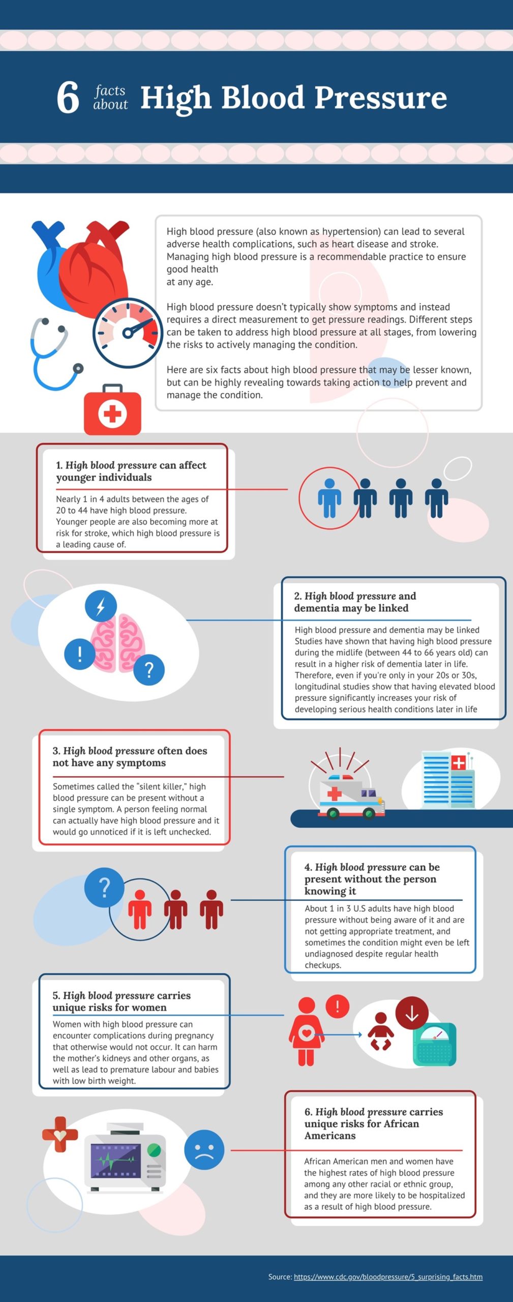
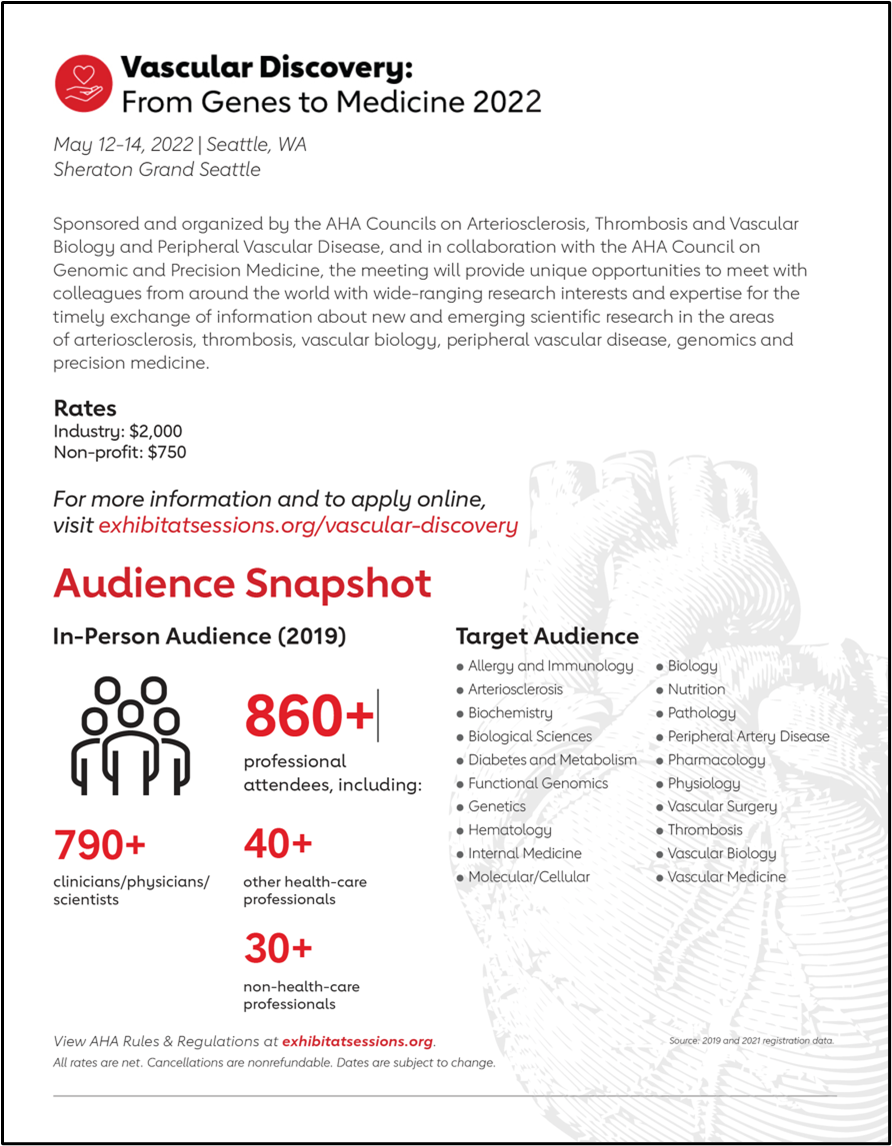
You might want to check out Vascular Discovery 2022, a 2 ½-day meeting, which is sponsored by the Council on Arteriosclerosis, Thrombosis and Vascular Biology, the Peripheral Vascular Disease Council, and the Council on Genomic and Precision Medicine, in cooperation with and the Society for Vascular Surgery’s Vascular Research Initiatives Conference (Flyer on the right).

What is the conference about?

The primary goal of the Vascular Discovery: From Genes to Medicine Scientific Sessions is to provide a forum for the timely exchange of information about new and emerging scientific research in lipids and lipoproteins, arteriosclerosis, thrombosis, vascular biology, genomics, precision medicine, peripheral vascular disease, and vascular surgery.

One of the long-standing objectives of this conference is to provide a unique platform for colleagues to build valuable networks, establish potential collaborations and promote early career scientists via providing role model inspirations and mentorship opportunities. The advantages of direct social interactions are indisputable, especially when it comes to build meaningful relationships. This year’s Vascular Discovery will be held in-person in Seattle. It will bring colleagues from worldwide, share groundbreaking science and bridge different disciplines in a classic old fashion way.

Who should attend?

This event will appeal to scientists and clinicians in cardiovascular medicine, cardiovascular research, thrombosis research, clinical cardiology, molecular/cellular biology, vascular biology, vascular medicine, vascular surgery, endocrinology, genetics, functional genomics, hematology, immunology, nutrition, and physiology.

Past highlights and feature events

While the planning committee is finalizing the abstracts and awardees, we can take a glimpse of what happened in 2021, which will shed some lights into the 2022 scientistic sessions. Vascular Discovery presented a virtual experience to the attendees in 2021, because of COVID pandemics. Since COVID has been the primary attention in public and scientific communities the past couple years, one of the important focuses of Vascular Discovery 2021 is on understanding how COVID-19 affects vascular systems, to identify who and when to treat patients in vascular research and clinical trials. Other important discussions are on identifying new approaches to understand the pathophysiology of atherosclerosis via risk factor identifications, and a distinguished lecture was presented by Marlene Rabinovitch, M.D. on multiple approaches of identifying a therapeutic target for treating pulmonary hypertension. More highlights are shown in the American Heart Association website1.

Are you excited about attending this year’s in-person Vascular Discovery: from Genes to Medicine Scientific Sessions in Seattle? Stay tuned on the latest programming of 2022, which hopefully will be finalized and released in early March.

Registration for the Vascular Discovery Conference is now open. Early Bird pricing closes on 3/31, and Advance pricing opens on 4/1.  Register now for the best price!

REFERENCE

1. American Heart Association. Science News 2021. https://professional.heart.org/en/meetings/vascular-discovery-from-genes

“The views, opinions, and positions expressed within this blog are those of the author(s) alone and do not represent those of the American Heart Association. The accuracy, completeness, and validity of any statements made within this article are not guaranteed. We accept no liability for any errors, omissions, or representations. The copyright of this content belongs to the author and any liability with regards to infringement of intellectual property rights remains with them. The Early Career Voice blog is not intended to provide medical advice or treatment. Only your healthcare provider can provide that. The American Heart Association recommends that you consult your healthcare provider regarding your health matters. If you think you are having a heart attack, stroke, or another emergency, please call 911 immediately.”

hidden

Insights About COVID-19 Health Outcomes in Smokers from Hospital Records

Early in the COVID-19 pandemic, clinicians noticed that some patients with pre-existing medical conditions were at higher risk of severe illness and death. Since then, many observational studies confirmed that people with diabetes, asthma, or cardiovascular disease had a substantially higher risk of COVID-19-related complications and death1,2. Such studies typically use hospital patient records to study relationships between individual risk factors, like age and sex, and long-term health outcomes. However, studies using hospital record data revealed that smokers may be underrepresented among patients with COVID-19, as highlighted by a recent review study of publications reporting smoking prevalence and clinical outcomes in patients diagnosed with COVID-193. The review highlights that only a single study out of 15 research articles examined had reported a prevalence of smokers among patients with COVID-19 that resembles the smoking prevalence of the general population. Such findings, raises the question of whether a smoker’s paradox exists with the COVID-19 pandemic. The smoker’s paradox refers to the observational phenomenon of smokers exhibiting improved prognosis and decreased short-term mortality, following cardiovascular events. This idea has been mostly debunked now as a bias in the analysis of observational data and smoking is not considered to be a causative factor that improves health outcomes4. So, what considerations should be taken when interpreting health records of patients in order understand whether smokers fare worse COVID-19 outcomes than non-smokers?

Questioning Data Quality and Biases in Patient Record Data

Smoking is associated with many immediate and long-term health consequences, and initiates disease promoting mechanism in cardiopulmonary tissues. A low representation of smokers in hospitalized COVID-19 patients may be due to biases in patient record data collection. A small percentage of people who perceive stigma associated with smoker status may actually conceal smoker status during a primary care visit5. Furthermore, smoker screening often does not include questions about smokeless tobacco, electronic cigarette use, and second-hand smoke use, despite the rise in popularity in electronic nicotine delivery systems and cigarette alternatives6. Standardization and improved tobacco-related electronic health record questionnaires may begin to address the question of how much tobacco smoke a person is exposed to by including questions that cover sources of exposure, quantity of use, and duration of exposure. Collecting patient data that covers a larger range of exposure possibilities including having someone else in the home that smokers, or individual behaviors of switching from traditional cigarettes to electronic cigarettes, smoking cessation patterns, and years of use may provide better insight into how smoking behaviors influence health outcomes. In the context of the COVID-19 pandemic, smokers’ status is difficult to ascertain in patients who are intubated, sedated, and unresponsive. Closely tracking individual smoker status over time is helpful in those situations when a patient’s care plan should include tobacco withdrawal symptom management.

Smoking directly influences cardiovascular and respiratory health outcomes and using hospital data to derive associations with COVID-19 health outcomes is prone to confounding bias, reverse causation, and inappropriate adjustments in analysis models. In the future, it be possible to use human biomarkers to uncover the specific health effects from smoking. This might include correlating urinary levels of nicotine and cotinine to understand the burden of different tobacco products. For now, research using animal models to assess the health effects of traditional smoking and electronic cigarettes provide insight into the short- and long-term consequences of smoking and elucidates the biochemical processes that exacerbate disease including tissue repair processes, inflammation, and oxidative stress. Understanding the mechanistic processes involved in the exacerbation of COVID-19 disease among smokers may ultimately help identify biomarkers of disease progression and pharmacological treatments for vulnerable populations.

References:

  1. Williamson EJ, Walker AJ, Bhaskaran K, et al. Factors associated with COVID-19-related death using OpenSAFELY. Nature. 2020;584(7821):430-436. doi:1038/s41586-020-2521-4
  2. Why lighting up and COVID-19 don’t mix. American Heart Association News. https://www.heart.org/en/news/2020/05/05/why-lighting-up-and-covid-19-dont-mix#:~:text=Studies%20from%20Wuhan%2C%20China%2C%20where,%2C%20compared%20to%20non%2Dsmokers.
  3. Usman MS, Siddiqi TJ, Khan MS, et al. Is there a smoker’s paradox in COVID-19? BMJ EBM. 2021;26(6):279-284. doi:1136/bmjebm-2020-111492
  4. Doi SA, Islam N, Sulaiman K, et al. Demystifying Smoker’s Paradox: A Propensity Score–Weighted Analysis in Patients Hospitalized With Acute Heart Failure. JAHA. 2019;8(23). https://www.ahajournals.org/doi/10.1161/JAHA.119.013056
  5. Stuber J, Galea S. Who conceals their smoking status from their health care provider? Nicotine & Tobacco Research. 2009;11(3):303-307. doi:1093/ntr/ntn024
  6. LeLaurin JH, Theis RP, Thompson LA, et al. Tobacco-Related Counseling and Documentation in Adolescent Primary Care Practice: Challenges and Opportunities. Nicotine & Tobacco Research. 2020;22(6):1023-1029. doi:1093/ntr/ntz076

“The views, opinions, and positions expressed within this blog are those of the author(s) alone and do not represent those of the American Heart Association. The accuracy, completeness, and validity of any statements made within this article are not guaranteed. We accept no liability for any errors, omissions, or representations. The copyright of this content belongs to the author and any liability with regards to infringement of intellectual property rights remains with them. The Early Career Voice blog is not intended to provide medical advice or treatment. Only your healthcare provider can provide that. The American Heart Association recommends that you consult your healthcare provider regarding your health matters. If you think you are having a heart attack, stroke, or another emergency, please call 911 immediately.”