hidden

An Interview with Dr. Ilana Kutinsky- Electrophysiologist and Cardiologist of the Apes

As a resident at Beaumont Hospital, I have the good fortune of working alongside some of the finest cardiologists in the country. I am constantly inspired by talent, innovation, and excellence in patient care. Dr. Ilana Kutinsky, associate professor at Oakland University William Beaumont School of Medicine, takes cardiology and commitment to health equity a step further by providing care to apes throughout the country.

Cardiovascular diseases are the leading cause of mortality not only in humans but also in the great ape species, including gorillas, chimpanzees, orangutans, and bonobos. Established in 2010, the Great Ape Heart Project (GAHP) aims to improve the understanding and management of cardiac disease in our cousin species. A cardiac electrophysiologist by training and profession, Dr. Kutinsky is one of the founders of the Great Ape Heart Project. I sat down with Dr. Kutinsky to learn more about her role as ‘cardiologist of the apes’.

DA: How did you get involved with the Great Ape Heart Project?

Dr. Kutinsky: Around 2000, I was a cardiology fellow at the University of Colorado. During my echo rotation, I picked up a call sitting in the darkroom. “We’re calling from the Denver Zoo. We need someone to come and do TEEs on our gorillas and orangutans. Would you be interested?” and I said “What?”. I talked to my attending and we decided to do it. So, we had Phillips loan us a giant TEE machine and bring it to the zoo. They anesthetized the gorilla and we did the TEE. At the time they were just realizing that captive gorillas die from heart diseases and there was no reference to what is normal. The next month, a female orangutan died from extremis from the anesthesia for the TEE. It was disconcerting to see an animal die for a test we did not even know how to interpret. I got in touch with Dr. Hayley Murphy, who had written a paper reporting echo findings from five gorillas in Boston. We shared our frustration and decided to write a grant to try to collect echos over the country and establish a normal. The grant was rejected but I continued to record readings on my own time by taking freehand measurements on VHS tapes. This is how the Gorilla Heart Project got started. Then it got bigger! We wrote a grant to the Institute of Museum and Library Sciences (IMLS) and got a half a million-dollar grant to set up the Great Ape Heart Project. We were able to hire primatologists, pathologists, veterinarians, and sonographers and set up a database. We have been able to establish normal, identify high-risk animals, and treat heart failure in gorillas. We have consulted on cases not only in the United States but all over the world. It’s a wonderful, although busy hobby for me!

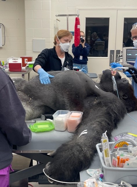
DA: How was your first experience as a cardiologist for the apes, the gorilla in Denver you did the TEE for?

Dr. Kutinsky: It was very similar to taking care of a human once the probe was inside the body. But the animals are enormous, between 350 to 580  pounds. Their chests are like barrels and their skulls are ginormous. To get the TEE probe down their mouths, you have to go past their incisors which are almost the size of my hand.

DA: Were you scared?

Dr. Kutinsky: No, you know they are under anesthesia. And someone is standing in the corner with a big gun. So, if they start to wake up, they will be immobilized. I wasn’t scared because I was so in awe that I was able to do something so amazingly cool. I remember calling my mom afterwards and telling her that every night I spent studying or being on call, all the effort made to get into medical school and cardiology was worth it that day.

DA: Were your co-fellows jealous of you?

Dr. Kutinsky: That’s a good question. Um, I think they thought I was crazy! Doing all that extra work on top of a busy fellowship schedule did not make sense to them.

DA: How is it working with a team of so many disciplines?

Dr. Kutinsky: It is amazing to work with a team of veterinarians, human cardiologists, veterinary cardiologists, sonographers, and pathologists. The vets are honestly some of the smartest people I know. They take care of a 500-pound gorilla with heart disease and then they attend to a tiny bird with an eye injury. Their breadth of knowledge is extraordinary. It has been incredibly rewarding experience working with everyone.

DA: What’s been your most interesting experience being a cardiologist for the apes?

Dr. Kutinsky: Going to Cameroon was one of my most interesting experiences. We were invited by the government of Cameroon to a sanctuary of rescued wild gorillas. We were able to see them up close and interact with them from across a mesh. We were there for 2 weeks, seeing them every day and feeling a connection with them. Usually, I don’t get called when they’re doing well. I’m called when they are sick and about to die, so they’re usually anesthetized. To see them awake, eating and going about their usual life was pretty cool.

DA: Do you have a favorite of the great ape species?

Dr. Kutinsky: Gorillas for sure. I like orangutans too but we don’t have any orangutans in the Detroit zoo so I don’t see them much. Chimpanzees are very naughty and a handful to work with.

DA: Have you become attached to any animal?

Dr. Kutinsky: I was very attached to Sunshine, a gorilla in the Detroit Zoo. He was an old, great animal with a bad heart. He died from influenza a few years ago. I was pretty tight with him. I like Mac in Columbus. He is a very handsome boy. There’s Tatu in Omaha, he’s a good boy. I’ll be sad when they die. Gorillas usually don’t live past 40 years and a lot of the ones with heart disease drop dead early. So we’re hoping to change that.

DA: Do you prefer treating humans or gorillas?

Dr. Kutinsky: Depends on the day! I love animals but I cherish the relationships with my patients. I love my job as an electrophysiologist. Everything I’ve learned as a human doctor I use to treat the gorillas. But if I won the lottery, I would probably devote all my time to the gorillas.

DA: Last question, has taken care of gorillas and other apes affected how you treat your patients? 

Dr. Kutinsky: That’s a great question. Hmm, I guess treating animals has made me a more compassionate doctor and person in general. Since the animals can’t complain or whine, you give them symptoms in your mind. You assume they are hurting or sad, and you try to take care of them. I think it has given me an extra bit of sympathy when I take care of my patients. And it makes me happier in general. When you have a passion, it enriches your whole life.

DA: I guess it was a lucky call that day in the echo lab.

Dr. Kutinsky: Absolutely! I was in the right place at the right time. I also think you have to be open to the universe and do things that are outside the box. I was willing to do the extra work outside of a busy fellowship. I was offered something special and I took it.


“The views, opinions, and positions expressed within this blog are those of the author(s) alone and do not represent those of the American Heart Association. The accuracy, completeness, and validity of any statements made within this article are not guaranteed. We accept no liability for any errors, omissions, or representations. The copyright of this content belongs to the author and any liability with regards to infringement of intellectual property rights remains with them. The Early Career Voice blog is not intended to provide medical advice or treatment. Only your healthcare provider can provide that. The American Heart Association recommends that you consult your healthcare provider regarding your health matters. If you think you are having a heart attack, stroke, or another emergency, please call 911 immediately.”

hidden

Taking the Guesswork out of HFpEF

With an aging population and a higher burden of comorbidities, the proportion of heart failure patients with a preserved ejection fraction, i.e. ejection fraction ≥ 50% is increasing.1 Heart failure with preserved ejection fraction (HFpEF) now accounts for more than half of all heart failure hospitalizations. Despite the increasing prevalence, HFpEF remains a nebulous entity. HFpEF is often alluded to without a complete understanding of the underlying pathophysiology. Diagnosing HFpEF can be challenging as opposed to heart failure with reduced ejection fraction (HFrEF). With a normal ejection fraction, attributing dyspnea to cardiac congestion without performing invasive hemodynamic testing requires good clinical suspicion and judgment. Moreover, euvolemic patients with compensated HFpEF can have elevated filling pressures and dyspnea only with exertion. Non-invasive measurement of cardiac pressures can be inconclusive and invasive cardiopulmonary exercise testing (CPET) is considered the gold standard in this population.2 However its routine use is not feasible as it is an invasive and technically complex procedure with limited availability. An algorithm incorporating clinical and non-invasive parameters can help stratify patients’ probability of having HFpEF. The utility of risk scores, such as the CHA2DS2-VASc, TIMI, and Wells’ scores, is well established in the field of cardiovascular disease. There has been research into developing similar algorithms/ prediction scores for the diagnosis of HFpEF. Here, we discuss 2 proposed scoring systems for HFpEF- H2FPEF and HFA-PEFF.

H2FPEF Score

Reddy and colleagues at Mayo Clinic, Rochester developed the H2FPEF score to help physicians discriminate HFpEF from non-cardiac causes of dyspnea in symptomatic patients without obvious fluid overload.3 The score was developed from a cohort of 414 patients with an ejection fraction ≥ 50%, who underwent invasive hemodynamic exercise testing for definitive evaluation of unexplained dyspnea. Different clinical and echocardiographic markers were evaluated through logistic regression to identify variables associated with HFpEF. Ultimately 6 routinely available variables (BMI > 30 kg/m2, atrial fibrillation, hypertension treated with ≥ 2 medications, pulmonary artery systolic pressure > 35 mmHg, age > 60 years, and E/e’ > 9) were used for the model. Each variable was assigned a point based on the strength of association observed with HFpEF diagnosed via invasive testing (Figure 1). The final score had good discriminatory power (area under the curve = 0.84) for differentiating HFpEF from other causes of dyspnea. As the score increased from 0 to 9, so did the probability of HFpEF. The robustness of the model was validated through sensitivity analyses and a test cohort of 100 patients. The authors proposed a Bayesian approach- using a low score (0-1) to rule out HFpEF, a high score (6-9) to make a diagnosis of HFpEF, and an intermediate score (2-5) to consider additional testing.

The major limitation of this important model is the setting of the study. It was conducted at a single institute serving as a referral center, which may not truly represent the general population. It is reassuring that the score has been validated in small external cohorts.4,5 Moreover, an analysis from the TOPCAT trial population showed that patients with a higher H2FPEF score had an increased risk of adverse outcomes, suggesting a prognostic value of the score.6

Figure 1. H2FPEF Score proposed by Reddy et al. https://doi.org/10.1161/CIRCULATIONAHA.118.034646

HFA-PEFF Score

In 2020, the Heart Failure Association (HFA) and the European Society of Cardiology (ESC) released a consensus recommendation for diagnosing HFpEF, proposing the stepwise HFA-PEFF algorithm.7 This recommendation centers around the use of the HFA-PEFF score. The proposed scoring system uses echocardiographic parameters and natriuretic peptide (BNP and NT-proBNP) levels. The variables are divided into major and minor criteria across 3 domains- functional, morphological, and biomarker (Figure 2). Parameters within a domain are not additive, hence the score can be used even when certain values are not available. Each domain can contribute a maximum of 2 points and the total score ranges from 0 to 6. A total score of 5-6 is considered diagnostic of HFpEF while a score of 0-1 makes HFpEF unlikely. An intermediate score of 2-4 warrants further testing with non-invasive or invasive functional testing.

Notably, the HFA-PEFF score did not utilize demographic and clinical parameters and the power of the score was not assessed. However, an independent study later demonstrated its validity in two separate cohorts.8

Figure 2. HFA-PEFF Score proposed by HFA and ESC. https://doi.org/10.1002/ejhf.1741

Both of the above scoring systems have been received with enthusiasm, given the lack of a clear definition and diagnostic framework for HFpEF. Studies evaluating the 2 scores have also been published. Parcha and colleagues studied the generalizability of the H2FPEF and HFA-PEFF scores in an analysis of participants with unexplained dyspnea from prior HFpEF trials and the Atherosclerosis Risk in Communities (ARIC) study.9 They found that both the scores could rule out HFpEF with a greater than 99% success rate but the H2FPEF score had a higher specificity than the HFA-PEFF score. Amanai and colleagues calculated H2FPEF and HFA-PEFF scores in patients with HFpEF referred for stress echocardiography.10 They found that both scores had similarly high positive and negative predictive values and a correlation with abnormal hemodynamics during exercise. Another study in the ARIC population also found that both high H2PEFF and HFA-PEFF scores were associated with increased risk of heart failure hospitalizations or death, suggesting the prognostic value of both.11

Both H2FPEF and HFA-PEFF are validated and easy-to-use scores using readily available clinical, laboratory, and echocardiographic parameters. The use of these scores in the appropriate patient and context can aid in the timely and accurate diagnosis of HFpEF. Growing recognition and emergence of effective therapies such as SGLT2 inhibitors are important strides for improving outcomes for patients with HFpEF.

References:

  1. Borlaug BA. Evaluation and management of heart failure with preserved ejection fraction. Nat Rev Cardiol. 2020;17(9):559-573. doi:10.1038/s41569-020-0363-2
  2. Sorajja P, Borlaug BA, Dimas VV, et al. SCAI/HFSA clinical expert consensus document on the use of invasive hemodynamics for the diagnosis and management of cardiovascular disease. Catheter Cardiovasc Interv. 2017;89(7):E233-E247. doi:10.1002/ccd.26888
  3. Reddy YNV, Carter RE, Obokata M, Redfield MM, Borlaug BA. A Simple, Evidence-Based Approach to Help Guide Diagnosis of Heart Failure With Preserved Ejection Fraction. Circulation. 2018;138(9):861-870. doi:10.1161/CIRCULATIONAHA.118.034646
  4. Sepehrvand N, Alemayehu W, Dyck GJB, et al. External Validation of the H2F-PEF Model in Diagnosing Patients With Heart Failure and Preserved Ejection Fraction. Circulation. 2019;139(20):2377-2379. doi:10.1161/CIRCULATIONAHA.118.038594
  5. Segar MW, Patel KV, Berry JD, Grodin JL, Pandey A. Generalizability and Implications of the H2FPEF Score in a Cohort of Patients With Heart Failure With Preserved Ejection Fraction. Circulation. 2019;139(15):1851-1853. doi:10.1161/CIRCULATIONAHA.118.039051
  6. Myhre PL, Vaduganathan M, Claggett BL, et al. Application of the H2 FPEF score to a global clinical trial of patients with heart failure with preserved ejection fraction: the TOPCAT trial. Eur J Heart Fail. 2019;21(10):1288-1291. doi:10.1002/ejhf.1542
  7. Pieske B, Tschöpe C, de Boer RA, et al. How to diagnose heart failure with preserved ejection fraction: the HFA-PEFF diagnostic algorithm: a consensus recommendation from the Heart Failure Association (HFA) of the European Society of Cardiology (ESC). Eur J Heart Fail. 2020;22(3):391-412. doi:10.1002/ejhf.1741
  8. Barandiarán Aizpurua A, Sanders-van Wijk S, Brunner-La Rocca HP, et al. Validation of the HFA-PEFF score for the diagnosis of heart failure with preserved ejection fraction. Eur J Heart Fail. 2020;22(3):413-421. doi:10.1002/ejhf.1614
  9. Parcha V, Malla G, Kalra R, et al. Diagnostic and prognostic implications of heart failure with preserved ejection fraction scoring systems. ESC Heart Fail. 2021;8(3):2089-2102. doi:10.1002/ehf2.13288
  10. Amanai S, Harada T, Kagami K, et al. The H2FPEF and HFA-PEFF algorithms for predicting exercise intolerance and abnormal hemodynamics in heart failure with preserved ejection fraction. Sci Rep. 2022;12(1):13. doi:10.1038/s41598-021-03974-6
  11. Selvaraj S, Myhre PL, Vaduganathan M, et al. Application of Diagnostic Algorithms for Heart Failure With Preserved Ejection Fraction to the Community. JACC Heart Fail. 2020;8(8):640-653. doi:10.1016/j.jchf.2020.03.013

“The views, opinions, and positions expressed within this blog are those of the author(s) alone and do not represent those of the American Heart Association. The accuracy, completeness, and validity of any statements made within this article are not guaranteed. We accept no liability for any errors, omissions, or representations. The copyright of this content belongs to the author and any liability with regards to infringement of intellectual property rights remains with them. The Early Career Voice blog is not intended to provide medical advice or treatment. Only your healthcare provider can provide that. The American Heart Association recommends that you consult your healthcare provider regarding your health matters. If you think you are having a heart attack, stroke, or another emergency, please call 911 immediately.”

hidden

PCSK9: From Bench to Bedside

(Image by sinclair.sharon28 from Creative Commons)

The proatherogenic role of low-density lipoprotein (LDL) cholesterol is well established and reduction of LDL cholesterol is one of the central pillars of management and prevention of atherosclerotic cardiovascular disease. The prevention of atherosclerotic events directly correlates with the reduction in LDL cholesterol and growing evidence supports the notion that the lower the LDL, the better the outcome.1 Statins are the backbone of lipid-lowering therapy and are indispensable for primary and secondary prevention. Despite trials showing no significant difference versus placebo in terms of serious adverse events, statin intolerance is commonly encountered in clinical practice.2 Additionally, patients on intensive statin therapy can have significant residual risk that can be minimized by additional lipid-lowering interventions.3 Hence, there was an urgent need for new strategies to lower LDL cholesterol levels.

In 2003, Abifadel and colleagues first reported a gain-of-function mutation in the proprotein convertase subtilisin/kexin type 9 (PCSK9) gene in a French family with autosomal dominant hypercholesterolemia, 4 a condition associated with significantly increased LDL cholesterol levels and risk of cardiovascular disease. Studies in mice subsequently delineated the underlying pathway: the PCSK9 gene encodes its namesake protein produced primarily in the liver.5 This protein binds to LDL receptors on the surface of hepatocytes, leading to their internalization and degradation. Reduced uptake of LDL particles by the hepatocytes leads to higher circulating levels of LDL cholesterol, increasing the risk of atherogenesis. Similar mechanisms are reported in humans. An analysis from the longitudinal Atherosclerosis Risk in Communities (ARIC) database showed that genetic variations in PCSK9 associated with lower levels of LDL cholesterol attenuated the risk of coronary heart disease.6 Studies also showed a reduction in triglyceride levels in patients with loss-of-function of PCSK9.7

These findings inspired the research and development of therapies targeting the PCSK9 pathway to achieve a reduction in LDL cholesterol levels. In 2009, Chan and colleagues developed a neutralizing antibody to PCSK9 that showed a significant reduction in LDL cholesterol in mice and monkeys.8 Within a decade of the discovery of the PCSK9 pathway, the race for human trials was on. In 2012, REGN727, a human monoclonal antibody, now known as alirocumab (Praluent®, by Regeneron and Sanofi) was shown to safely reduce LDL cholesterol levels.9  Similar results were seen with AMG145, later named evolocumab (Repatha®, by Amgen).10 Additional studies confirmed the ability of the two drugs to reduce LDL cholesterol by upto 60% without any serious adverse events. This led to FDA approval for both alirocumab and evolocumab as adjunct therapies to reduce LDL cholesterol in patients with (1) familial hypercholesterolemia and (2) established cardiovascular disease in 2015. Importantly, large randomized placebo-controlled trials, including the FOURIER and ODYSSEY trials, have shown remarkable reductions in the incidence of the composite clinical outcome of cardiovascular death, myocardial infarction, stroke, and hospitalization for acute coronary syndrome.11,12 Initial concerns of neurocognitive impairment with PCSK9 inhibitors have been allayed. The clinical use of PCSK9 inhibitors has been reassuring in terms of both safety and efficacy. The primary limitations to their use are high cost and the need for twice weekly or monthly subcutaneous injections. While the reduction in annual costs from around $14,000 to $5,800 is encouraging, high co-pays and frequent need for prior authorization continue to prevent many patients from reaping the benefits of PCSK9 inhibitors.

The recent FDA approval of inclisiran (Leqvio®, Novartis) was met with enthusiasm throughout the medical community, and rightly so. Inclisiran is a novel small interfering RNA (siRNA) based therapy that blocks the synthesis of PCSK9 from the liver. The ORION trials showed a 50% reduction in LDL cholesterol levels as compared to placebo in patients with atherosclerotic cardiovascular disease on maximally tolerated statin therapy.13 A major advantage of inclisiran is the biannual dosing as compared with the every 2-4 week dosing with PCSK9 inhibitors. Novartis, the company behind the drug is proposing a ‘buy-and-bill’ model where the drug will be administered in the office, which will likely improve adherence.14 The convenience factor might just provide inclisiran leverage over existing therapies. Many real-world challenges still need to be addressed including long-term safety data and ensuring cost-effective and equitable distribution of this potentially life-saving drug. Despite these concerns, the approval of inclisiran is an important landmark in the field of cardiovascular disease prevention. The trajectory of PCSK9 from a newly discovered molecule to a novel siRNA-based therapy in 20 years is inspiring and reinforces the importance of basic science and translational research.\

References:

  1. Koskinas KC, Siontis GCM, Piccolo R, et al. Effect of statins and non-statin LDL-lowering medications on cardiovascular outcomes in secondary prevention: a meta-analysis of randomized trials. Eur Heart J. 2018;39(14):1172-1180. doi:10.1093/eurheartj/ehx566
  2. Tobert JA, Newman CB. Statin tolerability: In defence of placebo-controlled trials. Eur J Prev Cardiol. 2016;23(8):891-896. doi:10.1177/2047487315602861
  3. Reith C, Armitage J. Management of residual risk after statin therapy. Atherosclerosis. 2016;245:161-170. doi:10.1016/j.atherosclerosis.2015.12.018
  4. Abifadel M, Varret M, Rabès JP, et al. Mutations in PCSK9 cause autosomal dominant hypercholesterolemia. Nat Genet. 2003;34(2):154-156. doi:10.1038/ng1161
  5. Wiciński M, Żak J, Malinowski B, Popek G, Grześk G. PCSK9 signaling pathways and their potential importance in clinical practice. EPMA J. 2017;8(4):391-402. doi:10.1007/s13167-017-0106-6
  6. Cohen JC, Boerwinkle E, Mosley TH, Hobbs HH. Sequence variations in PCSK9, low LDL, and protection against coronary heart disease. N Engl J Med. 2006;354(12):1264-1272. doi:10.1056/NEJMoa054013
  7. Handelsman Y, Lepor NE. PCSK9 Inhibitors in Lipid Management of Patients With Diabetes Mellitus and High Cardiovascular Risk: A Review. J Am Heart Assoc. 2018;7(13):e008953. doi:10.1161/JAHA.118.008953
  8. Chan JCY, Piper DE, Cao Q, et al. A proprotein convertase subtilisin/kexin type 9 neutralizing antibody reduces serum cholesterol in mice and nonhuman primates. Proc Natl Acad Sci U S A. 2009;106(24):9820-9825. doi:10.1073/pnas.0903849106
  9. Stein EA, Mellis S, Yancopoulos GD, et al. Effect of a monoclonal antibody to PCSK9 on LDL cholesterol. N Engl J Med. 2012;366(12):1108-1118. doi:10.1056/NEJMoa1105803
  10. Dias CS, Shaywitz AJ, Wasserman SM, et al. Effects of AMG 145 on low-density lipoprotein cholesterol levels: results from 2 randomized, double-blind, placebo-controlled, ascending-dose phase 1 studies in healthy volunteers and hypercholesterolemic subjects on statins. J Am Coll Cardiol. 2012;60(19):1888-1898. doi:10.1016/j.jacc.2012.08.986
  11. Sabatine MS, Giugliano RP, Keech AC, et al. Evolocumab and Clinical Outcomes in Patients with Cardiovascular Disease. N Engl J Med. 2017;376(18):1713-1722. doi:10.1056/NEJMoa1615664
  12. Schwartz GG, Steg PG, Szarek M, et al. Alirocumab and Cardiovascular Outcomes after Acute Coronary Syndrome. N Engl J Med. 2018;379(22):2097-2107. doi:10.1056/NEJMoa1801174
  13. Ray KK, Wright RS, Kallend D, et al. Two Phase 3 Trials of Inclisiran in Patients with Elevated LDL Cholesterol. N Engl J Med. 2020;382(16):1507-1519. doi:10.1056/NEJMoa1912387
  14. Pricey Inclisiran Is Rolling Out: a ‘Buy-and-Bill’ Model May Smooth Its Path. TCTMD.com. Accessed February 3, 2022. https://www.tctmd.com/news/pricey-inclisiran-rolling-out-buy-and-bill-model-may-smooth-its-path

“The views, opinions and positions expressed within this blog are those of the author(s) alone and do not represent those of the American Heart Association. The accuracy, completeness and validity of any statements made within this article are not guaranteed. We accept no liability for any errors, omissions or representations. The copyright of this content belongs to the author and any liability with regards to infringement of intellectual property rights remains with them. The Early Career Voice blog is not intended to provide medical advice or treatment. Only your healthcare provider can provide that. The American Heart Association recommends that you consult your healthcare provider regarding your personal health matters. If you think you are having a heart attack, stroke or another emergency, please call 911 immediately.”


hidden

A Reaffirmation on Medical Conferences

It is safe to say that AHA Scientific Sessions 2021, conducted virtually from November 13th to 15th was a big hit. The ease and flexibility of attending the Sessions from the comfort of my couch made me forget my initial concerns of missing out on the in-person experience. It will be interesting to see how the pandemic, and seeping of Zoom into our lives, change the future of medical conferences. Regardless of time and place, conferences will continue to be an important feature in medicine, especially in a constantly evolving field like cardiology. Here I discuss some of the reasons that drive us to attend medical conferences, and why we should continue to do so.

(Image from Creative Commons)

Present your work

Disseminating your research can be as important as the scientific process itself. Conferences are an excellent forum to present your findings in a timely manner, especially since publication can be a very long-drawn-out process. Presenting abstracts at conferences helps trainees build their CVs to reflect their interests and scholarly work. Although it can be nerve-wracking, presenting at conferences forces you to communicate about your work effectively. Addressing questions from experts and incorporating the feedback received can help with framing the manuscript for publication.

Keep up-to-date with the latest science

Undoubtedly the release of new study data creates the loudest buzz during medical conferences. Audiences gather to witness the release of trials that will influence clinical practice. You have the opportunity to interact with the investigators and address questions relevant to you and your patients. When not in the late-breaking sessions, there are poster/ presentation sessions to attend. You can also learn about the latest innovations from the industry and try your hand at new technologies showcased by vendors.

Get inspired

Even when not presenting, attending scientific conferences is an active process. Reviewing the latest trials and abstracts promotes critical thinking and sparks ideas. You notice the gaps in knowledge and may be inspired to address that in your research. You can learn from others’ successes and mistakes.

Learn/ refresh your knowledge

Medical conferences are a good place to catch up on the existing knowledge and earn CME points. Most conferences have workshops or skills-training sessions incorporated within the schedule. Lectures from distinguished speakers can provide you with a high-level review of a topic. Panel discussions bring together opinions from trainees, established clinicians, and researchers and can give a sense of what the coming years will bring to the field.

Form connections

Networking has become crucial for a career in medicine, whether in academia or private practice. Conferences are arguably the best place to establish and foster contacts with people at different stages of their careers. You have the opportunity to meet experts who you look up to, introduce yourself, and take advice. Your name might stick with potential employers, increasing your chances of landing a position in the future. For trainees, it may translate into gaining lifelong mentors or sponsors. Conferences are also a good place to meet peers, share ideas and experiences, and potentially set up collaborations.

Share the passion

Finally, there is a special joy in nerding along with others who nurture the same passion for medicine as you. People from different countries and at varying stages of their careers are brought together during the conferences. Knowing that there is a whole community of people with similar interests can motivate you for long after the conference is over.

All in all, attendees come out of a conference smarter, inspired, and excited for the next conference.

Below is a list of the upcoming major conferences in Cardiology. Hope to see you there!


“The views, opinions and positions expressed within this blog are those of the author(s) alone and do not represent those of the American Heart Association. The accuracy, completeness and validity of any statements made within this article are not guaranteed. We accept no liability for any errors, omissions or representations. The copyright of this content belongs to the author and any liability with regards to infringement of intellectual property rights remains with them. The Early Career Voice blog is not intended to provide medical advice or treatment. Only your healthcare provider can provide that. The American Heart Association recommends that you consult your healthcare provider regarding your personal health matters. If you think you are having a heart attack, stroke or another emergency, please call 911 immediately.”

hidden

BREAKING: Trials From the Realm of Cardiac Electrophysiology to Look Forward to in AHA Scientific Sessions 2021

The most anticipated cardiovascular conference of the year, AHA Scientific Sessions 2021 is upon us! In the days and hours leading upto the event, speakers have been revising their presentations, moderators going over their notes, and organizers working hard to prevent any glitches for this fully virtual experience. For many attendees, including myself, the most exciting part of the conference is the release of new trial data that may have a bearing on clinical practice and set the direction for future research. This year’s program is heavily ‘charged’ with incredible science in the field of cardiac electrophysiology. Below is a short guide to the studies from the field of electrophysiology being presented in the late breaking science (LBS) sessions.

CRAVE Trial- The Coffee and Real-time Atrial and Ventricular Ectopy (CRAVE) Trial

Presented by Gregory Marcus, MD

LBS.03 Prevention to Intervention in Atrial Arrhythmias, Sunday Nov 14th 8-9 AM EST

Many people report experiencing palpitations with higher caffeine intake and cutting down on coffee is a common advice heard in EP clinics. However, this anecdotal link is not backed by evidence with large population studies showing no association between caffeine intake and arrhythmias.1 Hence, the investigators of the CRAVE trial set out to investigate the effect of caffeine intake on cardiac ectopy.2 Based in University of California, San Francisco, this N-of-1 trial enrolled healthy volunteers and assigned them to 2-day blocks of coffee on-and-off days for 2 weeks. Continuous heart monitors were used to assess the primary outcome i.e., premature atrial and ventricular contractions. The results of this study will be interesting and may finally provide an answer to the important question- to bean or not to bean?

GIRAF Trial- Dabigatran Versus Warfarin on Cognitive Outcomes in Nonvalvular Atrial Fibrillation: Results of the GIRAF Trial

Presented by Bruno Caramelli, MD

LBS.03 Prevention to Intervention in Atrial Arrhythmias, Sunday Nov 14th 8-9 AM EST

Prevention of stroke is one of the pillars of management of atrial fibrillation (AF). However, even in the absence of stroke, patients with underlying AF have an increased risk of cognitive decline and dementia.3 Direct oral anticoagulants (DOACs) such as dabigatran have less food and drug interactions and better safety profiles compared to warfarin and are now considered the preferred agents for long-term stroke prevention in non-valvular AF. However, whether this consistent and stable anticoagulant effect offered by DOACs compared to warfarin translates into prevention or progression of dementia in the elderly patient with AF remains unknown. Investigators of the GIRAF trial conducted a prospective randomized controlled trial (N=200) comparing the effects of dabitran with warfarin on cognitive and functional outcomes at 2 years in an elderly population with AF.4 Clinicians from different fields will be craning their necks for the results of this important trial.

PALACS- Posterior Left Pericardiotomy Reduces Postoperative Atrial Fibrillation After Cardiac Surgery

Presented by Mario F Gaudino, MD

LBS.03 Prevention to Intervention in Atrial Arrhythmias, Sunday Nov 14th 8-9 AM EST

Post-operative AF plagues one-third of patients after cardiac surgery and is associated with longer hospital stays, increased risk of stroke, and higher mortality.5 Drainage of the pericardial cavity into the left pleural place by performing a posterior pericardiotomy has shown benefit in preventing post-operative AF in small studies. In the PALACS study, Dr. Gaudino and colleagues aimed to further assess the impact of posterior pericardiotomy during cardiac surgery on occurrence of post-operative AF.6 If this study also demonstrates a benefit with posterior pericardiotomy, the case for performing this relatively simple surgical procedure will be strengthened.

aMAZE Trial- Outcomes of Adjunctive Left Atrial Appendage Ligation Utilizing the LARIAT Compared to Pulmonary Vein Antral Isolation Alone: The aMAZE Trial

Presented by David Wilber, MD

LBS.03 Prevention to Intervention in Atrial Arrhythmias, Sunday Nov 14th 8-9 AM EST

Persistent symptomatic AF resistant to catheter ablation is an old nemesis of many cardiac electrophysiologists. Pulmonary vein antral isolation (PVI) has a lower success rate in maintaining sinus rhythm for persistent AF as compared with paroxysmal AF. In addition to harboring thrombi, the left atrial appendage (LAA) has been implicated in the maintenance of AF. The surgical Cox-Maze procedure, involving exclusion of the LAA and elimination of foci from in and around the LAA, has shown good results. The aMAZE trial is a randomized open-label trial that evaluated the safety and effectiveness of the transcatheter LARIAT System as an adjunct to PVI in patients with symptomatic persistent and long-standing persistent AF.7 The primary endpoint studied was freedom from episodes of AF > 30 seconds at 12 months. The results of this trial will provide further insight into the utility of LARIAT device LAA occlusion in conjunction with PVI.


I-STOP-Afib Trial- Testing Individualized Triggers of Atrial Fibrillation: A Randomized Controlled Trial

Presented by Gregory Marcus, MD

LBS.04 Information Overload? Striving to Improve Care Delivery Through Digital Health and Automated Data, Sunday Nov 14th 2.45-3.45 PM EST

A group of researchers at University of California, San Francisco have been working to study the triggers of AF in partnership with patients through the Health eHeart Study and StopAfib.org. In preparation for the I-STOP-Afib trial, they conducted a survey of 1,295 patients and found that almost three-quarters of patients self-identified triggers for episodes of AF, most commonly alcohol, caffeine, exercise, and sleep deprivation.8 The I-STOP-Afib study randomized patients to either trials of exposure and elimination of self-identified AF triggers for 6 weeks or symptom surveillance only.9 They used a smartphone application to direct the 2 groups, record daily AF episodes, and assess daily the quality of life at the end of 10 weeks. Regardless of the results, this study will set an example of using technology to empower patients to take charge of their own health.

Detection of Atrial Fibrillation in a Large Population Using Wearable Devices: The Fitbit Heart Study

Presented by Steven Lubitz, MD

LBS.04 Information Overload? Striving to Improve Care Delivery Through Digital Health and Automated Data, Sunday Nov 14th 2.45-3.45 PM EST

Over the past few years, wearable tech has been making waves in the field of EP. Smartwatch based continuous rhythm monitoring has shown promise in the detection of undiagnosed AF. The Fitbit Heart Study is a remote single-arm trial of 450,000 Fitbit device users in the US.10 It is designed to study the validity of a novel software algorithm for detecting AF. The study used compatible Fitbit devices using pulse photoplethysmography to detect irregular heart rhythms and followed abnormal readings with a week-long ECG patch. The strength of the Fitbit Heart Study over prior studies like the Apple Heart Study will be a large, predominantly female population using both iOS and Android smartphone platforms.

Log into AHA Scientific Sessions to livestream the release of these and other exciting trials. Keep buzzing #EPEEPS!

References:

  1. Voskoboinik A, Kalman JM, Kistler PM. Caffeine and Arrhythmias: Time to Grind the Data. JACC Clin Electrophysiol. 2018;4(4):425-432. doi:10.1016/j.jacep.2018.01.012
  2. https://clinicaltrials.gov/ct2/show/record/NCT03671759?view=record
  3. Santangeli P, Di Biase L, Bai R, et al. Atrial fibrillation and the risk of incident dementia: a meta-analysis. Heart Rhythm. 2012;9(11):1761-1768. doi:10.1016/j.hrthm.2012.07.026
  4. https://clinicaltrials.gov/ct2/show/NCT01994265
  5. Greenberg JW, Lancaster TS, Schuessler RB, Melby SJ. Postoperative atrial fibrillation following cardiac surgery: a persistent complication. Eur J Cardio-Thorac Surg Off J Eur Assoc Cardio-Thorac Surg. 2017;52(4):665-672. doi:10.1093/ejcts/ezx039
  6. Abouarab AA, Leonard JR, Ohmes LB, et al. Posterior Left pericardiotomy for the prevention of postoperative Atrial fibrillation after Cardiac Surgery (PALACS): study protocol for a randomized controlled trial. Trials. 2017;18(1):593. doi:10.1186/s13063-017-2334-4
  7. Lee RJ, Lakkireddy D, Mittal S, et al. Percutaneous alternative to the Maze procedure for the treatment of persistent or long-standing persistent atrial fibrillation (aMAZE trial): Rationale and design. Am Heart J. 2015;170(6):1184-1194. doi:10.1016/j.ahj.2015.09.019
  8. Groh CA, Faulkner M, Getabecha S, et al. Patient-reported triggers of paroxysmal atrial fibrillation. Heart Rhythm. 2019;16(7):996-1002. doi:10.1016/j.hrthm.2019.01.027
  9. https://clinicaltrials.gov/ct2/show/study/NCT03323099
  10. Lubitz SA, Faranesh AZ, Atlas SJ, et al. Rationale and design of a large population study to validate software for the assessment of atrial fibrillation from data acquired by a consumer tracker or smartwatch: The Fitbit heart study. Am Heart J. 2021;238:16-26. doi:10.1016/j.ahj.2021.04.003

“The views, opinions and positions expressed within this blog are those of the author(s) alone and do not represent those of the American Heart Association. The accuracy, completeness and validity of any statements made within this article are not guaranteed. We accept no liability for any errors, omissions or representations. The copyright of this content belongs to the author and any liability with regards to infringement of intellectual property rights remains with them. The Early Career Voice blog is not intended to provide medical advice or treatment. Only your healthcare provider can provide that. The American Heart Association recommends that you consult your healthcare provider regarding your personal health matters. If you think you are having a heart attack, stroke or another emergency, please call 911 immediately.”