hidden

Taking the Guesswork out of HFpEF

With an aging population and a higher burden of comorbidities, the proportion of heart failure patients with a preserved ejection fraction, i.e. ejection fraction ≥ 50% is increasing.1 Heart failure with preserved ejection fraction (HFpEF) now accounts for more than half of all heart failure hospitalizations. Despite the increasing prevalence, HFpEF remains a nebulous entity. HFpEF is often alluded to without a complete understanding of the underlying pathophysiology. Diagnosing HFpEF can be challenging as opposed to heart failure with reduced ejection fraction (HFrEF). With a normal ejection fraction, attributing dyspnea to cardiac congestion without performing invasive hemodynamic testing requires good clinical suspicion and judgment. Moreover, euvolemic patients with compensated HFpEF can have elevated filling pressures and dyspnea only with exertion. Non-invasive measurement of cardiac pressures can be inconclusive and invasive cardiopulmonary exercise testing (CPET) is considered the gold standard in this population.2 However its routine use is not feasible as it is an invasive and technically complex procedure with limited availability. An algorithm incorporating clinical and non-invasive parameters can help stratify patients’ probability of having HFpEF. The utility of risk scores, such as the CHA2DS2-VASc, TIMI, and Wells’ scores, is well established in the field of cardiovascular disease. There has been research into developing similar algorithms/ prediction scores for the diagnosis of HFpEF. Here, we discuss 2 proposed scoring systems for HFpEF- H2FPEF and HFA-PEFF.

H2FPEF Score

Reddy and colleagues at Mayo Clinic, Rochester developed the H2FPEF score to help physicians discriminate HFpEF from non-cardiac causes of dyspnea in symptomatic patients without obvious fluid overload.3 The score was developed from a cohort of 414 patients with an ejection fraction ≥ 50%, who underwent invasive hemodynamic exercise testing for definitive evaluation of unexplained dyspnea. Different clinical and echocardiographic markers were evaluated through logistic regression to identify variables associated with HFpEF. Ultimately 6 routinely available variables (BMI > 30 kg/m2, atrial fibrillation, hypertension treated with ≥ 2 medications, pulmonary artery systolic pressure > 35 mmHg, age > 60 years, and E/e’ > 9) were used for the model. Each variable was assigned a point based on the strength of association observed with HFpEF diagnosed via invasive testing (Figure 1). The final score had good discriminatory power (area under the curve = 0.84) for differentiating HFpEF from other causes of dyspnea. As the score increased from 0 to 9, so did the probability of HFpEF. The robustness of the model was validated through sensitivity analyses and a test cohort of 100 patients. The authors proposed a Bayesian approach- using a low score (0-1) to rule out HFpEF, a high score (6-9) to make a diagnosis of HFpEF, and an intermediate score (2-5) to consider additional testing.

The major limitation of this important model is the setting of the study. It was conducted at a single institute serving as a referral center, which may not truly represent the general population. It is reassuring that the score has been validated in small external cohorts.4,5 Moreover, an analysis from the TOPCAT trial population showed that patients with a higher H2FPEF score had an increased risk of adverse outcomes, suggesting a prognostic value of the score.6

Figure 1. H2FPEF Score proposed by Reddy et al. https://doi.org/10.1161/CIRCULATIONAHA.118.034646

HFA-PEFF Score

In 2020, the Heart Failure Association (HFA) and the European Society of Cardiology (ESC) released a consensus recommendation for diagnosing HFpEF, proposing the stepwise HFA-PEFF algorithm.7 This recommendation centers around the use of the HFA-PEFF score. The proposed scoring system uses echocardiographic parameters and natriuretic peptide (BNP and NT-proBNP) levels. The variables are divided into major and minor criteria across 3 domains- functional, morphological, and biomarker (Figure 2). Parameters within a domain are not additive, hence the score can be used even when certain values are not available. Each domain can contribute a maximum of 2 points and the total score ranges from 0 to 6. A total score of 5-6 is considered diagnostic of HFpEF while a score of 0-1 makes HFpEF unlikely. An intermediate score of 2-4 warrants further testing with non-invasive or invasive functional testing.

Notably, the HFA-PEFF score did not utilize demographic and clinical parameters and the power of the score was not assessed. However, an independent study later demonstrated its validity in two separate cohorts.8

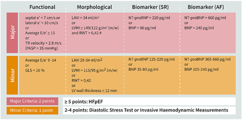
Figure 2. HFA-PEFF Score proposed by HFA and ESC. https://doi.org/10.1002/ejhf.1741

Both of the above scoring systems have been received with enthusiasm, given the lack of a clear definition and diagnostic framework for HFpEF. Studies evaluating the 2 scores have also been published. Parcha and colleagues studied the generalizability of the H2FPEF and HFA-PEFF scores in an analysis of participants with unexplained dyspnea from prior HFpEF trials and the Atherosclerosis Risk in Communities (ARIC) study.9 They found that both the scores could rule out HFpEF with a greater than 99% success rate but the H2FPEF score had a higher specificity than the HFA-PEFF score. Amanai and colleagues calculated H2FPEF and HFA-PEFF scores in patients with HFpEF referred for stress echocardiography.10 They found that both scores had similarly high positive and negative predictive values and a correlation with abnormal hemodynamics during exercise. Another study in the ARIC population also found that both high H2PEFF and HFA-PEFF scores were associated with increased risk of heart failure hospitalizations or death, suggesting the prognostic value of both.11

Both H2FPEF and HFA-PEFF are validated and easy-to-use scores using readily available clinical, laboratory, and echocardiographic parameters. The use of these scores in the appropriate patient and context can aid in the timely and accurate diagnosis of HFpEF. Growing recognition and emergence of effective therapies such as SGLT2 inhibitors are important strides for improving outcomes for patients with HFpEF.

References:

  1. Borlaug BA. Evaluation and management of heart failure with preserved ejection fraction. Nat Rev Cardiol. 2020;17(9):559-573. doi:10.1038/s41569-020-0363-2
  2. Sorajja P, Borlaug BA, Dimas VV, et al. SCAI/HFSA clinical expert consensus document on the use of invasive hemodynamics for the diagnosis and management of cardiovascular disease. Catheter Cardiovasc Interv. 2017;89(7):E233-E247. doi:10.1002/ccd.26888
  3. Reddy YNV, Carter RE, Obokata M, Redfield MM, Borlaug BA. A Simple, Evidence-Based Approach to Help Guide Diagnosis of Heart Failure With Preserved Ejection Fraction. Circulation. 2018;138(9):861-870. doi:10.1161/CIRCULATIONAHA.118.034646
  4. Sepehrvand N, Alemayehu W, Dyck GJB, et al. External Validation of the H2F-PEF Model in Diagnosing Patients With Heart Failure and Preserved Ejection Fraction. Circulation. 2019;139(20):2377-2379. doi:10.1161/CIRCULATIONAHA.118.038594
  5. Segar MW, Patel KV, Berry JD, Grodin JL, Pandey A. Generalizability and Implications of the H2FPEF Score in a Cohort of Patients With Heart Failure With Preserved Ejection Fraction. Circulation. 2019;139(15):1851-1853. doi:10.1161/CIRCULATIONAHA.118.039051
  6. Myhre PL, Vaduganathan M, Claggett BL, et al. Application of the H2 FPEF score to a global clinical trial of patients with heart failure with preserved ejection fraction: the TOPCAT trial. Eur J Heart Fail. 2019;21(10):1288-1291. doi:10.1002/ejhf.1542
  7. Pieske B, Tschöpe C, de Boer RA, et al. How to diagnose heart failure with preserved ejection fraction: the HFA-PEFF diagnostic algorithm: a consensus recommendation from the Heart Failure Association (HFA) of the European Society of Cardiology (ESC). Eur J Heart Fail. 2020;22(3):391-412. doi:10.1002/ejhf.1741
  8. Barandiarán Aizpurua A, Sanders-van Wijk S, Brunner-La Rocca HP, et al. Validation of the HFA-PEFF score for the diagnosis of heart failure with preserved ejection fraction. Eur J Heart Fail. 2020;22(3):413-421. doi:10.1002/ejhf.1614
  9. Parcha V, Malla G, Kalra R, et al. Diagnostic and prognostic implications of heart failure with preserved ejection fraction scoring systems. ESC Heart Fail. 2021;8(3):2089-2102. doi:10.1002/ehf2.13288
  10. Amanai S, Harada T, Kagami K, et al. The H2FPEF and HFA-PEFF algorithms for predicting exercise intolerance and abnormal hemodynamics in heart failure with preserved ejection fraction. Sci Rep. 2022;12(1):13. doi:10.1038/s41598-021-03974-6
  11. Selvaraj S, Myhre PL, Vaduganathan M, et al. Application of Diagnostic Algorithms for Heart Failure With Preserved Ejection Fraction to the Community. JACC Heart Fail. 2020;8(8):640-653. doi:10.1016/j.jchf.2020.03.013

“The views, opinions, and positions expressed within this blog are those of the author(s) alone and do not represent those of the American Heart Association. The accuracy, completeness, and validity of any statements made within this article are not guaranteed. We accept no liability for any errors, omissions, or representations. The copyright of this content belongs to the author and any liability with regards to infringement of intellectual property rights remains with them. The Early Career Voice blog is not intended to provide medical advice or treatment. Only your healthcare provider can provide that. The American Heart Association recommends that you consult your healthcare provider regarding your health matters. If you think you are having a heart attack, stroke, or another emergency, please call 911 immediately.”

hidden

New Treatment Options for Transthyretin Cardiac Amyloidosis – What do I need to know?

Clinical Features and Pathophysiology of Transthyretin Cardiac Amyloidosis (TTR-CA)

There has been an increasing awareness of the presence of cardiac amyloidosis (CA) in patients presenting with heart failure with preserved left ventricular ejection fraction (HFpEF). The clinical features suggestive of CA are recurrent heart failure with preserved ejection fraction (HFpEF) and restrictive left ventricular filling of unclear etiology. These patients characteristically have concentric left ventricular hypertrophy with wall thickness greater than 12 mm, biatrial enlargement, increased thickness of the interatrial septum and left atrial dysfunction even in the absence of atrial fibrillation or atrial flutter1. Typically, these patients have abnormal global longitudinal strain with “apical sparing” pattern2.

Amyloidoses are a group of protein-folding disorders in which more than one organ is infiltrated by proteinaceous deposits known as amyloid.  The deposits are derived from one of several amyloidogenic precursor proteins, and the prognosis of the disease is determined both by the organ(s) involved and the type of amyloid.  Amyloid involvement of the heart, cardiac amyloidosis (CA), carries the worst prognosis of any involved organ, and light-chain amyloidosis (AL-CA) is the most serious form of the disease (1). CA may be due to myocardial deposition of transthyretin protein derived from the liver known as transthyretin cardiac amyloidosis (TTR-CA) or may be due to AL-CA with myocardial deposition of immunoglobulin light chain proteins derived from a clone of plasma cells1. This blog will focus on the treatment of TTR-CA after a brief discussion about diagnosing this disease.


Diagnosing Transthyretin Cardiac Amyloidosis (TTR-CA)

TTR-CA is an underrecognized etiology for patients with recurrent exacerbations of HFpEF. However, the use of bone avid radiotracers such as 99m- technetium pyrophosphate (99m-Tc PYP) to diagnose TTR-CA have changed the diagnostic paradigm of this disease and have improved the ability to diagnose the disease readily1. Typically once the diagnosis is suspected clinically and by echocardiography or MRI, these patients should undergo clonal analysis with serum and urine free light chains and serum and urine immunofixation1. In patients who undergo 99mTc-PYP scans and have grade 2 or 3 myocardial uptake of the radiotracer on SPECT imaging, the positive predictive value of this finding with negative clonal analysis is close to 100% often deferring the need for myocardial biopsy for these patients3. It is important that once the diagnosis of TTR-CA is made that these patients undergo genotyping to determine if the mutant form (TTR-CAm) is present which is hereditary and presents earlier, usually 40+ years of age and has a slight male predominance compared with female1. In patients with TTR-CA who are genotype negative, these patients are defined as having the “wild type” sporadic form of TTR-CA (TTR-CAwt) and are usually older at 65+ years of age with a significant male predominance of 15:11.


Emerging Therapeutic Agents for Transthyretin Cardiac Amyloidosis

With the increasing awareness of TTR-CA and the ability to diagnose this disease that was once difficult to diagnose, there have been the development of various treatment options for these patients. There are three potential targets for treatment of patients with TTR-CA. These three potential targets include suppression of TTR synthesis, TTR stabilization and TTR fibril degradation and absorption4 Figure 1.

(i) Suppression of Transthyretin (TTR) synthesis

TTR synthesis can be suppressed by liver transplantation4. However there are 2 potential therapeutic medications that have been studied and have been shown to decrease TTR-Synthesis4. These 2 agents that have been shown to decrease TTR synthesis are patisiran5 and inotersen6.

Patisiran was shown in the APOLLO clinical trial to improve multiple clinical manifestations of hereditary transthyretin amyloidosis (TTR-CAm)5.

Patisiran is a RNA interference therapeutic agent that specifically inhibits hepatic synthesis of transthyretin. In the APOLLO clinical trial patients with TTR-CAm with polyneuropathy were studied and were treated with intravenous aptisiran 0.3 mg per kilogram of body weight once every 3 weeks after randomization and were compared with patients treated with placebo. Patisiran was shown at 18 months to result in a sustained and rapid decrease in transthyretin serum levels in patients treated with this agent with a 81% mean reduction in serum levels for all ages, gender and genotypes. Patisiran halted or reversed the progression of transthyretin amyloidosis and reduced the related neuropathic symptoms. This agent also improved the ability to ambulate with regards to improved gait speed and mobility and there was also an improvement in quality of life. The cardiac manifestation of the disease with regards to the echocardiographic measures of cardiac structure and function improved and there was also a reduction in NT-proBNP levels. The safety profile of the medication was good with the only side effects of the drugs being described as an increased incidence of peripheral edema and mild to moderate infusion related reactions with patisiran use5. There were no hematologic or nephrotoxic side effects of Patisiran noted during the study5.

Inotersen was shown in the international, randomized, double-blind, placebo-controlled NEURO-TTR study to improve the course of neurologic disease and quality of life in patients with TTR-CA6. Inotersen is a 2′-O-methoxyethyl–modified antisense oligonucleotide that inhibits hepatic production of transthyretin. In the NEURO-TTR study adults with stage 1 (patient is ambulatory) or stage 2 (patient is ambulatory with assistance) with TTR-CAm with polyneuropathy were included in the study and were randomized in 2:1 fashion to receive weekly subcutaneous injections of inotersen (300 mg) or placebo. The study period was 15 months. Over this study period the course of the neurologic symptoms related to TTR-CAm improved in addition to the quality of life. Steady state levels in reduction of circulating transthyretin protein was reached within 13 weeks and was sustained throughout the study period. The mean nadir in the decrease in circulating transthyretin from baseline levels in the inotersen group was a mean nadir of 74%6. The significant side effects of inotersen are thrombocytopenia and glomerulonephritis, therefore this should be managed with enhanced monitoring and treatment6.

Both patisiran and inotersen were approved by the Food and Drug Administration (FDA) for treatment of patients with TTR-CAm with evidence of neuropathy.

(ii) TTR Stabilization

Tafamidis is a benzoxazole derivative lacking nonsteroidal antiinflammatory drug activity that binds to the thyroxine-binding sites of transthyretin with high affinity and selectivity and inhibits the dissociation of tetramers into monomers thus stabilizing the transthyretin protein in TTR-CA. Tafamidis was shown in the multicenter, international, double-blind, placebo-controlled ATTR-ACT study to be a TTR stabilizer that decreased all-cause mortality, cardiovascular related hospitalization rate and the decline in functional capacity in patients with TTR-CA7.  In this study, patients were randomized in 2:1:2 fashion to 80mg of tafamidis or 20 mg of tafamidis or placebo over a 30 month period. Patients who received tafamidis had improved survival, had decreased cardiovascular (CV) related hospitalization rate and had less decline in functional capacity. This effect on decreased CV related hospitalization rate was seen amongst all patient treatment groups receiving tafamidis with the exception of those patients with New York Heart Association Class III heart failure. This was thought to be attributable to longer survival during the more severe stage of this disease process and highlights the importance of early diagnosis and treatment of this disease as it appears to have greater benefit when administered early in the course of the disease. No significant difference in clinical outcomes were seen between the 20 mg orally per day and the 80 mg orally per day dosing of tafamidis. Although the trial was designed with the requirement of tissue biopsy to make the diagnosis, the use of technetium labeled bone avid radiotracers in diagnosing TTR-CA have been validated as an accurate method for identifying these patients with high sensitivity and specificity3,8,9. This non-invasive method of diagnosing this disease leads to earlier identification and therefore earlier treatment of these patients with TTR-CA.  Tafamidis is the first drug approved by the Food and Drug Administration to treat patients with TTR-CA related cardiomyopathy, this drug was approved May 3, 2019.

Diflunisal is a non-steroidal anti-inflammatory drug that binds and stabilizes the transthyretin protein in TTR-CA against acid mediated fibril formation. Dosing is 250 mg po twice daily, side effects include cyclooxygenase (COX) enzyme related fluid overload, nephrotoxicity and gastrointestinal bleeding. This drug is still in trial phase and not yet approved by the FDA for use in patients with TTR-CA10.

(iii) TTR Fibril Degradation and Absorption

Fibril degradation and reabsorption in patients with TTR-CA can be achieved with several therapeutic agents such as doxycycline-tauroursodeoxycholic acid (TUDCA) and monoclonal anti-serum amyloid protein (SAP) antibody10.

Doxycycline-TUDCA removes amyloid protein that is already deposited and is administered orally as 100 mg BID/250 mg TID and is still under investigation10.

Monoclonal anti-serum amyloid protein (SAP) antibody works as an antibody against a normal non-fibrillar glycoprotein SAP and promotes a giant cell reaction that removes visceral amyloid deposits and is administered intravenously and is still under investigation. Potential side effects are infusion site reactions10.

Suppression TTR in reference 5 and 6. TTR stabilization and TTR fibril degradation/absorption in reference 10.

Monitoring Treatment:

With emerging therapeutic options for patients with TTR-CA, there is a need for a reliable method for detecting disease progression and monitoring improvement of the disease with treatment. While 99mTc-PYP has been shown to be able to accurately diagnose TTR-CA it has not been shown to be a useful tool to monitor disease progression. However, quantitative echocardiography and global longitudinal strain as well as cardiac magnetic resonance imaging (CMR) with tissue characterization  as well as amyloid imaging with Positron Emission Tomography (PET) radiotracers show promise as being potential tools to monitor disease progression and monitor treatment effects but are yet to be validated11.


Conclusion

The emergence of new treatment options for patients with TTR-CA provides a great degree of hope for these patients with a disease that was once thought to be very difficult to diagnose and even more difficult to treat. The FDA’s approval of patisiran, inotersen and most recently tafamadis as outlined previously is quite exciting news for patients with TTR-CA. However, it is hopeful that there will be an opportunity to have these medications be more affordable and accessible for these patients with TTR-CA so that they can all benefit from these therapies to improve their clinical outcomes.


References:

  1. Bullock-Palmer RP. Diagnosing cardiac amyloidosis: A wealth of new possibilities with nuclear cardiac imaging. J Nucl Cardiol. 2019 May 13. Doi: 10.1007/s12350-019-01740-w. [Epub ahead of print] PMID: 31087262
  2. Phelan D, Collier P, Thavendiranathan P, Popović ZB, Hanna M, Plana JC, et al. Relative apical sparing of longitudinal strain using two-dimensional speckle-tracking echocardiography is both sensitive and specific for the diagnosis of cardiac amyloidosis. Heart 2012;98(19):1442-8.
  3. Gillmore JD, Maurer MS, Falk RH, Merlini G, Damy T, Dispenzieri A, et al. Nonbiopsy diagnosis of cardiac transthyretin amyloidosis. Circulation 2016;133(24):2404-12.
  4. Castaño A, Drachman BM, Judge D, Maurer MS. Natural history and therapy of TTR-cardiac amyloidosis: Emerging disease-modifying therapies from organ transplantation to stabilizer and silencer drugs. Heart Fail Rev 2015;20(2):163-78.
  5. Adams D, Gonzalez-Duarte A, O’Riordan WD, Yang CC, Ueda M, Kristen AV, et al. Patisiran, an RNAi Therapeutic, for Hereditary Transthyretin Amyloidosis. N Engl J Med. 2018 Jul 5;379(1):11-21.
  6. Benson MD, Waddington-Cruz M, Berk JL, Polydefkis M, Dyck PJ, Wang AK, et al. Inotersen Treatment for Patients with Hereditary Transthyretin Amyloidosis. N Engl J Med. 2018 Jul 5;379(1):22-31.
  7. Maurer MS, Schwartz JH, Gundapaneni B, Elliott PM, Merlini G, Waddington-Cruz M, et al.Tafamidis Treatment for Patients with Transthyretin Amyloid Cardiomyopathy.. N Engl J Med. 2018 Sep 13;379(11):1007-1016.
  8. Castano A, Haq M, Narotsky DL, et al. Multicenter study of planar technetium 99m pyrophosphate cardiac imaging: predicting survival for patients with ATTR cardiac amyloidosis. JAMA Cardiol 2016;1:880-889.
  9. Bokhari S, Castaño A, Pozniakoff T, Deslisle S, Latif F, Maurer MS. (99m)Tc-pyrophosphate scintigraphy for differentiating light-chain cardiac amyloidosis from the transthyretin-related familial and senile cardiac amyloidoses. Circ Cardiovasc Imaging 2013;6:195-201.
  10. Castano A, Narotsky D and Maurer MS, Emerging Therapies for Transthyretin Cardiac Amyloidosis Could Herald a New Era for the Treatment of HFPEF. https://www.acc.org/latest-in-cardiology/articles/2015/10/13/08/35/emerging-therapies-for-transthyretin-cardiac-amyloidosis.
  11. Singh V, Falk R, Di Carli MF, Kijewski M, Rapezzi C, Dorbala S. State-of-the-art radionuclide imaging in cardiac transthyretin amyloidosis. J Nucl Cardiol 2019;26(1):158-73.