hidden

11 Women Cardiology Leaders – How to Overcome Adversity & Thrive

Summarized by Nidhi Madan MD Sarah Rosanel, MD; Cynthia Kos DO, Renee P. Bullock-Palmer MD, Kamala P. Tamirisa MD and Purvi Parwani MBBS MPH. Edited by Marissa Bergman.

Presented by the ACC Women in Cardiology (WIC) Section, AHA WIC Section and Women as One, this webinar highlighted a panel of female cardiologists with leadership roles in the field. The opportunity of gathering 11 female leaders of international Cardiology organizations comes rarely and the webinar was incredibly inspirational. It was co-moderated by ACC WIC Chair Dr. Toniya Singh, MD, Cardiologist at St. Louis Heart & Vascular and AHA WIC Chair, Dr. Annabelle Volgman, MD, Professor of Medicine, Rush College of Medicine;

The webinar focused on providing guidance, empowerment and optimism to women in cardiology through personal journeys and experiences. The presentations equipped attendees with the necessary skills and qualities to more than just survive, but, rather, thrive, during the ongoing pandemic and racial crisis.

Cindy Grines, MD, FACC, MSCAI

President of the Society of Cardiac Angiography & Interventions.

                                    “Accept the situation and have a game plan.”

Dr. Grines began the presentation with her personal journey. She had an extremely successful cardiology career in Michigan for over 25 years. Then, she decided to move, for family reasons, and began a new position as Academic Chair of Cardiology in New York. She was told during the interview process that her focus needed to be 90% on academics, research productivity, mentoring the faculty, and gaining the program a national presence. Over the next 1.5 years, she worked hard towards these goals and exceeded the expectations. Yet, despite going above and beyond in her professional duties, Dr. Grines was terminated from her position without a valid reason – with claims that it was a “business decision” and “trying to merge some roles.” She alluded to how she handled this unprecedented situation, and formulated a game plan. She negotiated a severance package and found her current position, with which she is very happy. Her presentation emphasized the importance of networking and  destigmatizing what might feel like a humiliating and isolating situation. Dr. Grines concluded with words of motivation:

“You need to pick yourself up, brush yourself off and get back in the saddle and ride that horse again. The bottom line is change is good and when these things happen to you it’s going to motivate you to do something different and to prove yourself.”

Roxana Mehran, MD, FACC, FACP, FCCP, FESC, FAHA, FSCAI  

 The Cofounder of “Woman As One.”

“Don’t give up on your goals.”

Dr. Mehran’s presentation started with a bang: “Celebrate Women!” She continued with powerful words, “When we focus on our goals, we can achieve everything and we should never give up on our goals. They are yours, cherish them, fight for it, you will achieve it.”

Dr. Mehran was born in Iran and she dreamt of being a doctor since she was quite young. Amidst the hostage crisis in Iran, her family immigrated to Queens, NY. Despite facing poverty and restarting her life as an outsider, she never lost sight of her aspirations and eventually became an interventional cardiologist. With her determination and strong will, Dr. Mehran was one of the first female fellows at Mount Sinai. She pursued her career and continued her mission to contribute to science and clinical outcomes. As a woman in a male dominated field, she felt the inequalities in interventional cardiology, and she made it her new goal to ensure women are heard. Ultimately, she co-founded “Women As One” to encourage women not to accept inequalities or harassment in any form. As she explained, “You just have to see it all, keep your eye on the ball just like they tell you in baseball and in tennis… and make sure you hit that bull’s eye. Work hard and it will come to you.”  She concluded with her favorite quote by Maya Angelou,

 “Do your best you can until you know better, then when you know better, do better.”

Athena Poppas, MD, FACC, FASE

President of the American College of Cardiology

 “Strategic Leadership & Change Management”

Strategic leadership has never been as important as it is during the challenging times of the pandemic. Dr. Poppas referred to the importance of influential leadership and emphasized that one does not need a title to lead. These times are an incredible opportunity for everyone to step up and contribute. She explained that strategic leadership is not linear, but mostly circular – anticipating, recognizing challenges, interpreting and making decisions, staying aligned but learning along the way. She then shared some of the key tools from her leadership toolbox:

  1. Authenticity is essential.
  2. Use influential skills rather than just telling someone what to do – utilize the tools of change management to bring people along.
  3. Manage conflict and work together.
  4. Realize one’s own strengths, be honest about those strengths and bounce ideas off friends and allies. Be cognizant about weaknesses with a goal to improve them.
  5. Put yourself out there and seize opportunities.

Dr. Poppas concluded by reiterating that change management and strategic leadership is a continuum and a continuous cycle of learning. At the same time, succession planning with mentoring and helping others is key, so that there is an entire group capable of replacing you.

Andrea Russo, MD, FHRS

Immediate past President of Heart Rhythm Society (HRS)

                                                               “Resilience”

 In Dr. Russo’s first week as President of HRS, a controversial topic of Maintenance od Certificate (MOC) surfaced. HRS was looking into ways to create a less disruptive and  more customizable educational program and certification. Therefore, HRS put together an MOC Task Force and conducted a member survey assessing the feasibility of other options. Throughout this battle, resilience helped her look into options that would be relevant to the HRS members. The COVID-19 pandemic put the annual HRS meeting in jeopardy. She led the team, which considered the safety of travel and alternate ways to deliver education. Arrhythmias related to the coronavirus needed attention with protocols; how to deliver EP care to patients in the COVID era while also protecting the EP team by reducing their exposure became a priority. To answer these questions, HRS put together a group called the COVID-19 Rapid Response Task Force to collate the major information and provide guidance. There was an outpouring of volunteers and these documents were prepared in record time. This experience emphasized  the resilience of a collective resolve from the volunteers who contributed to the HRS staff. Dr. Russo concluded by saying that COVID did jump start the utilization of online educational platforms and digital health to successfully deliver the HRS 2020 content online.   She explained that one of the most rewarding experiences of her presidency was the ability to share ideas, work together with leaders from around the globe and improve knowledge.

Christine Albert, MD, MPH, FHRS

President of Heart Rhythm Society

“Embrace Change, Be Creative”

 Dr. Albert’s advice is, when one cannot change the adversity, it is important to change gears and embrace the new opportunity. Listening to new suggestions, moving forward and ultimately bringing the group along as a leader are an integral part of being creative. Advances in digital forms of communication in COVID times are one such example of embracing the change.  She ended with these empowering words, “Don’t be afraid to forge ahead in adversity.”

                                                  Mariell Jessup, MD, FAHA

                Chief Science & Medical Officer of American Heart Association

                                               “Believe in your Capabilities”

 Dr. Jessup’s presentation focused on how it takes courage to overpower impostor syndrome and its nagging question, “Are you capable?” She pointed to Michelle Obama’s comments as a guiding example: “Am I good enough?” “Of course!” She argued that courage might not be easy to find every moment, and that friends and mentors play an important role against a doubtful mind.

She referred to Eleanor Roosevelt’s challenging life and quoted, “You gain strength, courage and confidence by every experience in which you really stop to look fear in the face. You are able to say to yourself, ‘I have lived through this horror. I can take the next thing that comes along.’ You must do the thing you think you cannot do.”

Dr. Jessup offered several more phrases and quotes to empower and remind women that it is vital to focus on courage to lift up mentees. She was reminded of Queen Elizabeth’s quote, “When life seems hard, the courageous do not lie down and accept defeat; instead, they are all the more determined to struggle for a better future.” Another voice of reason she found very relevant is Winston Churchill, regarding sharing courage “I never gave them courage; I was able to focus theirs.” She concluded her presentation on an uplifting note – “Have the courage!”

 Michelle Albert, MD, MPH, FAHA, FACC

President, Association of Black Cardiologists (ABC)

 “Remembering your purpose”

 Dr. Albert emphasized being innovative and creative while also being kind and compassionate in a society facing healthcare disparities. It is important to remember the purpose, when attempting to have an impact. She also emphasized harnessing one’s background to help focus on one’s individual passion and follow that purpose.

Raised by her grandparents, Dr. Albert witnessed hardship and segregation, and she perceived how the socioeconomic background of the patients influenced healthcare. As she explained, “The largest gap in healthcare is in cardiovascular medicine”.

Dr. Albert further highlighted the importance of appropriate support, including key mentorship and faith to overcome adversity. She stressed that being disciplined; bold, collaborative and always thinking outside of the box are key for achieving ultimate professional purpose.

She concluded by warning against transactional relationships or being predatory in the professional setting.

Chiara Bucciarelli-Ducci, MD, PhD, FESC, FRCP

CEO, Society of Cardiac Magnetic Resonance (SCMR)

What opportunities can this adversity bring?”

 Dr. Bucciarelli-Ducci believes there are endless opportunities and each challenge simply leads to more opportunities. She is a transformational leader, someone who tries to identify the need for change, create a vision, guide change through inspiration and work collaboratively. She always aspired to be that woman in cardiology and her experience has taught that with change always comes resistance. She stressed the importance of listening to all parties while honing the power of negotiation. She quoted Socrates, in emphasizing the power of a collaborative team, “The secret of change is to focus all of your energy, not on fighting the old, but on building the new.”

Her Italian background, upbringing and world history inspire her tremendously. To Dr. Bucciarelli-Ducci, the COVID-19 pandemic parallels what happened during World War II (WWII). Just like WWII, she believes that this pandemic is creating new ways of thinking, working and connecting with people across the globe.

Sharmila Dorbala, MD, MPH, FASNC

President, American Society of Nuclear Cardiology (ASNAC)

“Be Optimistic”

In Dr. Dorbala’s experience, “Optimism is one of the keys to success.” She believes that whether one looks at the glass as half-full or half-empty is a matter of perspective and choice. One can choose to be an optimist and train oneself to focus on the positives, and that optimism gives one confidence to take risks and then becomes contagious.

She provided an example of contrasting optimists and pessimists and how they view the world differently. Optimists see challenges as being temporary, something that can be conquered and used as a stepping-stone to better solutions, whereas pessimists view challenges as insurmountable obstacles. She referenced her research interest in cardiac amyloidosis to illustrate how optimism has influenced her own career. Dr. Dorbala actively chose to be optimistic and stayed in this field despite the hurdles she encountered. She always remained passionate about her field and confident that her hard work would lead to opportunities. She believes that the advances in medicine seen today are because the medical community chose to focus on the potential of the future.

Her overall advice for professional life is to have the integrity to do what is right, irrespective of the consequences, focus on excellence and be passionate about the cause. She reminds us to never underestimate the importance of having an optimistic outlook to gain confidence and to look for opportunities by embracing risks.

Judy Hung, MD, FASE

Incoming President, American Society of Echocardiography (ASE)

Forget the noise and forge ahead”

Dr. Hung emphasized that during one’s medical career there will be many instances of biases and inequality, intentional or unconscious. She advised that these injustices should not distract one from pursuing their goals.  To her, it is important to always stay in the lane. Dr. Hung explained that one could transform anger and sadness into positive energy, and make an impact professionally. Her strongest advice to women in cardiology is to stay focused and not let negative attributes of mental energy sway one away from their focus.

Biykem Bozkurt, MD, PhD, FHFSA, FACC, FAHA

President of Heart Failure Society of North America

“Create Change and acknowledge the ‘never-evers’ ”

 In a time that has left everyone grappling with unprecedented personal and professional challenges, how can do you thrive as leaders? Dr. Bozkurt argued, “most advancements come from acknowledgement of the ‘never-evers’”. “You have to face obstacles head on” or else face “stagnation and complacency.” She offered words of wisdom that adversity creates opportunity for resilience to get out of one’s comfort zone and create a meaningful change.

The COVID-19 pandemic has exacerbated a constant truth of the profession – doctors are witness to human suffering, but, at the same time, healing. “Do not sanitize suffering…learn from it… and teach the next generation,” said Dr. Bozkurt.  She cautioned against disinfecting the truth out of uncomfortable realities.  Amongst the suffering and sacrifice lies empathy, humility, and growth.

Dr. Bozkurt cited the story of Marguerite Matisse as a compelling example. Marguerite suffered from severe illness at a young age, requiring a tracheostomy. Despite poor health and a prominent scar, she became a lifelong muse for her father, the renowned artist Henri Matisse. As he once explained, “I don’t remember adversity, I remember resilience.” Dr. Bozkurt hopes that when the world looks back on the current healthcare, economic, racial, and political situations, Matisse’s quote will ring true.

Visit this website for access to this important webinar.

Summarized by Nidhi Madan MD Sarah Rosanel, MD; Cynthia Kos DO, Renee P. Bullock-Palmer MD, Kamala P. Tamirisa MD and Purvi Parwani MBBS MPH. Edited by Marissa Bergman.

“The views, opinions and positions expressed within this blog are those of the author(s) alone and do not represent those of the American Heart Association. The accuracy, completeness and validity of any statements made within this article are not guaranteed. We accept no liability for any errors, omissions or representations. The copyright of this content belongs to the author and any liability with regards to infringement of intellectual property rights remains with them. The Early Career Voice blog is not intended to provide medical advice or treatment. Only your healthcare provider can provide that. The American Heart Association recommends that you consult your healthcare provider regarding your personal health matters. If you think you are having a heart attack, stroke or another emergency, please call 911 immediately.”

hidden

The American Heart Association Annual Scientific 2019 Meeting- “An Engaging and Enlightening experience”

The recent American Heart Association Annual Scientific meeting held in Philadelphia, Pennsylvania was filled with many sessions centered around the area of Cardiac Imaging with emerging areas in Nuclear Cardiac imaging,Echocardiography, Cardiac CT as well as Cardiac MRI. There was also the long awaited results of the ISCHEMIA trial. In addition, the Women in Cardiology (WIC)  Committee had several sessions related to professional development including  several networking opportunities with the WIC networking luncheon as well as a networking WIC dinner that was sponsored by the University of Pennsylvania.   In this blog I will discuss several of these highlights.


Heart Disease in Women- Focus on Cardiac Imaging

Drs. Viviany Taqueti and Parham Eshtehardi moderated an excellent session on the multimodality assessment of microvascular coronary artery disease (CAD)  in women.

Invasive Assessment of Microvascular Coronary Artery Disease:

During this session Dr. Carl Pepine discussed invasive assessment of microvascular disease in women. He discussed the use of coronary TIMI frame count (cTFC) to predict adverse events in women with symptoms/signs of ischemia with no obstructive coronary artery disease (INOCA).  He discussed the results of a pilot study from the National Heart, Lung and Blood institute ( NHLBI) sponsored Women’s Ischemia Syndrome Evaluation (WISE) which showed that in women with INOCA, resting cTFC provided independent prediction of hospitalization for angina1. He also discussed the pros and cons of the three invasive methods for the assessment of coronary microvascular function: Thermodilution (IMR), Doppler wire (hMR) and Continuous Thermodilution (MVR). IMR being most user friendly of the 3 methods and has also been a method that has been validated against clinical outcomes. IMR is also feasible in every coronary anatomy. However, IMR is adenosine dependent. hMR and MVR both have reasonable reproducibility. hMR has also been validated against clinical outcomes while MVR provides a direct assessment on coronary blood flow and is adenosine free. However, IMR and hMR are dependent on the use of adenosine2. The indications for invasive coronary function testing (CFT) include (a) Evidence of ischemia with persistent chest pain and no obstructive coronary artery disease (CAD) (b) Chest pain refractory to medical management and (c) Preference for definitive diagnosis. Invasive CFT has excellent safety data with <0.6-0.7% serious adverse event rate (coronary dissection, myocardial infarction)3-5. Invasive diagnosis of coronary microvascular disease in women was associated with worse angina  and hospitalization free survival compared to women without evidence of microvascular CAD6.

Role of Positron Emission Tomography (PET) assessment of Microvascular CAD (CMD) in Women:

Dr. Sharmila Dorbala gave an excellent talk on the use of Cardiac PET to assess the presence of CMD with myocardial blood flow and coronary flow reserve assessment with robust evidence data with over 35 years of research data involving over 20,000 patients.  The presence of severely impaired coronary flow reserve (<2.0)  is associated with excess cardiovascular risk in women relative to men referred for coronary angiography7. She had also discussed the evidence showing that CMD diagnosed with PET was associated with an increased risk of heart failure with preserved ejection fraction (HFpEF). The future of myocardial blood flow assessment with Cadmium Zinc Telluride (CZT) SPECT cameras was also discussed. The key points for the diagnosis of CMD with nuclear perfusion imaging are the following:

  • Nuclear stress imaging is required to assess stress myocardial blood flow (MBF) and to assess myocardial flow reserve: stress MBF/rest MBF
  • The stress agent used for assessment of MBF is typically done with vasodilatory agents such as regadenoson, adenosine or dipyridamole. Cold pressor test is usually done in research labs to assess endothelial dysfunction. Exercise MBF is not very feasible.
  • Epicardial CAD has to be excluded
  • CMD is typically defined as MBF <2.

Quantitative Blood Flow assessment with Cardiac MRI (CMR):

Dr. Chiara Bucciarelli-Ducci gave a very informative talk on the use of CMR to assess myocardial blood flow. The general principles of quantification of blood flow requires knowledge of the amount of contrast agent in the myocardium and knowledge of the amount of contrast agent in the blood pool (arterial input function). The challenge in this assessment is the lack of linearity between signal and contrast concentration. The main sources of non-linearity and bias includes spatial signal variations (sensitivity profile of the surface coils), imperfect saturation of magnetization during contrast bolus passage, T2* decay and signal loss by contrast concentration in the blood pool and non-linear signal response (inherent to saturation recovery). Novel CMR techniques such as perfusion mapping and extracellular volume (ECV) assessment were also discussed. The assessment of cardiac perfusion imaging with inline quantitative flow mapping was also discussed, including the fact that this is a fully automated workflow without any user interaction.


Artificial Intelligence in Cardiac Imaging with Cardiac CT and Cardiac PET imaging

There was a very innovative session that discussed the use of machine learning and deep learning in FFR Cardiac CT (FFRct)  to improve diagnostic accuracy. Cardiac CTA has been shown to be an established diagnostic tool in clinical practice with FFRct offering functional information for intermediate and severe lesions. FFRct has also showed good correlation with invasive FFR. Machine Learning improves FFR-ct algorithms with improved accuracy, decreased analysis stime, potential to increase the availability of this FFRct technology and potential cost reduction. Deep Learning (DL) in automatic calcium scoring on cardiac CT using paired convolutional neural networks was also discussed. Assessment of ischemic myocardium with Cardiac CTA with DL was also discussed with the use of tissue segmentation and tissue characterization. The use of DL in cardiac PET was also discussed with regards to its utility in cardiovascular event prediction.


The ISCHEMIA trial

The long awaited ISCHEMIA trial results were released at the AHA 2019 and my take home points were outlined in my last blog, https://earlycareervoice.professional.heart.org/my-top-10-take-home-points-from-the-ischemia-trial/

ischemia trial

The ISCHEMIA trial is the largest trial studying an invasive versus conservative strategy for patients with stable ischemic heart disease8. The overall conclusions of the ISCHEMIA trial were:an initial invasive strategy compared with an initial conservative strategy did not demonstrate a reduced risk over a median follow up period of 3.3 years with regards to the primary endpoint of cardiovascular death, myocardial infarction, hospitalization for unstable angina and heart failure as well as with regards to the secondary endpoints of cardiovascular death or myocardial infarction. The probability of at least a 10% benefit of an invasive strategy on all cause mortality was < 10, based on pre-specified Bayesian analysis. The ISCHEMIA trial concluded that patients with stable CAD and moderate to severe ischemia had significant durable improvements in angina control and quality of life with an invasive strategy if they had angina occurring daily/weekly or monthly. Shared decision-making should be done to ensure alignment of treatment with patients’ goals and preferences for patients with angina. However, in patients without angina, an invasive strategy led to minimal symptom improvement or quality of life benefits as compared with a conservative strategy.  An early invasive strategy was not associated with a significant reduction in clinical events.


Imaging of Valvular Heart Disease

There was also an enlightening session on the role of cardiac imaging in the assessment of tricuspid valvular heart disease with a focus on severe secondary tricuspid regurgitation as well as congenital Ebstein’s anomaly of the tricuspid valve. The preoperative risk factors for significant post op tricuspid regurgitation that were noted were (a) preoperative tricuspid regurgitation of 2+ or more (b) atrial fibrillation and (c) huge left atrium9. Mitral valve surgery as well as double valve surgery including the aortic and mitral valve were associated factors for the development of late significant tricuspid regurgitation after left sided valve surgery10. Mild to moderate progressive functional  tricuspid regurgitation with tricuspid valve annular dilation of >40 mm or > 21 mm/m2 by 2D echocardiography has a class IIa indication for tricuspid valve repair at time of left sided valve surgery. In addition any symptomatic severe functional tricuspid regurgitation at the time of left sided valve surgery has a  class I indication for tricuspid valve repair or replacement11.

Ebstein’s anomaly involving the tricuspid valve accounts for <1% of congenital heart disease. The tricuspid valve malformation is often complex and typically involves the tricuspid valve leaflets, the chordal apparatus and the myocardium  of the right ventricle. The severity of tricuspid valve leaflet displacement on echocardiography has been identified as an independent predictor of cardiac mortality in patients with Ebstein’s anomaly12.

Primary tricuspid regurgitation may also result from lesions of the tricuspid valve apparatus itself such as in endocarditis, congenital disease or from mechanical trauma  to the leaflets, annulus and/or chordae. Echocardiography remains the primary imaging modality to diagnose the etiology and severity of tricuspid valve disease. Features on echocardiography that may indicate significant tricuspid regurgitation includes  tricuspid valve annular dilation of >40 mm or > 21 mm/m2, tethering distance >0.76 cm, tethering area >1.63 cm2 and right ventricular end systolic area of > 20 cm213. However, CMR and Cardiac CT are useful in some cases. CMR is considered the gold standard for the quantification of tricuspid regurgitation, quantification of right ventricular volumes, evaluation of right ventricular function, assessment of fibrosis and occasionally assessment of the etiology. Limitations for CMR includes presence of cardiac devices, arrhythmias, claustrophobia and renal failure (gadolinium). In special situations tricuspid regurgitation can be complex. 3D echocardiography provides useful information and it is sometimes necessary to assess tricuspid regurgitation with multimodality imaging as Cardiac CT can provide information about the anatomical regurgitant orifice area. Additionally, CMR and Cardiac CT can provide information about the dimensions of the tricuspid valve annular diameter, right heart volumes and function as well as vascular assessment13.  In the presence of significant tricuspid regurgitation, tricuspid valve repair should be performed whenever possible.


Multimodality imaging of Aortic Diseases

There was also an excellent session dedicated to the multi modality imaging with CT and MRI  of acute and chronic aortic diseases such as aortic intramural hematoma, aortic aneurysms,  aortic dissection and endovascular repair of the aorta (TEVAR) planning for aortic repair. 4D flow MRI was also discussed with regards to TEVAR planning.

With regards to aortic aneurysms, type B aortic dissection should be considered a chronic disease with complex pathophysiology with up to 75% of patients developing an aortic aneurysm14. Hemodynamic forces are believed to play a central role in this pathophysiology.

In the presence of type B intramural hematoma, tiny intimal disruptions/fenestrations (TID) does not confer poor prognosis as the risk of aorta related events are similar when compared to patient without TID. 14% of these patients progress to focal intimal disruption in a recent meta analysis15.  Focal intimal disruptions and ulcer like projections may cause large communications in intramural aortic hematoma with >3mm connection with the aortic lumen and are typically absent on the initial study. Limited intimal tears of the aorta although rare are an unquestionable cause of acute aortic syndromes and is considered a variant of aortic dissection. It is predominantly seen in type A intramural hematoma with an aneurysmal aorta and patients are older than patients with classic aortic dissection. Limited intimal tears can be seen on state of the art CT angiography and 3D volume rendering can make lesions more conspicuous.


Quality Cardiac Imaging

The last day of AHA 2019 was kicked off with an inspiring and informative session that discussed Quality in Cardiac Imaging that was led by several leaders in the field which included Dr. Pamela Douglas and Dr. Leslee Shaw. Dr. Ritu Sachdeva discussed strategies to maximize imaging information and outcomes by facilitating implementation by removing barriers and incentivizing schemes. She also discussed designing strategies to keep up with the pace of technology through improving quality, promoting innovation and research and focusing on adequate training in cardiac imaging. It was also mentioned that achieving excellence in imaging has to include collaboration between providers, professional societies, patients, payers and industry. The focus should move from “Volume Driven Healthcare” to “Value Driven Healthcare”.

The future of Cardiac Imaging in a value based healthcare system has to include definitions of the cardiovascular imager of the future, ensure robust innovation and research and maximizing imaging information and improving outcomes. This was outlined in the “Future of Cardiac Imaging Think Tank” article by Dr. Pamela Douglas16.


AHA Presidential Address

The AHA Opening session was a very energetic, encouraging and enlightening session. This session was opened by wonderful performances by Broadway’s Hamilton cast of actors.  The Presidential address by Dr. Robert Harrington (Figure 1)  was encouraging as he spoke on the importance of increasing diversity in the cardiology workforce as well as improving gender equality in the cardiology field.

Figure 1 President of the American Heart Association, Dr. Robert Harrington at the American Heart Association 2019 Annual Scientific Session Presidential Address

Figure 1 President of the American Heart Association, Dr. Robert Harrington at the American Heart Association 2019 Annual Scientific Session Presidential Address


AHA Women in Cardiology (WIC) Committee Events

  • Professional Development: There were many Women in Cardiology Professional Development sessions that covered a wide range of topics such as “Volunteerism to Advance Your Career” with Drs. Stacy Rosen and Michelle Albert, “Negotiations” with Drs. Sandra Lewis and Linda GIllam, “Time Management” with Drs. Toniya Singh and Gina Lundberg, “Self-Advocacy” with Drs. Elaine Tseng and Linda Shore, “Sponsor vs. Mentor” with Dr.  Emelia Benjamin and “Around the World with Go Red for Women”. These sessions were held in the Women in Science and Medicine Lounge in the expo hall and were very engaging and well attended sessions.


Networking Activities

WIC Networking Luncheon:

The AHA Women in Cardiology networking luncheon was an excellent session that provided an opportunity to network with other colleagues and AHA leadership. This session also hosted an excellent panel of speakers led by Dr. Laxmi Mehta who spoke on the topic of “Physician Burnout” and how to address this issue in Medicine. The other panel of speakers included Dr. Minnow Walsh who discussed the “Increased demand of the aging population and decreased supply of clinicians causing burnout – what can be done?”, Dr. Athena Poppas discussed “What can Cardiology leaders and Chiefs of Cardiology do to help decrease the stress from the clinical workload and increase career satisfaction?”, Dr. Sandy Lewis discussed “Legislative advocacy to decrease physician burnout​ – what can organizations do?” and Dr. Sherry Ann Brown discussed “What can cardiologists do to decrease their stress levels?” and she also discussed “What can fellowship program directors do to help fellows decrease burnout?” The attendees at the luncheon also added their own experiences relevant to this topic and also shared words of wisdom and advice.

The All Council Reception and Clinical Cardiology Council Dinner:

The All Council reception was very well attended with standing room only. It was also very lively with performance by the members from the Mummers Philadelphia group. This reception led into the Clinical Cardiology Council Dinner and during the dinner there were several awards given as well as recognition of the new Fellows of the American Heart Association. Dr. Sharon Reimold was the recipient of the AHA Women in Cardiology Mentoring Award.

The AHA WIC Networking dinner hosted by University of Pennsylvania:

The AHA Women in Cardiology committee was very grateful to the University of Pennsylvania led by Dr. Monika Sanghavi  who hosted a wonderful dinner that provided an opportunity for Women in Cardiology fellows in training (FIT) to network with several Women in Cardiology leaders in the field. We appreciated pearls of wisdom from leaders such as  Dr. Nanette Wenger, Dr. Sharonne Hayes, Dr. Minnow Walsh, Dr. Andrea Russo, Dr. Martha Gulati and Dr. Annabelle Volgmann. This was the second annual WIC event at the AHA Scientific meeting and we are hoping to continue this at future AHA Scientific meetings as these events have been very well received by our Women in Cardiology FITs.


AHA 2019 was an educational, engaging and exciting meeting where new scientific data relevant to Cardiology practice was presented. The event also offered many opportunities for networking with many leaders in the field. I look forward to the American Heart Association Scientific meeting in November 2020 in the beautiful city of  Dallas, Texas.


References:

  1. Petersen JW, Johnson BD, Kip KE, Anderson RD, Handberg EM, et al. (2014) TIMI Frame Count and Adverse Events in Women with No Obstructive Coronary Disease: A Pilot Study from the NHLBI-Sponsored Women’s Ischemia Syndrome Evaluation (WISE). PLoS ONE 9(5): e96630. doi:10.1371/journal.pone. 0096630
  2. Banning AP, De Maria GL.Measuring coronary microvascular function: is it finally ready for prime time? Eur Heart J. 2019 Jul 21;40(28):2360-2362. doi: 10.1093/eurheartj/ehz426. No abstract available. PMID: 31236565
  3. Wei J, Mehta PK, Johnson BD, Samuels B, Kar S, Anderson RD, Azarbal B, Petersen J, Sharaf B, Handberg E, Shufelt C, Kothawade K, Sopko G, Lerman A, Shaw L, Kelsey SF, Pepine CJ, Merz CN.Safety of coronary reactivity testing in women with no obstructive coronary artery disease: results from the NHLBI-sponsored WISE (Women’s Ischemia Syndrome Evaluation) study. JACC Cardiovasc Interv. 2012 Jun;5(6):646-53. doi: 10.1016/j.jcin.2012.01.023.PMID: 22721660
  4. Reriani M, Sara JD, Flammer AJ, Gulati R, Li J, Rihal C, Lennon R, Lerman LO, Lerman A.Coronary endothelial function testing provides superior discrimination compared with standard clinical risk scoring in prediction of cardiovascular events. Coron Artery Dis. 2016 May;27(3):213-20. doi: 10.1097/MCA.0000000000000347. PMID: 26882018
  5. Ong P, Athanasiadis A, Borgulya G, Vokshi I, Bastiaenen R, Kubik S, Hill S, Schäufele T, Mahrholdt H, Kaski JC, Sechtem U. Clinical usefulness, angiographic characteristics, and safety evaluation of intracoronary acetylcholine provocation testing among 921 consecutive white patients with unobstructed coronary arteries. Circulation. 2014 Apr 29;129(17):1723-30. doi: 10.1161/CIRCULATIONAHA.113.004096. Epub 2014 Feb 26. PMID: 24573349
  6. AlBadri A, Bairey Merz CN, Johnson BD, Wei J, Mehta PK, Cook-Wiens G, Reis SE, Kelsey SF, Bittner V, Sopko G, Shaw LJ, Pepine CJ, Ahmed B. Impact of Abnormal Coronary Reactivity on Long-Term Clinical Outcomes in Women. J Am Coll Cardiol. 2019 Feb 19;73(6):684-693. doi: 10.1016/j.jacc.2018.11.040. PMID: 30765035
  7. Taqueti VR, Shaw LJ, Cook NR, Murthy VL, Shah NR, Foster CR, Hainer J, Blankstein R, Dorbala S, Di Carli MF. Excess Cardiovascular Risk in Women Relative to Men Referred for Coronary Angiography Is Associated With Severely Impaired Coronary Flow Reserve, Not Obstructive Disease.’Circulation. 2017 Feb 7;135(6):566-577. Doi: 10.1161/CIRCULATIONAHA.116.023266. Epub 2016 Nov 14.PMID: 27881570
  8. https://www.google.com/url?q=http://www.ischemiatrial.org/&sa=D&ust=1576542824370000&usg=AFQjCNH06i1Ohvx0btiCyHrVJARGarMYOA
  9. Matsuyama K, Matsumoto M, Sugita T, Nishizawa J, Tokuda Y, Matsuo T. Predictors of residual tricuspid regurgitation after mitral valve surgery. Ann Thorac Surg. 2003 Jun;75(6):1826-8. PMID: 12822623
  10. Song H, Kim MJ, Chung CH, Choo SJ, Song MG, Song JM, Kang DH, Lee JW, Song JK.
    Factors associated with development of late significant tricuspid regurgitation after successful left-sided valve surgery. Heart. 2009 Jun;95(11):931-6. doi: 10.1136/hrt.2008.152793. Epub 2009 Mar 24. PMID: 19321491
  11. Nishimura RA, Otto CM, Bonow RO, Carabello BA, Erwin JP 3rd, Guyton RA, O’Gara PT, Ruiz CE, Skubas NJ, Sorajja P, Sundt TM 3rd, Thomas JD; ACC/AHA Task Force Members.2014 AHA/ACC Guideline for the Management of Patients With Valvular Heart Disease: a report of the American College of Cardiology/American Heart Association Task Force on Practice Guidelines.Circulation. 2014 Jun 10;129(23):e521-643. doi: 10.1161/CIR.0000000000000031. Epub 2014 Mar 3.
  12. Tobler D, et al. Tricuspid Valve Abnormalities. Congenital Heart Diseases in Adults Imaging and Diagnosis. Springer er. 2019
  13. Hahn RT, Thomas JD, Khalique OK, Cavalcante JL, Praz F, Zoghbi WA. Imaging Assessment of Tricuspid Regurgitation Severity. JACC Cardiovasc Imaging. 2019 Mar;12(3):469-490. doi: 10.1016/j.jcmg.2018.07.033. Review. PMID: 30846122
  14. Fattori R, Montgomery D, Lovato L, Kische S, Di Eusanio M, Ince H, Eagle KA, Isselbacher EM, Nienaber CA. Survival after endovascular therapy in patients with type B aortic dissection: a report from the International Registry of Acute Aortic Dissection (IRAD). JACC Cardiovasc Interv. 2013 Aug;6(8):876-82. doi: 10.1016/j.jcin.2013.05.003. PMID: 23968705
  15. Moral S, Cuéllar H, Avegliano G, Ballesteros E, Salcedo MT, Ferreira-González I, García-Dorado D, Evangelista A. Clinical Implications of Focal Intimal Disruption in Patients With Type B Intramural Hematoma. J Am Coll Cardiol. 2017 Jan 3;69(1):28-39. doi: 10.1016/j.jacc.2016.10.045. PMID: 28057247
  16. Douglas PS, Cerqueira MD, Berman DS, Chinnaiyan K, Cohen MS, Lundbye JB, Patel RA, Sengupta PP, Soman P, Weissman NJ, Wong TC; ACC Cardiovascular Imaging Council. The Future of Cardiac Imaging: a Report of a Think Tank Convened by the American College of Cardiology. JACC Cardiovasc Imaging. 2016 Oct;9(10):1211-1223. Doi: 10.1016/j.jcmg.2016.02.027. Review. PMID: 27712724


The views, opinions and positions expressed within this blog are those of the author(s) alone and do not represent those of the American Heart Association. The accuracy, completeness and validity of any statements made within this article are not guaranteed. We accept no liability for any errors, omissions or representations. The copyright of this content belongs to the author and any liability with regards to infringement of intellectual property rights remains with them. The Early Career Voice blog is not intended to provide medical advice or treatment. Only your healthcare provider can provide that. The American Heart Association recommends that you consult your healthcare provider regarding your personal health matters. If you think you are having a heart attack, stroke or another emergency, please call 911 immediately.

hidden

My Top 10 Take Home Points from The ISCHEMIA Trial

The long awaited results of the ISCHEMIA trial were presented at this weekend’s American Heart Association’s annual scientific meeting in Philadelphia, Pennsylvania by the Principal Investigator Dr. Judith Hochman (1). This study was a randomized parallel multicenter study that had an aim to compare outcomes of patients with stable ischemic heart disease and had moderate to severe ischemia on non-invasive stress testing who underwent optimal medical therapy (conservative arm) compared with those who underwent initial routine invasive therapy (invasive arm).


ischemia trial


The clinical and stress test eligibility criteria were:

  1.  Age > 21 years
  2. The presence of moderate or severe ischemia is defined as:
    1. Nuclear > 10% left ventricular ischemia (summed difference score of > 7)
    2. Stress echo with > 3 segments with stress induced moderate to severe hypokinesis or akinesis.
    3. CMR with
      1. Perfusion > 12% myocardial ischemia and/or
      2. Wall motion > 3/16 segments with stress induced severe hypokinesis or akinesis.
    4. Exercise tolerance testing (ETT) with > 1.5 mm ST depression in > 2 leads or >2 mm ST depression in single lead at <7 METS with angina.

Ischemia eligibility was determined by sites. All stress tests were interpreted at core labs.


The major clinical exclusion criteria were:

  1. New York Heart Association Class III-IV functional class
  2. Unacceptable angina despite medical therapy
  3. Left Ventricular Ejection Fraction (LVEF) <35%.
  4. Acute coronary syndrome within the last 2 months
  5. Percutaneous coronary intervention or coronary artery bypass graft within the last 1 year
  6. eGFR <30ml/min on dialysis


The coronary CT angiogram Eligibility Criteria were:

Inclusion Criteria:

  1. > 50% stenosis in a major epicardial vessel (stress imaging participants)
  2. > 70% stenosis in a proximal or mid vessel (ETT participants)


Major Exclusion Criteria:

  1. > 50% stenosis in unprotected left main disease.

This study initially enrolled 8518 patients of which 3339 were excluded due to screen failure due to insufficient ischemia (N=1350), nonobstructive coronary artery disease (CAD) (N=1218) and the presence of unprotected left main disease (N=434). The remaining 5179 patients were randomized either to the conservative arm (2591 patients) or to the invasive arm (2588 patients). The study follow up was over 3 years, the mean age of patients included in the study was 64  years and the number of women enrolled in the study was 23%. Forty-one percent of the study cohort had diabetes mellitus.


The primary outcomes of the study were:

  1. The primary outcome of cardiovascular death, myocardial infarction, resuscitated cardiac arrest or hospitalization for unstable angina or heart failure followed over a 3 year period occurred in 13.3% of the invasive group compared with 15.5% of the conservative arm (p=0.34) and this was seen across multiple sub-groups.


The secondary outcomes of the study were:

  1. Rates of cardiovascular death or myocardial infarction were similar in both the invasive and conservative arms  (11.7% vs. 13.9%, p=0.21).
  2. Rates of all-cause deaths were similar in both arms (6.4% in the invasive arm vs. 6.5% in the conservative arm, p=0.67).
  3. Invasive arm was associated with a higher rate of periprocedural myocardial infarction within the first 6 months post coronary revascularization (invasive/conservative hazard ratio [HR] 2.98, 95% confidence interval [CI] 1.87-4.74).
  4. There was a greater incidence of  spontaneous myocardial infarction in the conservative arm compared with the invasive arm that was seen after 3 years (invasive/conservative HR 0.67, 95% CI 0.53-0.83).

Improvement in quality of life with regards to anginal symptoms was observed only in patients with daily, weekly or monthly angina.


Study Limitations

The limitations of the study included the fact that this was an unblinded trial with no sham procedure.  Based on exclusion criteria the trial results are not applicable to patients with left ventricular ejection fraction less than 35%, significant (> 50%)  left main stenosis, very symptomatic patients and patients who have had acute coronary syndromes within the previous 2 months. Trial findings may not be extrapolated to centers with higher procedural complication rates. Completeness of revascularization has not yet been assessed. The other limitation was the limited amount of women enrolled in the study (23%) as many were excluded from randomization when compared to men due to less ischemia and more non-obstructive CAD.


Study Conclusion

The ISCHEMIA trial concluded that patients with stable CAD and moderate to severe ischemia had significant durable improvements in angina control and quality of life with an invasive strategy if they had angina occuring daily/weekly or monthly. Shared decision-making should be done to ensure alignment of treatment with patients’ goals and preferences for patients with angina. However, in patients without angina, an invasive strategy led to minimal symptom improvement or quality of life benefits as compared with a conservative strategy.  An early invasive strategy was not associated with a significant reduction in clinical events.


Based on these study findings my take home messages of the ISCHEMIA trial are that:

  1. This study validates the importance of Optimal Guideline Directed Medical Therapy (GDMT) and the need to control cardiovascular risk factors and optimize anti-anginal therapy in this population. This is a potential area for improvement in daily clinical practice in caring for patients with stable ischemic heart disease. The challenges with real world clinical practice is ensuring patient compliance with medications and the patient’s ability to afford and access medical therapy. This is particularly relevant with regards to cholesterol management in patients in whom statin therapy is not sufficient in lowering cholesterol and PCSK9 inhibitors may have to be considered. It is yet to be determined if outcomes would be different with achievement of optimal GDMT.
  2. Due to the known disparities in health care with regards to race and socioeconomic status, there is a need to determine if outcomes would be similar in minority as well as underserved patient populations.
  3. Only 23 % of the study population were females as many females were excluded due to non-obstructive coronary artery disease. Therefore I believe that it is uncertain that these study findings can be extrapolated to females.
  4. It is also unclear the impact of adding cardiac rehabilitation and exercise therapy to GDMT on this study population with regards to their overall clinical outcomes.
  5. It is encouraging that 80% of the patients in the invasive arm who had moderate to severe ischemia on non-invasive stress tests  were determined to require revascularization therapy due to significant CAD indicating good accuracy rates for stress testing. It would be interesting if this could be extrapolated to real world practice. This in my mind emphasizes the need for tools in stress testing to improve and maintain accuracy such as  attenuation correction and use of prone imaging with SPECT imaging, the use of solid state CZT cameras for SPECT imaging,  the value of cardiac positron emission tomography (PET) which has been shown to have greater accuracy when compared to SPECT (2) as well as the use of artificial intelligence to improve accuracy with nuclear stress testing (3).
  6. There was an increased incidence of periprocedural MI in the invasive arm which is not a surprising finding. However, I believe that this is thought provoking as it would be interesting to determine if these myocardial infarction events were due to in-stent restenosis or due to distal embolization within the stented vessel.
  7. While it may be reflexive to consider performing only Coronary CT angiogram in these patients with chest pain to rule out LM disease and deferring stress testing before determining management strategy,  I do believe that it is important to have objective evidence of ischemia before deciding to prescribe potentially lifelong anti-anginal therapy. This is relevant for each patient as this may not align with their desires.  Additionally, this would commit them and/or their insurers to this additional expense, therefore having a clinical indication for these medications is important.
  8. A longer period of follow up could potentially have different outcomes in the treatment arms of the study and it would be interesting to determine if there will be an ISCHEMIA Extend study to evaluate this further.
  9. The completeness of revascularization in the ischemic territory is an area of uncertainty based on these study findings. Therefore, further subgroup retrospective analysis of the invasive arm will hopefully be considered by the study investigators to further study this area.
  10. Patient centered shared-decision aid tools or applications will hopefully be developed to help the physician predict individual patient’s risks and benefits for each strategy. This will facilitate the patient -physician discussion to determine the patient’s overall desires and to determine treatment goals for the patient. This is important due to the fact that what may seem a reasonable management strategy to the physician may not be acceptable to the patient based on their desired lifestyle and/or treatment goals.

Overall, I believe the ISCHEMIA trial results validates the importance of optimal GDMT as well as the importance of shared decision making between the patient and the physician based on the overall clinical risk profile of each patient and the therapeutic goals for each patient.  Hopefully, these trial results will not necessarily lead to a paradigm shift in clinical practice but will result in clinical practice improvement in delivering customized patient care based on individual patient’s clinical risks, treatment goals and patient desires.

References:

  1. www.ischemiatrial.org
  2. Takx, RAP, Blomberg BA, El Aidi H, Habets J, de Jong PA, et al. Diagnostic Accuracy of Stress Myocardial Perfusion Imaging Compared to Invasive Coronary ANgiography with Fractional Flow Reserve Meta-Analysis. Circ Cardiovasc Imaging. 2015;8:e002666
  3. Slomka PJ,Betancur J, Liang JX, et al. Rationale and design of the REgistry of Fast Myocardial Perfusion Imaging with NExt generation SPECT (REFINE SPECT). J Nucl Cardiol 2018; Jun 19:[Epub ahead of print]


The views, opinions and positions expressed within this blog are those of the author(s) alone and do not represent those of the American Heart Association. The accuracy, completeness and validity of any statements made within this article are not guaranteed. We accept no liability for any errors, omissions or representations. The copyright of this content belongs to the author and any liability with regards to infringement of intellectual property rights remains with them. The Early Career Voice blog is not intended to provide medical advice or treatment. Only your healthcare provider can provide that. The American Heart Association recommends that you consult your healthcare provider regarding your personal health matters. If you think you are having a heart attack, stroke or another emergency, please call 911 immediately.

hidden

Cardiovascular Maternal Morbidity and Mortality In the United States – What is the Cardiovascular State of Health for Pregnant Women and What is the Role of the Cardiologist?

Introduction

Despite advances in health care in the United States (US) maternal morbidity and morbidity remains significantly higher in the US relative to other developed nations with a reported maternal mortality of 14 per 100,000 live births in 20151.  Unfortunately, maternal morbidity and mortality rate has steadily increased over the last 2 decades2. The Centers for Disease Control (CDC) implemented the Pregnancy Mortality Surveillance System. The CDC defines a pregnancy-related death as the death of a woman while pregnant or within 1 year of the end of a pregnancy – regardless of the outcome, duration or site of the pregnancy–from any cause related to or aggravated by the pregnancy or its management, but not from accidental or incidental causes2.  Although the maternal morbidity and mortality rate declined in the 20th century, recent statistics have shown that this rate has increased more than 2 fold as the number of reported pregnancy-related deaths in the United States steadily increased from 7.2 deaths per 100,000 live births in 1987 to 17.2 deaths per 100,000 live births in 2015. More recent date has suggested that this rate is even higher at 26.4 per 100,000 live births3. Cardiovascular disease (CVD) accounts for approximately a third of pregnancy related deaths and is the leading cause of maternal morbidity and mortality2.  According to the American College of Obstetrics and Gynecology (ACOG) acquired heart disease is thought to be the cause for the rising cardiovascular mortality in women with an increasing number of mothers entering  pregnancy with a greater burden of common risk factors for CVD such as age, obesity, diabetes and hypertension2,3.


Disparities in Outcomes

There are also significant racial and ethnic disparities seen in maternal morbidity and mortality rates in the US with Black women having  a greater than 3 fold higher rate compared to White, non-Hispanic women (42.8 per 100,000 vs. 13 per 100,000 live births)2. The lowest maternal morbidity and mortality rate is seen in Hispanic women with a rate of 11.4 per 100000 live births. This rate progressively increases with White Non Hispanic women having a rate of 13.0 per 100,000 live births followed by 14.2 per 100,000 in Asians/Pacific Islander, 32.5 in American Indian Alaskan Native, and is highest in Black Non-Hispanic Women of 42.5 per 100,000 live births2 Figure 1.

The cause of this disparity is multifold and may also be related to a higher prevalence of CVD risk factors such as obesity and hypertension in Black non-Hispanic women4. There may also be limited access to adequate postpartum care in this patient population. There has been some action taken by ACOG with regards to providing recommendations for addressing these disparities5,6. However, there is a lot of work left to be done in resolving these inequities in maternal healthcare.


Role of the Cardiologist

It is vital that mothers who are at increased risk for CVD or have established CVD be referred to a Cardiologist for cardiovascular assessment and management in the early postpartum period. Therefore, raising the awareness amongst the Obstetrics and Gynecology community of this necessity of cardiovascular care in these women is important. Additionally, for us in the Cardiology community it is important to recognize these female patients when they present to us for the first time for care. Their presentation may be in the antepartum or postpartum period. In the antepartum period it is vital for us to be able to differentiate pathologic cardiovascular signs and symptoms from the physiologic cardiovascular changes related to pregnancy. It is also important that if these women present to us in the antepartum or postpartum period that they have an adequate assessment of their cardiovascular risk. Key historical features to obtain includes a thorough obstetrics history as there are several pieces of the obstetric history that may indicate a higher cardiovascular risk such as preterm deliveries, pre-eclampsia and frequent first trimester miscarriages. A systematic review and meta-analysis published in Circulation in 2018 by Grandi S, et al analyzed 84 studies that included more than 28 million women and had indicated that women with placental abruption and stillbirth in addition to hypertensive disorders of pregnancy, gestational diabetes mellitus, and preterm birth are at increased risk of future cardiovascular disease7  Figure 2. In addition to an obstetrics history, a family history of heart disease particularly premature heart disease is also important. These women should also be assessed for common CVD risk factors such as obesity, hyperlipidemia, diabetes, hypertension, smoking and a sedentary lifestyle. These risk factors should be appropriately and intensively managed through a combination of therapeutic lifestyle changes and medications where appropriate.

In the prepartum period women intending to become pregnant should also be screened  with regards to their CVD risk assessment and these risk factors should be appropriately managed to improve their overall CVD health prior to becoming pregnant. This is especially so as pregnancy could be viewed as nature’s stress test and the more cardiovascularly healthy women are when they conceive the more likely they will have better cardiovascular outcomes in the postpartum period.

In unique cases of women with Congenital Heart disease, it is imperative that these patients are seen by an Adult Cardiologist with expertise in Adult Congenital heart disease before considering pregnancy as there may be cases where women with certain Adult Congenital heart diseases or pathology such as Eisenmenger’s syndrome should be advised to avoid pregnancy. Additionally, there may be cases where therapies or procedures may have to be considered prior to becoming pregnant such as women with Marfan’s syndrome with significant aortic root dilation.


Solutions to the Problem

The rise in maternal morbidity and mortality in the US has been attributed to acquired CVD1 and is therefore preventable. In order to address this problem the following should be considered:

  1. Recognition and management of CVD risk factors in the prenatal Period
  2. Appropriate cardiovascular assessment in the prenatal period for women with congenital heart disease to determine if pregnancy is contraindicated and if not contraindicated to determine suitable follow up of these women in the ante and postpartum period. Appropriate delivery plan should be outlined in an appropriate tertiary high Obstetrics risk center with appropriate cardiovascular and neonatal services available.
  3. Adequate cardiovascular follow up during the pregnancy and postpartum period for women with an intermediate as well as a high CVD risk.
  4. A multidisciplinary Pregnancy Heart Team approach is important for women with intermediate and high CVD risk in the antepartum and postpartum period.
  5. Early postpartum period cardiovascular assessment is important in the first 1-2 weeks post delivery for women with high CVD risk features such as women with placental abruption and stillbirth in addition to hypertensive disorders of pregnancy, gestational diabetes mellitus, and preterm births.
  6. Women with high CVD risk should have long term cardiovascular care not only in the first year postpartum but these women will likely require long term cardiovascular follow up even beyond a year to improve their lifelong cardiovascular risk.
  7. Removal of barriers to access to appropriate prenatal, antepartum and postpartum cardiovascular care is important for all women regardless of race or ethnicity.
  8. Raising awareness of the elevated maternal morbidity and mortality risk predominantly due to CVD is important in both the Cardiovascular and Obstetric Gynecology medical community so that as providers we can deliver the best possible care to these patients to improve their outcomes.


Future Directions

With the increasing maternal morbidity and mortality in the US that has been attributed to CVD there is a role for increased collaboration between the Cardiologist and the Obstetrician with regards to a Pregnancy Heart Team. The role of this team is vital in improving CVD outcomes in the antepartum and postpartum period for these women. Hopefully the research collaborative called the Heart Outcomes in Pregnancy: Expectations (HOPE) for Mom and Baby Registry which aims to address key clinical questions surrounding the preconception period, antenatal care, delivery planning and outcomes, and long-term postpartum care and outcomes of women will help to address the knowledge gaps and disparities in the care of women with heart disease in pregnancy8.

There is also a need for greater risk prediction tools with regards to assessing CVD risk in the prenatal, antenatal and postnatal period. The recently concluded Cardiac Disease in Pregnancy (CARPEG II) study indicated that there were 10 predictors that could be utilized to assess maternal CVD risk9. These 10 predictors include:

  1. 5 general predictors;
    1. Prior cardiac events or arrhythmias (3 points)
    2. Poor functional class or cyanosis (3 points)
    3. High-risk valve disease/left ventricular outflow tract obstruction (3 points)
    4. Systemic ventricular dysfunction (2 points)
    5. No prior cardiac interventions (1 point)
  2. 4 lesion-specific predictors:
    1. Mechanical valves (2 points)
    2. High-risk aortopathies (2 points)
    3. Pulmonary hypertension (2 points)
    4. Coronary artery disease (2 points)
  3. 1 delivery of care predictor (late pregnancy assessment) (1 point)

Patients with a higher CARPREG II score had a higher incidence of adverse cardiac events in pregnancy.

It is hopeful that these new initiatives will assist providers in improving their ability to appropriately risk stratify women in the prenatal, antepartum and postpartum period with regards to CVD risk. Additionally, it is hoped that  these initiatives will also improve care of these women through improved collaboration between the cardiologist and the obstetrician.


References:

  1. World Bank Statistics -2018 https://data.worldbank.org/indicator/SH.STA.MMRT?locations=FI-VE&year_high_desc=false Accessed July 28, 2019
  2. Centers for Disease Control Pregnancy Mortality Surveillance System. https://www.cdc.gov/reproductivehealth/maternalinfanthealth/pregnancy-mortality-surveillance-system.htm?CDC_AA_refVal=https%3A%2F%2Fwww.cdc.gov%2Freproductivehealth%2Fmaternalinfanthealth%2Fpmss.html Accessed July 28, 2019.
  3. American College of Obstetrics and Gynecologist (ACOG) Releases Comprehensive Guidance on How to Treat the Leading Cause of U.S. Maternal Deaths: Heart Disease in Pregnancy News Releases 2019. https://www.google.com/url?q=https://www.acog.org/About-ACOG/News-Room/News-Releases/2019/ACOG-Releases-Comprehensive-Guidance-on-How-to-Treat-Heart-Disease-in-Pregnancy?IsMobileSet%3Dfalse&sa=D&ust=1564343293391000&usg=AFQjCNGL5pYJww-2z_FrcgJuZhx4vTeRGA Accessed July 28, 2019.
  4. Heart Disease and Stroke Statistics-2019 Update: A Report From the American Heart Association. Benjamin EJ, Muntner P, Alonso A, Bittencourt MS, Callaway CW, Carson AP, Chamberlain AM, Chang AR, Cheng S, Das SR, Delling FN, Djousse L, Elkind MSV, Ferguson JF, Fornage M, Jordan LC, Khan SS, Kissela BM, Knutson KL, Kwan TW, Lackland DT, Lewis TT, Lichtman JH, Longenecker CT, Loop MS, Lutsey PL, Martin SS, Matsushita K, Moran AE, Mussolino ME, O’Flaherty M, Pandey A, Perak AM, Rosamond WD, Roth GA, Sampson UKA, Satou GM, Schroeder EB, Shah SH, Spartano NL, Stokes A, Tirschwell DL, Tsao CW, Turakhia MP, VanWagner LB, Wilkins JT, Wong SS, Virani SS; American Heart Association Council on Epidemiology and Prevention Statistics Committee and Stroke Statistics Subcommittee.Circulation. 2019 Mar 5;139(10):e56-e528. doi: 10.1161/CIR.0000000000000659
  5. American College of Obstetrics and Gynecology (ACOG) Committee Opinion No. 729: Importance of Social Determinants of Health and Cultural Awareness in the Delivery of Reproductive Health Care.Committee on Health Care for Underserved Women.Obstet Gynecol. 2018 Jan;131(1):e43-e48. doi: 10.1097/AOG.0000000000002459. Review.
  6. American College of Obstetrics and Gynecology (ACOG) Committee Opinion No. 649: Racial and Ethnic Disparities in Obstetrics and Gynecology.Obstet Gynecol. 2015 Dec;126(6):e130-4. doi: 10.1097/AOG.0000000000001213
  7. Grandi SM, Filion KB, Yoon S, Ayele HT, Doyle CM, Hutcheon JA, Smith GN, Gore GC, Ray JG, Nerenberg K, Platt RW. Cardiovascular Disease-Related Morbidity and Mortality in Women With a History of Pregnancy Complications. Circulation. 2019 Feb 19;139(8):1069-1079.
  8. Grodzinsky A, Florio K, Spertus JA, Daming T, Schmidt L, Lee J,
    Rader V, Nelson L, Gray R, White D, Swearingen K, Magalski
    A.Maternal Mortality in the United States and the HOPE Registry.
    Curr Treat Options Cardiovasc Med. 2019 Jul 25;21(9):42.
  9. . Silversides CK, Grewal J, Mason J, Sermer M, Kiess M, Rychel V,
    Wald RM, Colman JM, Siu SC. Pregnancy Outcomes in Women With
    Heart Disease: The CARPREG II Study J Am Coll Cardiol. 2018 May
    29;71(21):2419-2430


hidden

Balancing versus Integration of Motherhood and Your Career as a Female Cardiologist

Balancing versus Integration of Motherhood and Your Career as a Female Cardiologist

As we recently closed the academic year last month, I attended our graduating fellows’ dinner and I was reminded of the continued challenges of motherhood for many female cardiologists.  This reminder came in the form of a conversation I had with one of our Interventional Cardiology fellows who was completing her Interventional fellowship and we were discussing the challenges of being a mother and navigating a career in Cardiology. I gave her some pearls of wisdom from my own experiences that I have had so far in my career and am still learning myself.

These conversations took me back thirteen years ago when I started my fellowship as a general cardiology fellow and was entering into my ninth month of pregnancy. I still remember walking into the cardiology conference room on the first day of fellowship orientation and feeling all the doubts and fears of wondering if I would make it through those three years. Although I had completed my Internal Medicine residency and had garnered the recognition from my attendings and colleagues as a Chief Resident, my confidence was shaken as a young soon to be mother entering this challenging field. There are many pearls of wisdom I have learnt or have been taught along the way. In this month’s blog I will be discussing a few of these pearls of wisdom.

Before discussing these pearls I will delve into the statistics with regards to females and our experience in the Cardiology field.


Statistics on Gender gaps in Cardiology  and its Challenges for Mothers

Unfortunately there is still a scarcity of females in the Cardiology field.  Females represent only 13 % of Cardiologists in the United States (US)1. Female representation is even lower in the procedural fields of Cardiology such as Interventional Cardiology where only approximately 8% of interventional cardiologists are females and only 6% of electrophysiologists are females1. Among Cardiologists in the US, 72 percent of female cardiologists are mothers and 86 percent of male cardiologists are fathers. It is important to note that most of these fathers (57%) have a spouse who provides child care at home while only 13% of these mothers have similar support1. This poses a significant challenge for mothers in the field of Cardiology as most often there are long work hours in addition to overnight call particularly during fellowship training. There is also a significant lack of scheduling flexibility during these training years and also in practice. These challenges often result in reliance on extended family members, colleagues,  or hired help to assist with child care.

Another challenge in our field is the concern with regards to radiation exposure particularly during procedural rotations and for proderural specialties such as Interventional Cardiology, Electrophysiology as well as the emerging field of Structural Cardiac Imaging. This poses challenges for mothers who are considering pregnancy or who are pregnant.

Generally, the Cardiology field is perceived as a very difficult field with long and grueling work hours. This perception along with the very unfortunate fact that it is still a male predominant field with potential gender bias and discrimination has resulted in many very talented females avoiding this field altogether.

As mothers in Cardiology we are pulled in 2 different directions, one direction with regards to our patient care and professional duties and responsibilities and the other direction with regards to our duties and responsibilities as a mother. While this seems daunting, there are several actions that can be taken to mitigate these challenges so that we can feel fulfilled both as a mother and as a Cardiologist.


Pearls of Wisdom

Build Your Support System- Your “Village”

Regardless of whether you are in training or in practice as a Mother in Cardiology, one thing will remain true throughout your career, you will need to create and build your “village” of support. This village of support will be a necessity particularly with regards to child care during long days and long nights at work. This village of support involves your spouse, extended family members, your colleagues and/or hired help.  This is a must, you will not be able to do this all on your own as much as we may have that “superwoman” mentality. Once you have created and built this village of support you should show your appreciation for each member of this village. This appreciation will go a long way especially when they may have to be called upon in the middle of the night or on weekends to provide child care when you have patient care duties to attend to.


Focus on Quality rather than Quantity of time with our Children:

Time with our children is precious, therefore focusing on making that time quality time is what is most important and will be the most memorable. Therefore, on weekends or days when you are away from work spending time with your children doing activities that are engaging, meaningful and fun is important.


Time Management

Planning your time both at work and at home with your children is a key factor. A family calendar is very useful in planning and managing time with your kids to ensure that there are no work scheduling conflicts. The weekend is a good time to reset, recover and plan for the week ahead. This may mean that meal plans are created for the week and food may need to be prepped ahead on the weekends so that preparing dinner in the week can be less daunting. This also applies to lunches for the children in the week.Grocery shopping should also be planned to alleviate that additional stress of getting this done in the week. Grocery delivery services may also be useful in this regard. Time for household chores should also be planned and if you are able to outsource some of these tasks to your spouse, older aged children, extended family members or hired help this is recommended.

Time management is also important at work to minimize any unnecessary distractions or interruptions so that we can perform our patient care duties in a safe and time efficient manner. Taking work home should be minimized as this often robs us of precious time that should be spent with our children and has the potential to result in professional burnout.

Time management is also vital with regards to professional activities such as board exam preparation and research activities. My advice is to start planning for these early in your training to give yourself enough lead time to be well prepared for the exam and with regards to research, enough lead time to complete your research activity during your training. As a fellow and even as a practicing cardiologist, a study guide should be created early so that you spend some time each day studying for board exams if applicable and reading the medical literature to keep yourself up to date in the field. Audio files are very useful especially during your commute to and from work as this will readily facilitate board exam preparation and even keeping up with the medical literature.


Prioritization

Making a “to do” list and prioritizing this list is important. You cannot do it all at the same time. There are times that less valuable activities with regards to motherhood or with regards to your profession may have to be placed on the “back burner” and revisited at another time when you may be available. There are times when these less valuable activities may even have to be deferred.


Finding Your “Me Time” is Important.

It is important to find the time to recharge and take care of yourself. Self care involves small things from finding the time at work to eat healthfully and rehydrate throughout the day. Self care also involves making the time to participate in an activity that takes you away from the throws of the day. This activity may be a hobby such as arts and crafts, gardening, playing an instrument, playing a sport or exercising.  Making the time to exercise is also vital to maintain not only your health but to maintain your physical and mental endurance. Getting adequate sleep is another important part of self care as sleep deprivation results in increased risk of professional burnout and not being able to function at your best throughout  the day. The emphasis is on making this time as often times it is far easier to have an excuse of not having the time to do these activities and it may create guilt as a mother. However, it is important to remember that if you are not happy and energized then this may be reflected at work and at home and in the long run will likely lead to a lack of fulfillment as a mother and as a cardiologist.

If you are feeling overwhelmed, it is important to seek assistance with your responsibilities if possible. Sometimes this assistance may have to be hired help. There are many services in the market that assist not only in child care but also provides assistance with other responsibilities such as professional cleaning services and laundry service. There are also tutoring services available to assist  school  aged children to not only ensure that they are completing assignments but to also ensure that they are keeping up with the school syllabus throughout the school year.


Finding a Mentor

Finding a mentor is important as this person not only provides guidance with regards to your career but could also be an advocate for you during your fellowship training. Developing and maintaining this mentorship relationship is invaluable and may evolve as you progress in your career. Having more than one mentor is often recommended as no one mentor will be able to provide guidance on every aspect of your career. For more ideas on the value of mentorship in Cardiology see my earlier blog on this topic.2


Negotiate for A Flexible Work Schedule

If it is possible it can be useful if a flexible schedule could be negotiated with your fellowship program director. An example of this would be allowing for time off for maternity leave after delivery of your child with the understanding that your fellowship completion date would be delayed to ensure that the 36 months of fellowship training is completed. Another example is re-arranging the fellowship rotation schedule to avoid exposure to radiation during cardiac cath rotations in your pregnancy. Therefore, during the pregnancy period rotations could be limited to those outside of the cardiac catheterization lab.

As a practicing cardiologist there may be more flexibility for negotiating with your practice group if in private practice or the Cardiology Chair if you are working in a hospital based academic setting to allow for an extended maternity leave  or to have the ability to go part time during the pregnancy. There are unique challenges to these arrangements in a relative value unit (RVU) based productivity model. However, it is useful to try to negotiate for these arrangements to ensure that you have the time that you need to take care of your child after the delivery as those moments are precious and also to ensure that you are able to have the greatest chance for a healthy pregnancy with regards to a reasonable work schedule.


Change the things you can and accept the things you cannot change

As a working mother in a demanding job as a Cardiologist it is important to remember that you cannot do it all and you are not perfect. This is a struggle for most of us to remember as many of us are high achieving women. However, as mothers and as physicians we should strive to do our best for our children and the best for our patients with the understanding that there are times the outcome may not be what we hoped for. During these moments we have to realize that we are also human and we can only be expected to change the things we can and accept the things that we cannot change.


Work-Life Integration vs. Work Life Balance

Over the last 5 to 10 years the term “work life integration” has been seen as a more realistic goal for working mothers rather than “work life balance” as the latter is seen as more of a myth and a rather unrealistic goal for many. The boundaries between your professional life and your personal life is often blurred especially in a demanding field such as Cardiology. This is even more true in today’s practice environment with electronic medical records and constant connectivity between emails and texts. It is most desirable to unplug when we are away from work, however this is not always possible. We also have to embrace the fact we are working mothers in a demanding field that we can find fulfilling but is sometimes daunting. This means that achieving a balance between your professional life and your personal life is often impossible and many times we may have to incorporate the two roles in a more adaptive work-life integration model. This may mean that there are times you may have to take the kids along with you to a scientific medical meeting and this could be made possible particularly if your spouse or family member is able to attend with you to take care of the children while you are attending the sessions at the meeting. This integration of both of your roles allow for a happier situation both for yourself as a mother not having to spend a prolonged time away from your children  and a happy situation for the children who often see this as vacation time with you. In fact several cardiology professional meetings have made accommodations for nursing mothers with areas designated at the meeting for nursing. This has a far reaching and positive impact for female cardiologists as this encourages attendance to these meetings even if nursing.


Conclusion

Being a female cardiologist and a mother, I consider to be a blessing. Our children often look up to us as positive role models as we navigate through a challenging and demanding Cardiology field to take care of our patients while also being able to take care of our children and provide the best life possible for them. I also believe that being a mother teaches us many skills that we often even subconsciously apply to our  jobs as physicians to make us better listeners to our patients and better communicators with our patients. Being a mother also makes us more efficient with our time and more productive at work3. Our children also learn the values of hard work, dedication, compassion and empathy from our role as a Cardiologist. Embracing our roles of motherhood and a practicing female cardiologist can be fulfilling as we have the privilege of having a meaningful positive impact on the health of our patients while taking the best care of our children and “yes” we can have the best of both worlds.


References:

  1. Lewis SJ, Mehta LS, Douglas PS, Gulati M, Limacher MC, Poppas A, Walsh MN, Rzeszut AK, Duvernoy CS; American College of Cardiology Women in Cardiology Leadership Council. Changes in the Professional Lives of Cardiologists Over 2 Decades.J Am Coll Cardiol. 2017 Jan 31;69(4):452-462. doi: 10.1016/j.jacc.2016.11.027. Epub 2016 Dec 21. Review.
  2. Bullock-Palmer RP. The Invaluable Importance of Mentorship Throughout Your Career as a Female Cardiologist. American Heart Association Early Career blog site. February 25, 2019. https://earlycareervoice.professional.heart.org/the-invaluable-importance-of-mentorship-throughout-your-career-as-a-female-cardiologist/
  3. Krapf M, Ursprung HW, and Zimmermann C. Parenthood and Productivity of Highly Skilled Labor: Evidence from the Groves of Academe January 11, 2014
hidden

New Treatment Options for Transthyretin Cardiac Amyloidosis – What do I need to know?

Clinical Features and Pathophysiology of Transthyretin Cardiac Amyloidosis (TTR-CA)

There has been an increasing awareness of the presence of cardiac amyloidosis (CA) in patients presenting with heart failure with preserved left ventricular ejection fraction (HFpEF). The clinical features suggestive of CA are recurrent heart failure with preserved ejection fraction (HFpEF) and restrictive left ventricular filling of unclear etiology. These patients characteristically have concentric left ventricular hypertrophy with wall thickness greater than 12 mm, biatrial enlargement, increased thickness of the interatrial septum and left atrial dysfunction even in the absence of atrial fibrillation or atrial flutter1. Typically, these patients have abnormal global longitudinal strain with “apical sparing” pattern2.

Amyloidoses are a group of protein-folding disorders in which more than one organ is infiltrated by proteinaceous deposits known as amyloid.  The deposits are derived from one of several amyloidogenic precursor proteins, and the prognosis of the disease is determined both by the organ(s) involved and the type of amyloid.  Amyloid involvement of the heart, cardiac amyloidosis (CA), carries the worst prognosis of any involved organ, and light-chain amyloidosis (AL-CA) is the most serious form of the disease (1). CA may be due to myocardial deposition of transthyretin protein derived from the liver known as transthyretin cardiac amyloidosis (TTR-CA) or may be due to AL-CA with myocardial deposition of immunoglobulin light chain proteins derived from a clone of plasma cells1. This blog will focus on the treatment of TTR-CA after a brief discussion about diagnosing this disease.


Diagnosing Transthyretin Cardiac Amyloidosis (TTR-CA)

TTR-CA is an underrecognized etiology for patients with recurrent exacerbations of HFpEF. However, the use of bone avid radiotracers such as 99m- technetium pyrophosphate (99m-Tc PYP) to diagnose TTR-CA have changed the diagnostic paradigm of this disease and have improved the ability to diagnose the disease readily1. Typically once the diagnosis is suspected clinically and by echocardiography or MRI, these patients should undergo clonal analysis with serum and urine free light chains and serum and urine immunofixation1. In patients who undergo 99mTc-PYP scans and have grade 2 or 3 myocardial uptake of the radiotracer on SPECT imaging, the positive predictive value of this finding with negative clonal analysis is close to 100% often deferring the need for myocardial biopsy for these patients3. It is important that once the diagnosis of TTR-CA is made that these patients undergo genotyping to determine if the mutant form (TTR-CAm) is present which is hereditary and presents earlier, usually 40+ years of age and has a slight male predominance compared with female1. In patients with TTR-CA who are genotype negative, these patients are defined as having the “wild type” sporadic form of TTR-CA (TTR-CAwt) and are usually older at 65+ years of age with a significant male predominance of 15:11.


Emerging Therapeutic Agents for Transthyretin Cardiac Amyloidosis

With the increasing awareness of TTR-CA and the ability to diagnose this disease that was once difficult to diagnose, there have been the development of various treatment options for these patients. There are three potential targets for treatment of patients with TTR-CA. These three potential targets include suppression of TTR synthesis, TTR stabilization and TTR fibril degradation and absorption4 Figure 1.

(i) Suppression of Transthyretin (TTR) synthesis

TTR synthesis can be suppressed by liver transplantation4. However there are 2 potential therapeutic medications that have been studied and have been shown to decrease TTR-Synthesis4. These 2 agents that have been shown to decrease TTR synthesis are patisiran5 and inotersen6.

Patisiran was shown in the APOLLO clinical trial to improve multiple clinical manifestations of hereditary transthyretin amyloidosis (TTR-CAm)5.

Patisiran is a RNA interference therapeutic agent that specifically inhibits hepatic synthesis of transthyretin. In the APOLLO clinical trial patients with TTR-CAm with polyneuropathy were studied and were treated with intravenous aptisiran 0.3 mg per kilogram of body weight once every 3 weeks after randomization and were compared with patients treated with placebo. Patisiran was shown at 18 months to result in a sustained and rapid decrease in transthyretin serum levels in patients treated with this agent with a 81% mean reduction in serum levels for all ages, gender and genotypes. Patisiran halted or reversed the progression of transthyretin amyloidosis and reduced the related neuropathic symptoms. This agent also improved the ability to ambulate with regards to improved gait speed and mobility and there was also an improvement in quality of life. The cardiac manifestation of the disease with regards to the echocardiographic measures of cardiac structure and function improved and there was also a reduction in NT-proBNP levels. The safety profile of the medication was good with the only side effects of the drugs being described as an increased incidence of peripheral edema and mild to moderate infusion related reactions with patisiran use5. There were no hematologic or nephrotoxic side effects of Patisiran noted during the study5.

Inotersen was shown in the international, randomized, double-blind, placebo-controlled NEURO-TTR study to improve the course of neurologic disease and quality of life in patients with TTR-CA6. Inotersen is a 2′-O-methoxyethyl–modified antisense oligonucleotide that inhibits hepatic production of transthyretin. In the NEURO-TTR study adults with stage 1 (patient is ambulatory) or stage 2 (patient is ambulatory with assistance) with TTR-CAm with polyneuropathy were included in the study and were randomized in 2:1 fashion to receive weekly subcutaneous injections of inotersen (300 mg) or placebo. The study period was 15 months. Over this study period the course of the neurologic symptoms related to TTR-CAm improved in addition to the quality of life. Steady state levels in reduction of circulating transthyretin protein was reached within 13 weeks and was sustained throughout the study period. The mean nadir in the decrease in circulating transthyretin from baseline levels in the inotersen group was a mean nadir of 74%6. The significant side effects of inotersen are thrombocytopenia and glomerulonephritis, therefore this should be managed with enhanced monitoring and treatment6.

Both patisiran and inotersen were approved by the Food and Drug Administration (FDA) for treatment of patients with TTR-CAm with evidence of neuropathy.

(ii) TTR Stabilization

Tafamidis is a benzoxazole derivative lacking nonsteroidal antiinflammatory drug activity that binds to the thyroxine-binding sites of transthyretin with high affinity and selectivity and inhibits the dissociation of tetramers into monomers thus stabilizing the transthyretin protein in TTR-CA. Tafamidis was shown in the multicenter, international, double-blind, placebo-controlled ATTR-ACT study to be a TTR stabilizer that decreased all-cause mortality, cardiovascular related hospitalization rate and the decline in functional capacity in patients with TTR-CA7.  In this study, patients were randomized in 2:1:2 fashion to 80mg of tafamidis or 20 mg of tafamidis or placebo over a 30 month period. Patients who received tafamidis had improved survival, had decreased cardiovascular (CV) related hospitalization rate and had less decline in functional capacity. This effect on decreased CV related hospitalization rate was seen amongst all patient treatment groups receiving tafamidis with the exception of those patients with New York Heart Association Class III heart failure. This was thought to be attributable to longer survival during the more severe stage of this disease process and highlights the importance of early diagnosis and treatment of this disease as it appears to have greater benefit when administered early in the course of the disease. No significant difference in clinical outcomes were seen between the 20 mg orally per day and the 80 mg orally per day dosing of tafamidis. Although the trial was designed with the requirement of tissue biopsy to make the diagnosis, the use of technetium labeled bone avid radiotracers in diagnosing TTR-CA have been validated as an accurate method for identifying these patients with high sensitivity and specificity3,8,9. This non-invasive method of diagnosing this disease leads to earlier identification and therefore earlier treatment of these patients with TTR-CA.  Tafamidis is the first drug approved by the Food and Drug Administration to treat patients with TTR-CA related cardiomyopathy, this drug was approved May 3, 2019.

Diflunisal is a non-steroidal anti-inflammatory drug that binds and stabilizes the transthyretin protein in TTR-CA against acid mediated fibril formation. Dosing is 250 mg po twice daily, side effects include cyclooxygenase (COX) enzyme related fluid overload, nephrotoxicity and gastrointestinal bleeding. This drug is still in trial phase and not yet approved by the FDA for use in patients with TTR-CA10.

(iii) TTR Fibril Degradation and Absorption

Fibril degradation and reabsorption in patients with TTR-CA can be achieved with several therapeutic agents such as doxycycline-tauroursodeoxycholic acid (TUDCA) and monoclonal anti-serum amyloid protein (SAP) antibody10.

Doxycycline-TUDCA removes amyloid protein that is already deposited and is administered orally as 100 mg BID/250 mg TID and is still under investigation10.

Monoclonal anti-serum amyloid protein (SAP) antibody works as an antibody against a normal non-fibrillar glycoprotein SAP and promotes a giant cell reaction that removes visceral amyloid deposits and is administered intravenously and is still under investigation. Potential side effects are infusion site reactions10.

Suppression TTR in reference 5 and 6. TTR stabilization and TTR fibril degradation/absorption in reference 10.

Monitoring Treatment:

With emerging therapeutic options for patients with TTR-CA, there is a need for a reliable method for detecting disease progression and monitoring improvement of the disease with treatment. While 99mTc-PYP has been shown to be able to accurately diagnose TTR-CA it has not been shown to be a useful tool to monitor disease progression. However, quantitative echocardiography and global longitudinal strain as well as cardiac magnetic resonance imaging (CMR) with tissue characterization  as well as amyloid imaging with Positron Emission Tomography (PET) radiotracers show promise as being potential tools to monitor disease progression and monitor treatment effects but are yet to be validated11.


Conclusion

The emergence of new treatment options for patients with TTR-CA provides a great degree of hope for these patients with a disease that was once thought to be very difficult to diagnose and even more difficult to treat. The FDA’s approval of patisiran, inotersen and most recently tafamadis as outlined previously is quite exciting news for patients with TTR-CA. However, it is hopeful that there will be an opportunity to have these medications be more affordable and accessible for these patients with TTR-CA so that they can all benefit from these therapies to improve their clinical outcomes.


References:

  1. Bullock-Palmer RP. Diagnosing cardiac amyloidosis: A wealth of new possibilities with nuclear cardiac imaging. J Nucl Cardiol. 2019 May 13. Doi: 10.1007/s12350-019-01740-w. [Epub ahead of print] PMID: 31087262
  2. Phelan D, Collier P, Thavendiranathan P, Popović ZB, Hanna M, Plana JC, et al. Relative apical sparing of longitudinal strain using two-dimensional speckle-tracking echocardiography is both sensitive and specific for the diagnosis of cardiac amyloidosis. Heart 2012;98(19):1442-8.
  3. Gillmore JD, Maurer MS, Falk RH, Merlini G, Damy T, Dispenzieri A, et al. Nonbiopsy diagnosis of cardiac transthyretin amyloidosis. Circulation 2016;133(24):2404-12.
  4. Castaño A, Drachman BM, Judge D, Maurer MS. Natural history and therapy of TTR-cardiac amyloidosis: Emerging disease-modifying therapies from organ transplantation to stabilizer and silencer drugs. Heart Fail Rev 2015;20(2):163-78.
  5. Adams D, Gonzalez-Duarte A, O’Riordan WD, Yang CC, Ueda M, Kristen AV, et al. Patisiran, an RNAi Therapeutic, for Hereditary Transthyretin Amyloidosis. N Engl J Med. 2018 Jul 5;379(1):11-21.
  6. Benson MD, Waddington-Cruz M, Berk JL, Polydefkis M, Dyck PJ, Wang AK, et al. Inotersen Treatment for Patients with Hereditary Transthyretin Amyloidosis. N Engl J Med. 2018 Jul 5;379(1):22-31.
  7. Maurer MS, Schwartz JH, Gundapaneni B, Elliott PM, Merlini G, Waddington-Cruz M, et al.Tafamidis Treatment for Patients with Transthyretin Amyloid Cardiomyopathy.. N Engl J Med. 2018 Sep 13;379(11):1007-1016.
  8. Castano A, Haq M, Narotsky DL, et al. Multicenter study of planar technetium 99m pyrophosphate cardiac imaging: predicting survival for patients with ATTR cardiac amyloidosis. JAMA Cardiol 2016;1:880-889.
  9. Bokhari S, Castaño A, Pozniakoff T, Deslisle S, Latif F, Maurer MS. (99m)Tc-pyrophosphate scintigraphy for differentiating light-chain cardiac amyloidosis from the transthyretin-related familial and senile cardiac amyloidoses. Circ Cardiovasc Imaging 2013;6:195-201.
  10. Castano A, Narotsky D and Maurer MS, Emerging Therapies for Transthyretin Cardiac Amyloidosis Could Herald a New Era for the Treatment of HFPEF. https://www.acc.org/latest-in-cardiology/articles/2015/10/13/08/35/emerging-therapies-for-transthyretin-cardiac-amyloidosis.
  11. Singh V, Falk R, Di Carli MF, Kijewski M, Rapezzi C, Dorbala S. State-of-the-art radionuclide imaging in cardiac transthyretin amyloidosis. J Nucl Cardiol 2019;26(1):158-73.


hidden

Importance of Physician Well Being in Today’s Practice Environment

Strategies to decrease physician burnout

Introduction

Each year in the United States, we lose talented physician colleagues to suicide1 and we are left asking ourselves how could this have been avoided. Over the last few years there has been an increasing focus on physician well being and avoiding physician burnout within the medical community. This endeavor in raising awareness within the medical community has extended to national medical meetings with focused sessions on physician well being.  It is often perceived that this problem is not extensive and does not impact patient care. However, research in this area has provided data that dispels these myths. It has been shown that physician burnout had reached an all time high in 2014 affecting over 50% of physicians in some series and data has actually shown that physician burnout negatively affects patient outcomes2.

In this month’s blog, I will outline the latest statistics on the prevalence of physician burnout, the causes of physician burnout, signs of physician burnout, the impact of physician burnout on the healthcare system and will conclude on proposed methods for avoiding physician burnout.


Prevalence of Physician Burnout

The prevalence of physician burnout has been quoted as being as high as 54% in 2014 in a study led by Shanfelt, et al2. Burnout also affects female physicians greater than their male counterparts3. The problem with burnout is not confined to attending physicians but starts early in medical school and continues through residency and fellowship training3. In a study led by Shanafelt, et al it was shown that burnout was most prevalent in the 55-64 year old age group and least prevalent in the > 65 year old age group4. With regards to physician specialty, it was found that burnout was most prevalent in specialty physicians compared with primary care providers4. It is also more prevalent for physicians in surgical specialties versus non-surgical specialties4. Physician burnout is also highly prevalent for physicians on the frontlines in specialties such as Internal Medicine, Emergency Medicine, Family Medicine and Obstetrics and Gynecology3.


Definition and Signs of Physician Burnout

In 1974 Clinical Psychologist, Herbert Freudenberger defined burnout as exhaustion resulting from “excessive demands on energy, strength, or resources” in the workplace, characterizing it by a set of symptoms including malaise, fatigue, frustration, cynicism, and inefficacy3. Freudenberger further expanded on this definition to state that burnout usually occurs in the context of an increased demand on personal involvement and empathy with increased susceptibility in very dedicated and committed individuals3. This suggests that our very best physicians who are very conscientious about their work are susceptible to burnout which has great implications with regards to very talented physicians leaving their institutions and sometimes even exiting the field all together. There may be physical manifestations of burnout such as extreme emotional responses to feeling overwhelmed such as crying or yelling with extreme anger3. Other symptoms of burnout may also be persistent headaches, insomnia and resulting sleep deprivation and its consequences, lingering cold like symptoms, gastrointestinal symptoms, extreme exhaustion and shortness of breath3.

More recently, Christina Maslach developed a model of burnout which consists of three components, which include emotional exhaustion, depersonalization, and a diminished sense of personal accomplishment3. The Maslach Burnout Inventory (MBI), was later proposed and is composed as three subscales to measure the extent of an individual’s symptoms along each of the three components3.  The MBI is the most commonly used tool to assess physician burnout3.


Causes of Physician Burnout

Surveys such as the Physician Life Inventory have identified three main contributors to physician burnout3, The top three contributors are:

  1. Increasing work hours On average US physicians spend 51 hrs/week at work and 25% of US physicians spend as much as 60 hrs/week at work3. This has negative implications on work life balance and leads to job dissatisfaction and burnout.
  2. Increased administrative bureaucratic tasks– these include confusing, misaligned and burdensome regulatory tasks3. These tasks rob US physicians of quality time during the work week that could be spent attending to their patients clinically. In fact for every 1 hour of clinical time a physician spends with their patient there are 2 hrs gained in bureaucratic tasks3. Additionally, US physicians spend over 2 ½ hours per week just completing these tasks3.
  3. Increased Electronic Medical Related (EMR) related tasks in Practice- Electronic Medical Records (EMRs) and their related tasks3 is another significant contributor to physician burnout. These tasks include clinical chart reviews and completion of documentation, billing and coding, order entry and inbox task completion. Ironically, although EMRs were introduced into healthcare as a tool to help providers streamline workflow and reduce their clerical tasks, it has been shown that physicians have become increasingly burdened by EMRs. Primary care providers spend more than half of an 11.4 hr workday on EMR related tasks with 1.5 hrs of their day spent at home after clinic, oftentimes labeled as “pajama time”3. Therefore, physicians are essentially spending more of their time during their workday completing EMR related tasks rather than direct patient care3.


Impact of Physician Burnout on the Healthcare System

Physician burnout has significant negative implications on the healthcare system. The gravest implication is loss of very talented physicians to suicide. Physician suicide affects female physicians to a greater degree compared to their male colleagues5. The suicide rate among male physicians is 1.41 times higher than the general male population and among female physicians, the relative risk is greater with a  2.27 times greater risk  than the general female population5.

Burnout among physicians has negative consequences for patients with association with increased occurrence of major medical errors which also includes burnout among surgeons with a positive correlation of degree of burnout with increased rate of medical errors3.


At an institutional level increasing rates of physician burnout not only leads to increased job dissatisfaction among physicians and high staff turnover rate with loss of very talented physicians but also has negative consequences with decrease in patient satisfaction scores3. This increased staff turnover rate and decreased patient satisfaction scores could potentially lead to loss of revenue due to loss of productivity due to decrease in physician staff and also due to loss of business with patients seeking medical care elsewhere.


Avoiding Physician Burnout

There are several initiatives that have been proposed to decrease physician burnout rates. These initiatives include involvement of leadership and administrators to combat this problem3. A few institutions have included a Chief Wellness Officer whose focus is to improve job satisfaction among physician staff through promoting and fostering an environment that supports physician wellness. It is vital that leadership recognizes burnout as a true problem and prioritize addressing this issue proactively from the top leadership of an institution. This is a key component of combating this problem.

Another initiative to combat burnout is choosing physician incentives wisely3. Many incentives that are chosen by hospitals focus on increased salary through increased productivity based on relative value units (RVU). This often leads to greater physician demands with regards to greater work hours which leads to burnout. However, choosing incentives that are quality based rather than productivity based may have better consequences not only on positively impacting patient care but also reduced physician demands with regards to work hours.

A work environment that fosters and prioritize a healthy work life balance is also important to combat burnout3. This prioritization of work life balance includes an allowance for flexible work schedules. It has been shown that physicians who are able to spend at least 20% of their time on the part of work they find most fulfilling significantly lower their chances of burning out3. In addition, helping physicians to improve their time management may also improve work life balance.

Setting boundaries between work and personal life activities is vital to maintaining a healthy and happy work life balance. Taking work home or completing EMR related tasks at home erodes this balance and should be minimized and ideally avoided. Finding and pursuing hobbies not related to work is also another great way to minimize stress and burnout as well and this should be encouraged in the work environment.

Institutions should also consider providing resources for self care and mental health services to combat burnout3. Physicians could be provided with protected time to seek mental health care confidentially to assist physicians who may have depression or anxiety that requires mental healthcare, Self care tools that could be provided include healthy meal options for physicians in the cafeteria, on site gyms for physicians or providing discounts at local gyms to encourage and promote exercise and self care.

An environment that encourages peer interaction and support can also assist in combating burnout3. Many institutions no longer have physician lounges which was an area where physicians could congregate and discuss the work day and foster a sense of camaraderie3. This is unfortunate as this often leads to reduced social interactions with physician peers and leads to a greater sense of isolation. Institutions should consider providing physician lounges. However, even a small coffee/tea and snack station strategically placed in areas where physicians tend to gather can go a long way to promote social interaction among physicians to build a sense of community. This area has been studied and it has been shown that encouraging physician solidarity reduces burnout and that when physicians are engaged in small group discussions every other week they experienced significant reductions in depersonalization and emotional exhaustion3. Some institutions such as Stanford have found creative ways to promote physician social interaction such as paying for small group physician dinners at local restaurants throughout the work year3.

Targeting and addressing burnout in medical school is vital and has far reaching positive implications as medical students are given or taught the tools necessary to minimize stress, anxiety and depression early in their career. Some medical schools have even revised their curriculum to make it more medical student friendly to decrease stress and burnout and this has been accomplished without sacrificing academic performance3.


Conclusion

Physician burnout remains an increasingly relevant issue within the medical community. There has been a heightening of the awareness of this problem and an attempt in addressing this issue. Although it has been shown that there has been some improvement in work life integration among physicians4, physicians remain at increased risk for burnout compared to individuals in other fields4. It is therefore important that the medical community continue to shine the light on this issue to raise awareness of this problem. It is also important for institutions and medical societies to focus on physician wellness to combat the issue of physician burnout. Happy and fulfilled doctors will most likely result in happy and engaged patients and improved patient care.


References:


  1. Center, C., Davis, M., Detre, T., Ford, D. E., Hansbrough, W., Hendin, H., Laszlo, J., Litts, D.A., Mann, J., Mansky, P.A., Michels, R., Miles, S.H., Proujansky, R., Reynolds, C.F. 3rd, Silverman, M. M. (2003). Confronting Depression and Suicide in Physicians. JAMA, 289(23), 3161. doi:10.1001/jama.289.23.3161
  2. Shanafelt TD, Hasan O, Dyrbye LN, et al. Changes in burnout and satisfaction with work-life balance in physicians and the general US working population between 2011 and 2014 [published correction appears in Mayo Clin Proc. 2016;91(2):276]. Mayo Clin Proc. 2015;90(12):1600-1613.
  3. Reith T P (December 04, 2018) Burnout in United States Healthcare Professionals: A Narrative Review. Cureus 10(12): e3681. DOI 10.7759/cureus.3681
  4. Shanafelt TD, West CP, Sinsky C, Trockel M, Tutty M, Satele DV, Carlasare LE, Dyrbye LN. Changes in Burnout and Satisfaction With Work-Life Integration in Physicians and the General US Working Population Between 2011 and 2017. Mayo Clin Proc. 2019 Feb 13. pii: S0025-6196(18)30938-8. doi: 10.1016/j.mayocp.2018.10.023. [Epub ahead of print]
  5. Schernhammer, E. S., & Colditz, G. A. (2004). Suicide Rates Among Physicians: A Quantitative and Gender Assessment (Meta-Analysis). American Journal of Psychiatry AJP, 161(12), 2295-2302. doi:10.1176/appi.ajp.161.12.2295
hidden

What Are the 7 Steps to A Heart Healthy Lifestyle

The key ‘take home’ points for patients based on the latest 2019 American College of Cardiology (ACC)/American Heart Association (AHA) Guidelines on Primary Prevention of Cardiovascular disease.

Introduction

Following the theme of Preventive Lifestyle from the 2018 American Heart Association meeting, EPI | Lifestyle 19,  the Cardiology community eagerly awaited the release of the highly anticipated 2019 American College of Cardiology (ACC)/American Heart Association (AHA) Guidelines on Primary Prevention of Cardiovascular disease1. These guidelines were released at the recent ACC annual scientific meeting in New Orleans, Louisiana March 16-18, 2019. There is an increased focus on the importance of lifelong adherence to a heart healthy lifestyle of eating heart healthily and maintaining a physically active lifestyle. There were also new recommendations with regards to use of aspirin for primary prevention. These recommendations have caused some anxiety with regards to use of aspirin, a common drug used by many persons over the last several decades hoping to prevent heart disease. Cardiologists have already received questions from their patients regarding aspirin use and the recently release prevention guidelines. In this blog I will focus on the key take home messages for patients from these prevention guidelines and the seven steps to heart healthy living outlined in the guidelines.


Where should I begin?

A heart healthy lifestyle is one that is important to start at any age, and the earlier this is started in life, the better the degree of prevention. Living a heart healthy lifestyle should first begin with an assessment of your cardiovascular (CV) risk which is defined as the probability/chance of an individual having a cardiovascular event, such as a heart attack or stroke, over the next 10 years. CV risk is based on family history of premature heart disease, age, gender, ethnicity, history of tobacco smoking, level of physical activity, diet, the presence of diabetes, hypertension and/or hyperlipidemia.

Your CV risk should be assessed by your physician. Based on your history, physical exam and blood testing, a CV risk profile can be assessed and calculated based on the ACC AHA CV risk calculator. After your risk is calculated, your physician can customize their recommendations based on your CV risk profile. Most times further testing may not be necessary. However, for individuals with an elevated CV risk score further testing may be recommended. These tests may include a Cardiac CT scan without contrast to assess for the presence and degree of calcification of the blood vessels of the heart, which suggests the presence of hardening of the blood vessels known as atherosclerosis. This atherosclerosis indicates a high CV risk as it is a usual precursor for heart attacks and strokes and for patients with this finding further treatment and/or testing may be recommended by your physician.


Next steps

There are 7 main take home messages for healthy individuals preventing heart disease, the first three steps are focused on living a healthy lifestyle. The last 4 steps focuses on recommendations related to medical therapy and should be actively discussed with your provider to customize recommendations based on your CV risk profile.


Step 1 – Heart Healthy Diet 

A diet that is focused on eating fresh fruits, vegetables, legumes, nuts and whole grains is recommended. Sweetened drinks, processed foods, foods with a high content of sodium, and foods containing trans fats and saturated fats should be avoided.


Step 2 – Physically Active Lifestyle 

Maintaining a physically active lifestyle is also recommended with at least 150 minutes a week of moderate intensity exercise such as a brisk walk or 75 minutes a week of high intensity exercise such as playing basketball, rowing, et cetera. Generally, maintaining physical activity should be a daily regimen rather than focused on 1 or 2 days a week which was emphasized in the 2018 updated second edition of the Physical Activity guidelines that were released by the Department of Health and Human services2.


Step 3 – Cessation of Tobacco Smoking 

Tobacco smoking is the single most potent reversible risk factor for cardiovascular disease. It is recommended that tobacco smoking is avoided to prevent the development of cardiovascular disease. This recommendation is relevant for all age groups.


Step 4 – Maintaining Healthy Cholesterol Levels 

Your cholesterol levels should be checked by your physician on a regular basis as determined by your provider and latest guidelines. Based on your individual CV risk, your physician may opt to start medical therapy to manage your cholesterol or may opt to perform further testing such as a non-contrast Cardiac CT to determine calcifications in the blood vessels of the heart reported as a “CAC score.” This CAC score will assist your physician to determine the need for medical therapy and/or further testing.


Step 5 – Maintaining a Healthy Blood Pressure 

Achieving and maintaining a healthy blood pressure of <130/80 is recommended. This may or may not require medical therapy as determined by your physician. A physically active lifestyle, low sodium diet and a diet rich in fruits and vegetables are helpful in maintaining a healthy blood pressure.


Step 6 – Maintaining a Healthy Glucose level and Adequate Control of Type 2 Diabetes Mellitus (DM)  

Adequate control of type 2 DM is important to prevent cardiovascular disease. A heart healthy diet as outlined previously in this blog along with one that is low in sugar and processed foods, as well as maintaining a physically active lifestyle, are vital in controlling DM. Additionally for diabetic patients on medications, Metformin is a primary line of treatment while newer drugs such as SGLT-2 inhibitor and GLP-1 receptor agonist are secondary line of treatment options for these patients to prevent the development of CV disease.


Step 7 – Aspirin Use

For decades aspirin has been useful in individuals with established CV disease to decrease risk of future cardiac events such as a heart attack. However, there is an increased risk of bleeding associated with aspirin use. For healthy individuals without established CV disease who have a low CV risk profile the increased risk of bleeding with aspirin use outweighs the benefit of cardiovascular disease prevention. For this reason it is recommended that use of aspirin for primary prevention of CV disease should be reserved only for selected patients with a high CV risk profile. Use of aspirin should therefore be discussed with your physician prior to considering starting or stopping an aspirin regimen.


Conclusion – Putting it all together!

The 2019 ACC AHA Primary Prevention guideline1 focuses on a heart healthy lifestyle and focuses on a patient centered approach that emphasizes active engagement and discussion between patient and physician to determine the best customized approach and recommendations based on an individual’s CV risk profile.

There are several patient related resources such as:

References:

  1. WRITING COMMITTEE MEMBERS, Arnett DK, Blumenthal RS, Albert MA, Michos ED, Buroker AB, Miedema MD, Goldberger ZD, Muñoz D, Hahn EJ, Smith Jr SC, Himmelfarb CD, Virani SS, Khera A, Williams Sr KA, Lloyd-Jones D, Yeboah J, McEvoy JW, Ziaeian B, ACC/ AHA TASK FORCE MEMBERS, O’Gara PT, Beckman JA, Levine GN, Chair IP, Al-Khatib SM, Hlatky MA, Birtcher KK, Ikonomidis J, Cigarroa JE, Joglar JA, Deswal A, Mauri L, Fleisher LA, Piano MR, Gentile F, Riegel B, Goldberger ZD, Wijeysundera DN, 2019 ACC/AHA Guideline on the Primary Prevention of Cardiovascular Disease, Journal of the American College of Cardiology (2019), doi: https://doi.org/10.1016/j.jacc.2019.03.010.
  2. The Physical Activity Guidelines for Americans: THe HHS Roadmap for an Active Healthy Nation. Second Edition. ADM Brett P. Giroir, MD


hidden

The Invaluable Importance of Mentorship Throughout Your Career as a Female Cardiologist

Featuring an Interview with Dr. Stacey Rosen recipient of the American Heart Association (AHA) 2018 “Women in Cardiology Mentoring Award”


Lack of Females in the Cardiology Field

Over 36% percent of Internal Medicine residents are females. Despite this fact, females account for less than 20% of the Adult Cardiologist workforce in the United States and account of less than 10% of Interventional Cardiologists in the United States1. Recruitment and retention of many talented female cardiologists remain a constant challenge and is due to a variety of reasons. Some of these reasons include the thought that cardiology is a grueling field that does not allow for work life balance and is often inhospitable for females desiring to start a family. There is also difficulty in retaining females in the field due to increased gender discrimination in the field1. In fact the Professional Life Survey conducted by the American College of Cardiology had reported that many female cardiologists in the field report a high level of career satisfaction which has not changed over the last 20 years1. However, there are many challenges that have remained the same for female cardiologists over the last 2 decades, such as gender discrimination, the need to arrange for paid or unpaid childcare, being single and not having any children1. In addition, there has been aging of the workforce and there are increasingly more female cardiologists practicing in an academic and/or hospital employed setting rather than in private practiceand therefore having less autonomy over their work schedule and environment.


Need for Effective Mentorship For Female Cardiologists and the American Heart Association Women in Cardiology Mentoring Award


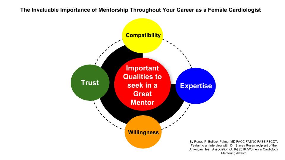
There is an ever increasing need to not only recruit more females in the field of Cardiology, but to also retain many talented female cardiologists in the field. Finding a good mentor and fostering good mentorship is invaluable for many females throughout their career in Cardiology. The Women in Cardiology Committee of the American Heart Association (AHA) values the importance of good mentorship and as such bestows the Women in Cardiology Mentor Award that is sponsored by the AHA Council on Clinical Cardiology to Cardiologists who have been recognised as having an outstanding record of effectively mentoring and supporting female cardiologists. Dr. Stacey E. Rosen,  Endowed Chair and Vice President for the Katz Institute for Women’s Health at Northwell Health, Partners Council Professor of Women’s Health at Hofstra North Shore-LIJ School of Medicine at Hofstra University and Professor of Cardiology at the Donald and Barbara Zucker School of Medicine at Hofstra/Northwell is the recipient of the 2018 AHA Women in Cardiology Mentoring Award. She has mentored and supported numerous female cardiologists, and in the following interview, I had the honor of discussing the following questions with Dr. Rosen.

Courtney could we add a hyperlink to the AHA WIC mentor award nomination site?


What attracted you to the field of Cardiology?

Dr. Rosen: “I am the first member of my family to become a doctor. I am the daughter of educators and while in high school , I volunteered at Memorial Sloan Kettering in the pediatric child-life center. I thought that the medical field would allow me to pursue various professional options including clinical care, education, advocacy and research. I felt one could never be bored in medicine! As a student in the 6-year medical program at Boston University, we dissected a bovine heart and I was so amazed by the “simplicity” of the structure. Nothing else seemed intriguing after that – my only important decision was between pediatric cardiology and adult cardiology.”

Who were the inspirational persons that influenced this decision?

Dr. Rosen: “The Division of Cardiology at Boston University School of Medicine in the 1980’s – and ever since – was extraordinary. The faculty at Boston City Hospital (BCH) demonstrated commitment and passion for those in the underserved neighborhoods near BCH and the faculty at University Hospital were national respected clinicians , investigators and educators.”

Who were your mentors in Cardiology and how did they contribute to the advancement of your career?

Dr. Rosen: I completed Internal Medicine residency and a chief resident year at Montefiore Medical Center in the Bronx, and was privileged to work with an impressive Division of Cardiology. Hildrud S. Mueller, MD and James Scheuer , MD taught me the importance of rigor and attention to detail in both clinical work and research. I presented my first oral presentation with John Fisher, MD – the recently retired division chief – and learned to perform a complete and thorough cardiac exam with Mark Menegus, MD.

As a fellow at Cornell – New York Hospital, Mary Roman, MD, Peter Okin, MD, Paul Kligfiled, MD, and Richard Devereux, MD fostered my fascination with imaging and non-invasive cardiology. Doppler physics was replacing hemodynamic assessment in the cardiac catheterization lab and I was hooked!

Finally, my first faculty position was at Mount Sinai Medical Center. Working in the echo lab with Martin Goldman, MD was an extraordinary opportunity. He inspired me to view echocardiography as a critical tool to enhance optimal patient care and helped me to develop the skills to become a lab director in my next position.

You have mentored many colleagues in the Cardiology field and have been valued by many as a great mentor, which led to your selection for the 2018 AHA Women in Cardiology Mentorship Award. What are the factors that mentees should consider when selecting a mentor?

Dr. Rosen:Mentees should consider several critical factors:

  • Compatibility – Choose someone with whom you are compatible but not someone who is a “mini me“ of yourself. You do want a mentor who will challenge you, be comfortable providing feedback, and teach you to internalize and utilize feedback to advance your goals.
  • Trust – You want a mentor whom you can trust – you will likely be sharing important and perhaps confidential conversations. It is also critical to know that you must earn a sense of trust from your mentor.
  • Expertise – Your mentor does not have to have the most senior titles or positions, but should have the requisite expertise to help you advance your career and help navigate challenges.
  • Willingness – A great mentor is one who is devoted to helping you develop a vision and is delighted to share knowledge and wisdom. It should be someone who is a good listener and has sufficient time to commit to the relationship.


Would you recommend having more than one mentor?

Dr. Rosen:  “Absolutely! But do remember that the mentee needs to commit sufficient time to each relationship in order to optimize the value of the partnership


How can mentees truly harness the power of strong mentorship?

Dr. Rosen:First – choose your mentor wisely and respect the relationship. Meet with individuals who you think may be good mentors and ask questions that will help you make a decision and get advice from friends and colleagues. Networking is often the key to identifying good choices.

Second – do the work and always respect the relationship! Discuss the goals and expectations of the partnership, as well as the process for communication, meetings and feedback. Be respectful of your mentor’s time and build trust immediately.


What are the differences between mentorship and sponsorship?

Dr. Rosen: Simply put – mentors advise you, while sponsors advance your career. A mentor is someone who can offer support, guidance and feedback, and allow you to develop your personal vision. A sponsor is earned – not chosen. A sponsor connects us to opportunities and advocates for our career advancement. It has been said that women are over-mentored and under-sponsored.


How has the experience of being a great mentor for so many colleagues contributed to your own career and personal growth?

Dr. Rosen: My late father was a middle school principal and my mother is a retired school teacher who taught in an underserved community in Brooklyn, NY. I learned from them the deep satisfaction one gets from teaching and advancing someone’s skills and abilities. I am also so grateful to MY mentors and get great satisfaction from “paying it forward.”  I am certain that I have learned as much from my mentors as they have learned from me!

What have been the 3 most rewarding experiences you have had throughout your career?

Dr. Rosen: As the director of the fellowship program at Northwell, I have had the privilege to work with dozens of trainees. I have helped impact their professional success and hopefully, the joy they find from practicing cardiology.

As chief of cardiology at Long Island Jewish Medical Center, I was able to rebuild a division that had weathered a hospital merger. Together with my colleagues, we completely revamped the division.

As a lifelong advocate for women’s heart health, I get enormous pride seeing the impact we have had on women’s health through improved clinical care, advances in gender-specific investigation and through advocacy and changes in policy. I know that our work is not done and that we still need to continue to advance women’s heart health agenda.

How has the field of Cardiology evolved over the duration of your career with regards to gender diversity and inclusivity of women in Cardiology?

Dr. Rosen: Unfortunately, our field has not advanced sufficiently when it comes to gender diversity and inclusion in Cardiology. Currently, fewer than 25% of cardiology fellows are female and fewer than 15% of board-certified cardiologists are women. Now that women are 50% of medical school graduates, the importance of developing a strategic approach to this lack of diversity is critical, or we will see a true talent drain in the near future. The good news is that both the American Heart Association and the American College of Cardiology have focused on lessening this disparity by better understanding the barriers facing female cardiologists and by making changes that will encourage young women to choose Cardiology.

What advice would you give to females considering a career in Cardiology?

Dr. Rosen: I can honestly think of no better choice! As clinicians, we can develop long-term longitudinal relationships and have an enormous impact on health and longevity. I believe that cardiology combines the best features of primary care and subspecialty medicine. As investigators, we can have a lasting impact on individuals and communities. Cardiology is also a field that is perfect for those who enjoy advocacy and advancing health policy improvements. I urge women NOT to eliminate Cardiology as a possibility because of concern about the challenges. Find the area of the field that you love, without fear or compromise, and then make decisions that will allow you to fulfill your vision of work – life integration.

References:

  1. Lewis SJ, Mehta LS, Douglas PS, et al. Changes in the Professional Lives of
    Cardiologists Over 2 Decades on behalf of the American College of Cardiology Women in Cardiology Leadership Council. J Am Coll Cardiol 2017


hidden

The Powerful Role of Social Media in the Field of Cardiology

The growth and use of social media have grown exponentially over the last decade with an eight fold increase since 20051. Social media is generally defined as an Internet-based platform that allows individuals and/or communities to gather virtually to communicate ideas, collaborate, share information, share pictures and videos, either as a direct message or general post in real time1. There are several social media platforms that healthcare professionals may use, such as Twitter, LinkedIn, Doximity, and Facebook. Twitter has been a very popular platform in the field of Cardiology with many Cardiologists, Cardiovascular research scientists, Cardiology providers, professional journals and Cardiology-based professional societies using this platform to expand their reach to their colleagues, professional society members, and the public in an effort to educate, advocate and raise awareness. There are several powerful roles that social media serves in the Cardiology field. These include networking, sharing meaningful opinions, fostering educational discussions centered around a cardiology topic or paper of interest, promoting or raising awareness of the latest research or guideline publication, promoting a professional meeting or event, promotion of healthy initiatives, collaboration among colleagues and support of colleagues.


Beneficial Uses of Social Media

a) Networking

Social media platforms allow many professionals in the cardiology field to connect with other colleagues and follow prominent cardiologists and research scientists. This connection transcends geographic borders, and therefore allows users to extend their networking reach internationally. This ability to network provides a sense of community and serves as one’s professional village where colleagues are able to share their professional ideas and share opinions on various topics.

b) Sharing important opinions and educational discussions on topics or publications relevant to Cardiology

Twitter also allows users to discuss topics and publications relevant to cardiology. Many times these are threads of a conversation joined by several colleagues. However, several professional organizations such as the American Society of Echocardiography (ASE) and the American College of Cardiology (ACC) have virtual tutorials, called “tweetorials,” which allows users to present and discuss a topic of interest in real time. These discussions can be very educational and serve as great learning tool. These discussions may also include reference to relevant publications and allows users to stay up to date with the scientific literature, as well.

c) Promotion of professional meetings and events

Many professional societies, such as the American Heart Association (AHA), American College of Cardiology (ACC), American Society of Echocardiography (ASE), American Society of Nuclear Cardiology (ASNC), Society of Nuclear Cardiology (SCCT), and the Society of of Cardiac Magnetic Resonance Imaging (SCMR), use social media platforms to promote their annual scientific meetings and events at these meetings. In fact, many of these meetings have social media (SoMe) ambassadors to help in promoting their meeting and to share important educational slides and messages from the meeting with other social media users, which is an excellent educational tool in getting important points out to the the rest of the cardiology community and the public.  Additionally, these professional meetings allow for cardiology colleagues who have connected virtually on Social Media to meet in person, as well. A hashtag (#) is a metadata tag that is used on social media platforms that allows posted content associated with a specific theme or content to be easily found2. Useful and popular hashtags used in the field of cardiology on Twitter are #CardioTwitter and #Cardiology. In fact many of the annual scientific meetings for several professional organizations will use hashtags for their meeting to allow social media users to readily identify posted social media content related to the meeting. This usually generates a significant degree of social media traffic and commentary related to the meeting and this further promotes the meeting and the professional organization globally. In fact at the 2018 annual American Heart Association’s Scientific Sessions, (#AHA18) there were over 300 million impressions generated globally2 on Twitter using the #AHA18 hashtag.

d) Discussion and promotion of latest research papers and guidelines

Many professional medical journals post important publications such as research papers and guidelines on social media to assist in promotion of important educational documents. In addition social media users also post their latest research papers and invited talks to help in promoting their scientific work and in the sharing of important educational information. With regards to posted research papers, it has been suggested that citations of research papers on Twitter can increase the citation rate of the paper and can also increase the impact factor of the the publication journal2. A prior analysis3 has shown that social media activity related to a publication paper increases the citation rate of the paper and therefore helps to promote published academic work. In fact the latest 2018 AHA/ACC Cholesterol management guidelines4, as well as the latest Physical Activity guidelines5, were released at the recently concluded American Heart Association meeting (#AHA18) and there was a significant amount of social media activity and discussion related to these two manuscripts. This therefore assisted to raise awareness of these guidelines within the cardiovascular community.

e) Starting healthy initiatives and sharing health promoting information with peers and the public

Promotion of healthy initiatives, such as heart healthy eating and increasing physical activity, have also been done on social media. Many cardiologists have used social media to share health educational material with the public and their colleagues.

f) Collaboration with and Providing Support for colleagues

Social media, especially Twitter, can help to create your professional community with colleagues who have similar professional interests. It provides a platform for collaboration with peers for various initiatives and opens the door for opportunities to collaborate with colleagues on research projects. This social media village creates a network that can be supportive with regards to helping to promote your professional interests and your academic publications through retweets and commentary.


Responsible use of social media

Responsible use of social media is very important, Always ensure that there is adherence to patient privacy regulation and ensure that social media posts are free of any patient identifying information. It is also vital that you maintain a high level of professionalism and avoid posting any social media information or pictures that can be professionally and ethically compromising for both yourself and others. It is very important not to tarnish your professional brand.6


Conclusion

The benefits of social media platforms such as Twitter are numerous and proves to be an increasingly relevant  learning tool that assists in keeping one abreast of the medical literature. Twitter is also very useful for one’s career  growth and provides a great opportunity for networking with peers globally. Social media helps in building your professional brand.


References:

  1. Ventola CL. Social Media and Health Care Professionals: Benefits, Risks, and Best Practices. P T. 2014 Jul; 39(7): 491-499, 520.
  2. American Heart Association Scientific Sessions 2018 meeting metrics provided by the AHA
  3. Eysenbach G. (2011) Can Tweets predict citations? Metrics of social impact based on Twitter and correlation with traditional metrics of scientific impact. J Med Internet Res 13:e123.
  4. Grundy SM, Stone NJ, Bailey AL, Beam LT, Birtcher KK, et al. 2018AHA/ACC/AACVPR/AAPA/ABC/ACPM/ADA/AGS/APhA/ASPC/NLA/PCNA Guideline on the Management of Blood Cholesterol. JACC Nov 2018, 25709; DOI: 10.1016/j.jacc.2018.11.003
  5. The Physical Activity Guidelines for Americans: THe HHS Roadmap for an Active Healthy Nation. Second Edition. ADM Brett P. Giroir, MD
  6. Bullock-Palmer RP. You Are Now a Board-Certified Cardiologist and Cardiac Imager…Now What? The Importance of Lifelong Learning and Career Growth
    May 2018 https://www.acc.org/membership/sections-and-councils/imaging-section/section-updates/2018/05/17/09/44/you-are-now-a-boardcertified-cardiologist-and-cardiac-imager