hidden

#BlackCardioInHistory: Daniel Hale Williams – Pioneer in open-heart surgery in the United States

This is part of the #BlackCardioInHistory series.  #AHAEarlyCareerVoice is partnering with #BlackInCardio to feature a series of profiles of black/African American Cardiologists for #BlackInCardioWeek2020.  For more information: blackincardio.com


Daniel Hale Williams III was born to a family that owned a barber business and worked in the Equal Rights League in Hollidaysburg, Pennsylvania, in 1856 (1). Young Daniel moved to Baltimore as a young boy to become a shoemaker’s apprentice when his father died but did not like the work (1). His family had moved to Illinois, so he moved back with them and began barbering with the long-term goal of pursuing his education (1).

Daniel Hale Williams III apprenticed with Dr. Henry Palmer, who was an accomplished surgeon. From there, Daniel attended the Chicago Medical College (1).

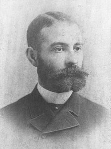
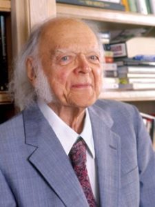
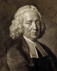
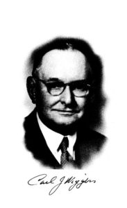
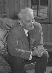
(2) Daniel Hale Williams (Pre 1923 photograph, public domain). https://en.wikipedia.org/wiki/File:Daniel_Hale_Williams.jpg From Wikipedia page: Daniel Hale Williams.

After completing medical school, Dr. Williams began to practice on Chicago’s south side while teaching anatomy at the Chicago Medical College (1). He was an early adopter of Louis Pasteur’s sterilization to prevent transmission of infection (1).

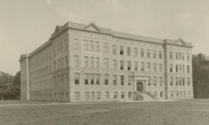
In the late 1880s in America, Black Americans were prevented from being admitted to hospitals and could not be hired at hospitals (1). While Black women had a long history of working as nurses, they were often denied opportunities for formal training (3). Dr. Daniel Hale Williams III disagreed with this practice and opened the Provident Hospital and Training School for Nurses which was the first hospital in the United States that was racially integrated (1).

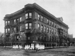
(3) Provident Hospital and Training School for Nurses (36th and Dearborn Streets, Chicago). Chicago History Museum (ICHi-040212). https://www.chicagohistory.org/provident-hospital/. Brittany Hutchinson.

A few years later, in 1893, Dr. Williams treated a man with a significant stab wound to the chest, named James Cornish (1). Dr. Williams successfully sutured the man’s pericardium allowing Cornish to live for many years. Only 3 other surgeons, Dr. Francisco Romero, Dr. Henry Dalton, and Dr. Dominique Jean Larrey, have been credited as performing open-heart surgery previously (1,4). One year later, in 1894, Dr. Williams was appointed as Chief Surgeon of the Freedmen’s Hospital, which has seen a high relative mortality rate. He moved back to Chicago upon marrying Alice Johnson, where he continued working for Provident Hospital (1).

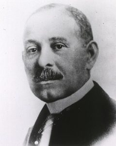
(2) Daniel Hale Williams (National Library of Medicine believes this to be public domain). https://commons.wikimedia.org/wiki/File:Daniel_H._Williams.jpg From Wikipedia page: Daniel Hale Williams.

In 1897 Dr. Williams was appointed to the Illinois Department of Public Health to increase medical standards in hospitals (2). In 1913, Dr. Williams was the only Black American member of the American College of Surgeons (5). Dr. Williams died in 1931 in Idlewild, Michigan from a stroke (2). Some of Dr. Williams’s honors include membership in the Chicago Surgical Society and American College of Surgeons, which were both uncommon for Black Americans at the time (2). Dr. Williams also received honorary degrees from Howard University in Washington DC and Wilberforce University in Wilberforce, Ohio (2).


Reference

  1. Biograhy.com Daniel Hale Williams Biography. 6/5/2020. https://www.biography.com/scientist/daniel-hale-williams. Accessed 10-19-2020.
  2. Daniel Hale Williams (Pre 1923 photograph, public domain). https://en.wikipedia.org/wiki/Daniel_Hale_Williams#cite_note-EB-6 From Wikipedia page: Daniel Hale Williams
  3. Provident Hospital and Training School for Nurses (36th and Dearborn Streets, Chicago). Chicago History Museum (ICHi-040212). https://www.chicagohistory.org/provident-hospital/. Brittany Hutchinson.
  4. Wikipedia.com Henry Dalton. 4/4/2020. https://en.wikipedia.org/wiki/Henry_Dalton. Accessed 10-19-20
  5. Daniel Hale Williams American Physician. Encyclopedia Britannica. 2018.


“The views, opinions and positions expressed within this blog are those of the author(s) alone and do not represent those of the American Heart Association. The accuracy, completeness and validity of any statements made within this article are not guaranteed. We accept no liability for any errors, omissions or representations. The copyright of this content belongs to the author and any liability with regards to infringement of intellectual property rights remains with them. The Early Career Voice blog is not intended to provide medical advice or treatment. Only your healthcare provider can provide that. The American Heart Association recommends that you consult your healthcare provider regarding your personal health matters. If you think you are having a heart attack, stroke or another emergency, please call 911 immediately.”


hidden

#BlackCardioInHistory: Dr. Edward William Hawthorne

This is part of the #BlackCardioInHistory series.  #AHAEarlyCareerVoice is partnering with #BlackInCardio to feature a series of profiles of black/African American Cardiologists for #BlackInCardioWeek2020.  For more information: blackincardio.com


Edward William Hawthorne was born near Port Gibson, Mississippi as the son of a minister and teacher. He suffered from polio at the age of 7 (1). He graduated high school in Washington DC and began his undergraduate study at Fisk University but later transferred to Howard University (1). It was at Howard University where Hawthorne spent most of the rest of his life.

(2) https://www.acc.org/latest-in-cardiology/articles/2018/02/13/14/42/harold-on-history-black-history-month-and-pioneering-african-american-physicians. Photo Credit: Howard University.

Hawthorne earned his bachelor’s in science degree in 1941 and completed his medical degree in 1946 (1). Hawthorne was an intern at Freedmen’s Hospital from 1946-1947 and developed an interest in research, especially in the cardiovascular field (1). Dr. Hawthorne completed a Masters in Science in physiology in 1949, and then a Ph.D. in physiology in 1951, both from the University of Illinois in Chicago (1). This would be Hawthorne’s only stint away from Washington DC.

In 1951 Dr. Hawthorne moved back to Washington DC as he was appointed to faculty at Howard University(1). Dr. Howard helped organize masters and doctoral graduate programs in physiology at Howard and a laboratory focusing on cardiovascular research(1). Dr. Hawthorne was the head of the physiology department until 1969(1). All the while Dr. Hawthorne was moving ranks of the administration. He was assistant Dean of the College of Medicine from 1962-1967, associate Dean of the College of Medicine until 1970, and Dean of the Graduate School of Arts and Sciences in 1974. During these appointments, he was also Chairmen of the department of physiology and biophysics in 1969-1974(1).

Hawthorne referred to his research as “a personal vendetta against ignorance”(1). Dr. Hawthorne was a leader in Renal physiology research and utilized animal models ranging from rats, rabbits, dogs, and horses(1). Dr. Hawthorne was a pioneer in using techniques to measure heart function and size in conscious animals(1). In 3 papers from 1959 and 1962, both published in Circulation Research, Hawthorne examines hypertension and left ventricle size in dogs (3, 4, 5). Dr. Hawthorne pioneered an experimental hypertension model in dogs consisting of constricting the brachiocephalic and left subclavian arteries surgically(5).

Figure 1 from Chronic experimental hypertension in dogs after constriction of brachiocephalic and left subclavian arteries. HAWTHORNE EW, et al. Circ Res. 1962. PMID: 13905535.

Dr. Hawthorne would go on to publish many papers on cardiovascular physiology, which ultimately led to his election as a fellow of the American College of Cardiology in 1969 and vice president of the American Heart Association from 1969-1972(1). He was also active in predominantly Black associations, including the John A. Andrew Clinical Society, Alpha Omega Alpha, Alpha Phi Alpha, and Association of Former Interns and Residents of Freedmen’s Hospital. In 1980 he was elected to the prestigious Institute of Medicine of the National Academy of Sciences(1). Dr. Hawthorne died in October 1986, only 4 months after his final manuscript was published(6). Dr. Hawthorne is remembered as a pioneer for cardiovascular physiology research and education and a pioneer in the field of hypertension.


Reference

  1. Hawthorne, Edward William. https://doi-org. /10.1093/anb/9780198606697.article.1201084 American National Biography. Accessed 10-19-2020
  2. https://www.acc.org/latest-in-cardiology/articles/2018/02/13/14/42/harold-on-history-black-history-month-and-pioneering-african-american-physicians. Photo Credit: Howard University.
  3. Instantaneous dimensional changes of the left ventricle in dogs. HAWTHORNE EW. Circ Res. 1961. PMID: 13712425.
  4. Telemetering of ventricular circumference in dogs. HAWTHORNE EW, et al. J Appl Physiol. 1961. PMID: 13905534.
  5. Chronic experimental hypertension in dogs after constriction of brachicephalic and left subclavian arteries. HAWTHORNE EW, et al. Circ Res. 1962. PMID: 13905535.
  6. Estimation of left ventricular mass in conscious dogs. B Coleman, L N Cothran, E L Ison-Franklin, E W Hawthorne. Among authors: hawthorne ew. Am J Physiol. 1986. PMID: 3789168

“The views, opinions and positions expressed within this blog are those of the author(s) alone and do not represent those of the American Heart Association. The accuracy, completeness and validity of any statements made within this article are not guaranteed. We accept no liability for any errors, omissions or representations. The copyright of this content belongs to the author and any liability with regards to infringement of intellectual property rights remains with them. The Early Career Voice blog is not intended to provide medical advice or treatment. Only your healthcare provider can provide that. The American Heart Association recommends that you consult your healthcare provider regarding your personal health matters. If you think you are having a heart attack, stroke or another emergency, please call 911 immediately.”

hidden

#BlackCardioInHistory: Dr. Marie Maynard Daly

This is part of the #BlackCardioInHistory series.  #AHAEarlyCareerVoice is partnering with #BlackInCardio to feature a series of profiles of black/African American Cardiologists for #BlackInCardioWeek2020.  For more information: blackincardio.com


Photo credit: Ted Burrows, Archives of the Albert Einstein College of Medicine (https://www.sciencehistory.org/historical-profile/marie-maynard-daly)

Dr. Marie Maynard Daly (1921-2003) became the first African American woman to receive a PhD in Chemistry. Dr. Daly was born in Queens, New York. She had the ambition to become a chemist through the influences of her father, who was on track to graduate from Cornell with an undergraduate degree in Chemistry when he was forced to drop out because of financial circumstances. Dr. Daly graduated magna cum laude from Queens College in New York with a bachelor’s degree in Chemistry. She then enrolled in a Master’s program in Chemistry at New York University and graduated in 1 year while working at Queens College as a part-time laboratory assistant.

Marie M. Daly Biography. Biography.com/scientist/marie-m-daly 08/26/2020

From NYU, soon to be Dr. Daly enrolled in a doctoral program at Columbia. Her dissertation was titled A Study of the Products Formed by Action of Pancreatic Amylase on Corn Starch. Dr. Daly received her PhD in Chemistry in 1947 and was the first African American woman to receive a PhD in chemistry in the United States. Dr. Daly went on to teach at Howard University and then began researching at the Rockefeller Institute in New York as a post-doctoral fellow. After 7 years at the Rockefeller Institute, Dr. Daly started to teach at the College of Physicians and Surgeons at Columbia University in New York in 1955. She continued her research while at Columbia studying arterial metabolism.

Dr. Daly made seminal findings in arterial metabolism. In particular, she published findings of how respiration and cytochrome oxidase activity are altered in rat aortas during hypertension which was published in the Journal of Experimental Medicine in 1958. In 1960 Dr. Daly moved to Albert Einstein College of Medicine at Yeshiva University and from 1958 to 1963, Dr. Daly was an investigator for the American Heart Association.

M.M. DALY, E.G. GURPIDE. J Exp Med. 1959 Feb 1;109(2):187-95. doi: 10.1084/jem.109.2.187. PMID: 13620848

In 1963 Dr. Daly published another paper, this time examining the concentration of cholesterol and cholesterol synthesis in hypertensive rats, which she published in the Journal of Clinical Investigation. In 1970 Dr. Daly published a methods paper on how to isolate intimal-medial tissues in arteries from rabbits. Dr. Daly was an integral part of the early molecular study using small mammal models to examine atherosclerosis and hypertension and was a pioneer in cardiovascular research.

M.M. DALY, Q.B. DEMING, V.M. RAEFF, L.M. BRUN. J Clin Invest. 1963 Oct;42(10):1606-12. doi: 10.1172/JCI104845. PMID: 14074354

H. WOLINSKY, M.M. DALY. Proc Soc Exp Biol Med. 1970 Nov;135(2):364-8. doi: 10.3181/00379727-135-35052. PMID: 4921030

WOLINSKY, M.M. DALY. Proc Soc Exp Biol Med. 1970 Nov;135(2):364-8. doi: 10.3181/00379727-135-35052. PMID: 4921030

Thus, it is not surprising that Dr. Daly was a member of the board of governors of the New York Academy of Sciences along with being a fellow of the American Cancer Society, American Association for the Advancement of Science, New York Academy of Sciences, and Council of the American Heart Association. In 1999, just 4 years before her death, the National Technical Association recognized Dr. Daly as one of the top 50 women in Science, Engineering, and Technology. Finally, in 2016, a new elementary school was named The Dr. Marie M. Daly Academy of Excellence after Dr. Daly and her many achievements.


“The views, opinions and positions expressed within this blog are those of the author(s) alone and do not represent those of the American Heart Association. The accuracy, completeness and validity of any statements made within this article are not guaranteed. We accept no liability for any errors, omissions or representations. The copyright of this content belongs to the author and any liability with regards to infringement of intellectual property rights remains with them. The Early Career Voice blog is not intended to provide medical advice or treatment. Only your healthcare provider can provide that. The American Heart Association recommends that you consult your healthcare provider regarding your personal health matters. If you think you are having a heart attack, stroke or another emergency, please call 911 immediately.”

hidden

Sensing oxygen – Leland Clark and the origins of direct oxygen measurement in blood

As we’ve all been back from AHA19 for a few weeks now, I began thinking about the research presented. Throughout the conference I tried to focus on topics of aging and cardiovascular disease, which is my current research focus. I kept finding myself in front of posters or at talks on metabolism and my personal favorite organelle, the mitochondria.

I’ve been fascinated by mitochondria since graduate school where I ran ~300 Seahorse assays on clinical samples among other mitochondrial measures. To be clear, I was a bit burnt out on the Seahorse after my PhD but that feeling has faded. These oxygen consumption assays to measure mitochondrial function have gained popularity in recent years, which got me thinking about the origins of these devices.

The first measures of oxygen tension in blood were developed by Leland Clark1. A major limitation of measuring oxygen tension in solutions was that the liquid needed to either be quiescent or constantly stirred, which limited the use in vivo1.

Dr. Leland Clark

Dr. Leland Clark2 (https://en.wikipedia.org/wiki/Leland_Clark#/media/File:Dr._Leland_C._Clark_Jr_2005.jpg)

Measurements I blood, which would be clinically useful, were difficult because tissues were constantly moving. Leland Clark overcame this by developing the polarographic electrode, now termed the Clark Electrode1. This became the foundation for measures of oxygen consumption rate that is the gold standard measure of mitochondrial function in isolated tissue. These are now used in the Seahorse respirometer and the Oroboros oxygraph, the 2 most popular commercial respirometers.

Polarographic oxygen electrode

Polarographic oxygen electrode1 (https://www.physiology.org/doi/pdf/10.1152/jappl.1953.6.3.189)

The development of the Clark Electrode allowed surgeons to have immediate measurements of blood oxygen during surgery, which allowed for open heart surgeries. Dr. Clark not only developed the Clark Electrode, he helped to develop the glucose monitor, which is still critical for many diabetic patients to monitor their care. He also helped to develop the first heart-lung machine helped pave the way for one of the first intensive care units in the world3.

While Dr. Clark was more concerned with measuring oxygen saturation in blood to improve direct patient care, he didn’t realize that he would also greatly advance the field of metabolism by allowing for accurate measurements of mitochondrial function. I personally owe Dr. Clark a debt of gratitude because I wouldn’t be where I am today without his pioneering inventions.


References:

  1. https://www.physiology.org/doi/pdf/10.1152/jappl.1953.6.3.189
  2. https://en.wikipedia.org/wiki/Leland_Clark#/media/File:Dr._Leland_C._Clark_Jr_2005.jpg
  3. https://www.ysi.com/about/history


The views, opinions and positions expressed within this blog are those of the author(s) alone and do not represent those of the American Heart Association. The accuracy, completeness and validity of any statements made within this article are not guaranteed. We accept no liability for any errors, omissions or representations. The copyright of this content belongs to the author and any liability with regards to infringement of intellectual property rights remains with them. The Early Career Voice blog is not intended to provide medical advice or treatment. Only your healthcare provider can provide that. The American Heart Association recommends that you consult your healthcare provider regarding your personal health matters. If you think you are having a heart attack, stroke or another emergency, please call 911 immediately.

hidden

Finding your way at a large national conference: tips and tricks

Attending a scientific conference can be daunting, especially for trainees. You’re still getting your sea legs so-to-speak with regard to your science, and on top of that, you may be presenting a poster or talk to an audience of the most distinguished researchers in your field. Many of us feel the same way, and it can be difficult to muster the courage to go up to someone and introduce yourself. But this is the perfect time to do it!

To really make the most out of your conference experience, especially at a large meeting like AHA Scientific Sessions, it is important to prepare ahead of time! Yes, you will need to book your flight and hotel and register for the conference itself, but you also need to consider how you’re getting to the airport or how you’re getting from the airport to your hotel. Do you know if there is a special lot for rideshare pickups or are you getting a shuttle? Does your hotel offer early check-in? These are all important questions, and at least in my experience, are often overlooked. Plan ahead for the little details, so your brain can be as stress-free as possible on the day of your travel.

Next, to the conference itself. Plan which sessions that you want to go to, but don’t overbook yourself. Scientific conferences often have some downtime, but perhaps you want to get a little exercise while you’re away or if you plan to get a late dinner with colleagues, you might want to skip the early-morning session. Find out what works best for you, so that you can focus on the science and network without feeling completely drained.

Planning which sessions you want to attend can be extremely important for poster presentations. Often, there are so many posters that you won’t have time to wander up and down the entire exhibit hall perusing at your leisure. Search the program using keywords and make a list of a handful of posters you’d like to see. Or if you have a colleague presenting a poster, do them a favor a pay a visit. If they have legions of people trying to get a glimpse of their work, then move on, but if they’re waiting and nobody is talking with them, go up and talk. It can be extremely isolating to stand next to a poster waiting anxiously for someone to come by and show interest.

One great way to get to know more people in any scientific society is to get involved. If you’re a trainee, there is usually some standard way that you can volunteer to join a committee. In AHA, you can apply to be an early career blogger! This is a surefire way to meet people. This serves many purposes. It helps the society in various ways. It also helps your CV. Finally, you are forced to meet and interact with people, many of whom may be in leadership roles. Knowing people at the conference you’re attending is a great way to boost the number of people that come to your poster or presentation and generally improves your conference experience.

My last tip is to have fun. If you use your time wisely, you can come away from a conference with a head full of new ideas in addition to some new friends.


The views, opinions and positions expressed within this blog are those of the author(s) alone and do not represent those of the American Heart Association. The accuracy, completeness and validity of any statements made within this article are not guaranteed. We accept no liability for any errors, omissions or representations. The copyright of this content belongs to the author and any liability with regards to infringement of intellectual property rights remains with them. The Early Career Voice blog is not intended to provide medical advice or treatment. Only your healthcare provider can provide that. The American Heart Association recommends that you consult your healthcare provider regarding your personal health matters. If you think you are having a heart attack, stroke or another emergency, please call 911 immediately.

hidden

Haemostatics: Stephen Hales and the first direct measurement of blood pressure

Blood pressure lowering medications are among some of the most effective and useful medications currently used in medicine. In 2017 the ACC/AHA released new guidance for the evaluation and management of high blood pressure in adults.1 This guidance outlines what is considered normal, elevated, and hypertension in adults.1 We now understand the great importance of blood pressure in health but this wasn’t always the case.

As far as we know, blood pressure was first mentioned by G. Harveo (1628) where he warned surgeons that blood could “jet out” of the artery.2 What I was interested in was the first measurement of blood pressure as we know it. That first measurement was done by Reverend Stephen Hales in 1733.2

I remember learning this during undergrad, and the image is striking. It is a picture of Hales and his associate with a horse laying on its side. They inserted a brass tube into the femoral artery connected to a glass tube running vertically out of the horse’s neck.3 The blood reached 8 feet 3 inches.4 Further work by Hales and others went on to describe blood pressures in different species and different vascular beds, but I haven’t been able to get the image of this horse out of my head.2

`

3(http://www.epi.umn.edu/cvdepi/wp-content/uploads/2011/05/Hales-Horse.jpg)

3(http://www.epi.umn.edu/cvdepi/wp-content/uploads/2011/05/Hales-Horse.jpg)

As my last post for the AHA Early Career Bloggers, I wanted to look into Stephen Hales. Who was he? Why did he do this experiment in the horse? And what else did he do? Hales was a Reverend in Middlesex who dabbled in many scientific pursuits. What I was interested in were those pertaining to the cardiovascular system. I went directly to the source: Statical Essays Containing Haemastatics.4

4 https://books.google.com/books?id=uDQ-AAAAcAAJ (eBook)

4 https://books.google.com/books?id=uDQ-AAAAcAAJ (eBook)


To get some context, this is what was thought about the cardiovascular system of arteries and veins at the time, “As an animal body consists not only of a wonderful texture of solid parts, but also of a large proportion of fluids, which are continually circulating and flowing, through and inimitable embroidery of blood vessels…it has, ever since the important discovery of the circulation of the blood, been looked upon as a matter well worth the inquiring into, to find the force and velocity with which these fluids are impelled…”4

This book doesn’t have any illustrations, unfortunately, but there are descriptions of the many experiments Hales performed. He repeated his experiment on pressures in the horse several times. In fact, the famous image of Hales with the horse was from his 3rd horse experiment. Eventually Hales started collecting the blood and determining how much there was in addition to pouring wax into the empty heart to make a cast and determine the chamber volumes. He compiled this into a pair of tables. 4

4 https://books.google.com/books?id=uDQ-AAAAcAAJ (eBook)


Based on Hales’ measures, he calculated that “a quantity [of blood] equal to the dog’s weight, will pass through the heart in 11.9 minutes”. You’ll notice that Hales made calculations for “Man” but these were derived from data from the other animals and he never performed these experiments on humans.4

Stephen Hales contributed to science in many ways including in other fields. He is famous for inventing a ventilator that circulated air in ships, prisons, and mines which likely saved many lives. Hales had no medical training. He obtained a bachelors degree in divinity and a Master of Arts. Hales is a reminder that great discoveries can be made by anyone with a curious mind.5

4 https://books.google.com/books?id=uDQ-AAAAcAAJ (eBook)

4 https://books.google.com/books?id=uDQ-AAAAcAAJ (eBook)


Don’t forget to register for #Hypertension19 happening this September 5-8 in New Orleans! 


References

  1. https://www.heart.org/-/media/data-import/downloadables/hypertension-guideline-highlights-flyer-ucm_497841.pdf
  2. The history of blood pressure measurement: from Hales to our days. V.A. Tsyrlin, M.G. Pliss, N.V. Kuzmenko. April 2016. Arterial Hypertension.
  3. http://www.epi.umn.edu/cvdepi/wp-content/uploads/2011/05/Hales-Horse.jpg (http://www.epi.umn.edu/cvdepi/essay/reverend-stephen-hales-on-blood-pressure/)
  4. Stephen Hales. Statical Essays Containing Haemastatics. (eBook: https://books.google.com/books?id=uDQ-AAAAcAAJ
  5. https://en.wikipedia.org/wiki/Stephen_Hales


hidden

Carl Wiggers – A Pioneering Figure in Cardiovascular Physiology

In my previous post about the history of AHA research grants, I became interested in another early figure within the AHA, Carl J. Wiggers. He was an Albert Lasker Award winner as well as the first editor-in-chief of the AHA journal Circulation Research. He was a fascinating figure.

Carl Wiggers

Source: National Academy of Sciences1: http://www.nasonline.org/publications/biographical-memoirs/memoir-pdfs/wiggers-carl.pdf


Dr. Wiggers was born in Davenport, Iowa, USA in 1883 and showed promise in academics early. His high school teachers prodded him to pursue medicine. He couldn’t afford 4-years of college, so he chose to attend the University of Michigan over Johns Hopkins Medical School. At the time U of M only required a high school diploma, while JHU required a college degree. This is the Medical School building at the time (now the School for Natural Resources and the Environment).1

medical school

“Medical School; BL004458.” http://quod.lib.umich.edu/b/bhl/x-bl004458/bl004458. University of Michigan Library Digital Collections. Accessed: July 15, 2019.


During medical school, Dr. Wiggers was offered a paid student assistantship in physiology to perform research, and upon graduation from medical school in 1906, he was promoted to an instructor in physiology.1 In 1912, Wiggers traveled to German to study with Prof. Otto Frank.

Wiggers apparently smuggled some of Frank’s designs back to his lab in the USA, now at Cornell.2 Wiggers wanted to improve the measurement of blood pressure and intended to use Otto Frank’s reflecting mirror monometer, eventually developing a portable version to bring to the patient’s bedside.1

Although World War I was raging, in 1918, Carl Wiggers was persuaded to accept a position at Western Reserve (now Case Western Reserve) University where he remained throughout his career. Here he could establish his own cardiovascular research center.1

In 1952, the American Heart Association asked Wiggers to organize and edit a new journal focused on basic research on fundamental studies of the cardiovascular system, Circulation – Research.1 The first issue was published in 1953 as Circulation Research (no hyphen) and quickly positioned the journal as a leader in cardiovascular research.3


Circulation Research

Origin and Early Years. Arnold M. Katz. Circulation Research.3 2001;88:1105–1111. https://doi.org/10.1161/hh1101.091991


At Western Reserve, Wiggers published over 400 original manuscripts and was instrumental in the career of many individuals including Nobel Prize winner Corneille Heymans in 1930. He was committed to training so much so that he often granted sole authorship to early investigators to further their careers.1

Wiggers is famous for his Wiggers Diagram as well as being a member of the National Academy of Sciences, an AHA Gold Hear Award recipient, and Lasker Award winner.1 He was a pioneer in physiology who continues to have a lasting impact today.

aortic blood momentum

Wikipedia.4 Modified from ugawara M, Uchida K, Kondoh Y, et al. Aortic blood momentum–the more the better for the ejecting heart in vivo? Cardiovasc Res 1997; 33(2): 433–46)


References:

  1. Eugene M. Landis. 1976. Carl John Wiggers 1883-1963. National Academy of Sciences www.nasonline.org/publications/biographical-memoirs/memoir-pdfs/wiggers-carl.pdf
  2. Am J Physiol. 1998 Apr;274(4):L467-74. doi: 10.1152/ajplung.1998.274.4.L467.
  3. Origin and Early Years. Arnold M. Katz. Circulation Research. 2001;88:1105–1111. https://doi.org/10.1161/hh1101.091991
  4. Wikipedia. Modified from ugawara M, Uchida K, Kondoh Y, et al. Aortic blood momentum–the more the better for the ejecting heart in vivo? Cardiovasc Res 1997;
hidden

The History of the AHA

Following up on an earlier post about the history of the NIH R01 grant, which morphed into a history of the NIH and the National Cancer Institute, I wanted to find out more about the history of the American Heart Association and the first AHA research awards.

This was much easier information to obtain! It is on the AHA website. The first award went to biochemist, Dr. Albert Szent-Gyorgyi, who was fascinating and possibly the topic of a future post. He received the first AHA grant in 1948 while he was at the Marine Biological Laboratory in Woods Hole, MA. He primarily contractile components of muscle including the heart.

Dr. Albert Szent-Gyorgyi (https://en.wikipedia.org/wiki/Albert_Szent-Gy%C3%B6rgyi)


The first AHA logo

The first AHA logo (https://www.heart.org/en/about-us/history-of-the-american-heart-association)

Easily finding this answer led to another question. When and how did the AHA begin in the first place? The public and physicians knew little about cardiovascular diseases in the early 1900’s when the AHA was founded. Heart disease was thought to be a slow, drawn-out death sentence. A group of 6 physicians believed that with scientific research, a cure could be found.

One of the first AHA meetings.

One of the first AHA meetings.(https://www.heart.org/en/about-us/history-of-the-american-heart-association)


The 6 founding members of the AHA were: Drs. Lewis A. Conner, Robert H. Halsey, Paul D. White, Joseph Sailer, Robert B. Preble, and Hugh D. McCulloch. Since the founding in 1924, the AHA’s has been primarily a scientific association. After awarding the first research grant in 1948, AHA began publishing its first journal Circulation in 1950.

From that point on, the AHA was instrumental in funding research that linked smoking and saturated fats to heart disease. AHA research was instrumental in the development of implantable pacemakers, CPR, artificial heart valves, statins, and AEDs. They also established a personal favorite of mine, the Jump Rope for Heart, which I proudly participated in as a child.

There were several changes geared toward unifying the AHA’s objectives, research standards, and guidelines through the years. In 1995, the AHA declared its strategic driving force: Providing credible heart disease and stroke information for effective prevention and treatment. The AHA still functions as a scientific organization but one that faces first and foremost on the public. The AHA’s driving force guides the scientific efforts of AHA members to public benefit.


Are you an AHA member and interested in applying for a research program or award?AHA research funding opportunities are listed at https://professional.heart.org/en/research-programs/application-information

hidden

The History of the Western Blot

Everybody has a western blot horror story. The history of the western blot. What has been your worst western blot experience?

Most basic scientists have done countless western blots in the lab. The western blot, or SDS PAGE and immunoblot, technique is indispensable in most labs as a method to measure relative expression level of your protein of interest. As an early career scientist, I’ve done hundreds of western blots, and I often wonder who came up with this technique.

When I was learning this technique in grad school, I was told that it is called a “western” blot because the precursor techniques for detecting DNA and RNA in gels were called the “southern” blot and “northern” blot. The southern blot being named after Edwin Southern.

This was actually very easy to find out. I went to the references section of the “Western Blot” Wikipedia page and found the first citation for the technique (not yet called a western blot). There were two papers published in 1979 on the technique by different groups. The first was by Jaime Renart, Jakob Reiser, and George Stark in July. The second paper by Harry Towbin, Theophil Staehelin, and Julian Gordon was published in September, and both papers were published in the Proceedings of the National Academy of Sciences of the USA.1,2

According to PNAS, the Towbin paper has over 43,000 citations while the Renart paper has only 493. Both papers undoubtedly have had a much wider impact but most current papers do not cite these originals anymore. A pubmed search for the terms “western blot” or “protein immunoblot” returns a combined 130,000 papers.1,2

The main difference between these two manuscripts was that the Renart technique transferred the proteins from the SDS gel into paper whereas the proteins were transferred into nitrocellulose in the Towbin paper. Now nitrocellulose is used and many use PVDF membranes; however, different labs have different preferences.

The original assembly from the Towbin paper was very simple and not much different from the more sophisticated apparatus marketed by biotech companies today.

Towbin H, Staehelin T, Gordon J. Proc Natl Acad Sci U S A. 1979 Sep;76(9):4350-4.

Towbin H, Staehelin T, Gordon J. Proc Natl Acad Sci U S A. 1979 Sep;76(9):4350-4.

The Renart paper shows blots probed with specific antisera and they look remarkably like blots shown in manuscripts today. The Towbin paper is remarkable because the authors then go on to show a variety of ways this technique can be used. They ran a 1-dimensional gel and stained all of the protein, in this case 60-S ribosomal proteins from chicken liver. Then they ran a 2-dimensional gel (shown below) of the same 60-S ribosomal proteins from chicken liver. Finally, they used antibodies to detect specific proteins and they demonstrated this using both horseradish peroxidase (HRP) and fluorescein (FITC).

Renart J, Reiser J, Stark GR. Proc Natl Acad Sci U S A. 1979 Jul;76(7):3116-20.

Renart J, Reiser J, Stark GR. Proc Natl Acad Sci U S A. 1979 Jul;76(7):3116-20.


Towbin H, Staehelin T, Gordon J. Proc Natl Acad Sci U S A. 1979 Sep;76(9):4350-4.

Towbin H, Staehelin T, Gordon J. Proc Natl Acad Sci U S A. 1979 Sep;76(9):4350-4.

This technique was officially coined “western blotting” in 1981 in a paper by W. Neal Burnette.3 While the name of the western blot is not very important, the technique is obviously one of the most valuable tools for molecular biologists, and one that provides constant amusement (or frustration) for graduate students and post-docs worldwide.

I have many of my own tips and tricks for western blotting but I won’t list them here. Everyone has their own western blot horror stories. One of my worst experiences is mixing up the red and black wires and seeing my precious samples running up and out of the wells, and if you use pre-cast gels which are available from several suppliers, please be kind to them. Don’t squeeze them:


Daniel Tyrrell, 2018, LC3 I and II western blot

Daniel Tyrrell, 2018, LC3 I and II western blot


References:

  1. Renart J, Reiser J, Stark GR. Proc Natl Acad Sci U S A. 1979 Jul;76(7):3116-20.
  2. Towbin H, Staehelin T, Gordon J. Proc Natl Acad Sci U S A. 1979 Sep;76(9):4350-4.
  3. Burnette WN. Anal Biochem. 1981 Apr;112(2):195-203.


hidden

The First Funded R01 Ever

The first two extramural grants for a research project (the precursor to today’s R01) were given on March 21, 1938 to Dr. John Bittner at Jackson Laboratory (then, the Roscoe B. Jackson Memorial Laboratory) in Bar Harbor, Maine for his work on breast cancer1,2.

Bittner J. Mammary Tumors In Mice In Relation to Nursing. Cancer Research. 1937;30(3).

Bittner J. Mammary Tumors In Mice In Relation to Nursing. Cancer Research. 1937;30.

One grant for $3,200 was awarded directly to Dr. Bittner, and the other for $9,900 was awarded to the Roscoe B. Jackson Memorial Laboratory by the National Cancer Institute. These first “R01-type” awards were given by the NCI from 1938-1943, and in that time, 65 grants-in-aid were paid2.

Marshino O. Administration of the National Cancer Institute Act, August 5, 1937, to June 30, 1943. JNCI: Journal of the National Cancer Institute. 1944:429-43.

Marshino O. Administration of the National Cancer Institute Act, August 5, 1937, to June 30, 1943. JNCI: Journal of the National Cancer Institute. 1944:429-43.

In the early days of extramural grants in the USA through the 1970’s, most awards were Program Projects, Centers, and Development grants. The NIH preferred to grant awards for multidisciplinary centers to carry out a range of projects in a certain area of interest. It wasn’t until after Watergate and Nixon’s resignation that Investigator-Initiated Research Projects (R01) began to dominate large Program-Project grants (P01)3.

To understand the path from these first grants to today’s R01’s, it is important to understand the history of the NIH. The Hygienic Laboratory became the NIH in 1930 with the passage of the Ransdell Act, and the NCI began with the National Cancer Institute Act in 1937. The NCI could administer extramural grants for research on cancer, but the NIH at large could not3.

The federal government greatly increased research spending in medical research during World War II, and when the war was over, both the government and scientists wanted to continue federally funded research. The NIH couldn’t legally administer extramural grants until the passage of the Public Health Service Act in 1944 which consolidated the NCI and NIH under one roof under the Surgeon General.

In 1946 the Research Grants Office was created to administer extramural research grants and fellowships awards. Congressional funding of the NIH increased steadily, and all the while the NIH was adapting its branding by renaming institutions (i.e. changing “National Microbiological Institute” and “Experimental Biology and Medicine Institute” to flashier names like “National Institute of Arthritis and Metabolic Diseases” and “National Institute of Allergy and Infectious Diseases,” which further enticed congressional funding). From 1946 to 1953, the number of funded projects increased from 80 to 2,000 and the funds increased from $780,000 to over $20,500,0003.

Mandel R. A Half Century of Peer Review, 1946-1996: Division of Research Grants, National Institutes of Health. Division of Research Grants, National Institutes of Health; 1996.

Mandel R. A Half Century of Peer Review, 1946-1996: Division of Research Grants, National Institutes of Health. Division of Research Grants, National Institutes of Health; 1996.

In 1946, the NIH Syphilis Study Section had its inaugural meeting. This was the first study section meeting at the NIH4. From this point, more and more study sections met to discuss the state of science and to review grant applications. In 1950, the standardization of study sections began with the assignment of priority scores from 1 to 5. This made it possible to rank grants based on merit for funding.

Mandel R. A Half Century of Peer Review, 1946-1996: Division of Research Grants, National Institutes of Health. Division of Research Grants, National Institutes of Health; 1996.

Mandel R. A Half Century of Peer Review, 1946-1996: Division of Research Grants, National Institutes of Health. Division of Research Grants, National Institutes of Health; 1996.

These changes lessened the workload of study sections initially, but as research design became more sophisticated, more and more of the meeting time was devoted to project discussions3. Eventually, some within the research community became dissatisfied with the closed-door process of grant review. In 1973, the Office of Management and Budget called for the abolishment of study sections, but the acting NIH Director, Dr. John Sherman, defended peer review as the highest priority.

Political events at the time caused turmoil within the NIH. A combination of budget constraints due to the war in Vietnam along with Watergate and procedural confusion and secrecy regarding the peer review process led to a reorganization at NIH. A familiar name, Ruth Kirschstein, director of the NIGMS in 1974, stated, “How can a system, devised in an era of elitism, of secrecy, and of economic growth…be adopted to an era in which stress in on equal opportunity, openness, and limited availability of funds?3.

Mandel R. A Half Century of Peer Review, 1946-1996: Division of Research Grants, National Institutes of Health. Division of Research Grants, National Institutes of Health; 1996.

Mandel R. A Half Century of Peer Review, 1946-1996: Division of Research Grants, National Institutes of Health. Division of Research Grants, National Institutes of Health; 1996.

There was rampant favoritism and bias in the review process, and over the next decade, the NIH worked to standardize peer review practices. This included extending project length beyond 3 years in 1984, scoring grants by percentiles in 1987. In the late 1980’s and through the 1990’s, grant applications could be submitted on discs rather than paper and a centralized database of applications was developed.

While the first grant application wasn’t submitted electronically using the PHS-398 form, it was the first time an NIH institute, the NCI, funded an extramural grant for an individual research project, which we now refer to as an R01.


References:

  1. Bittner J. Mammary Tumors In Mice In Relation to Nursing. Cancer Research. 1937;30(3).
  2. Marshino O. Administration of the National Cancer Institute Act, August 5, 1937, to June 30, 1943. JNCI: Journal of the National Cancer Institute. 1944:429-43.
  3. Mandel R. A Half Century of Peer Review, 1946-1996: Division of Research Grants, National Institutes of Health. Division of Research Grants, National Institutes of Health; 1996.
  4. Pederson T. The “study” role of past National Institutes of Health study sections. Mol Biol Cell. 2012;23(17):3281-4.