hidden

A new and evolving health struggle for Heart failure patients: COVID-19

It’s safe to say we are not living in normal times.  This is Heart Failure (HF) in the time of the coronavirus disease-2019 (COVID-19). Patients with COVID-19 and preexisting cardiovascular disease (CVD) are at an increased risk of severe disease and death. Moreover, infection has also been associated with cardiac injury such as acute myocardial infarction (AMI), myocarditis, and stress-induced cardiomyopathy leading to subsequent cardiogenic shock (CS) requiring advanced heart failure therapies. There is a bidirectional relationship between viral upper respiratory traction infection(URI) and worsening HF with an increase in hospital re-admission rate as previously noted with influenza. Patients with HF are especially susceptible to influenza-related complications, including acute decompensated HF and secondary pneumonia. Furthermore, HF is associated with greater in-hospital mortality and adverse clinical outcomes. With around 1 million confirmed COVID-19 cases and counting in the US, one would expect an increase in heart failure admissions. Over the past several weeks as the number of COVID-19 admissions increase, the number of patients admitted with heart failure admissions have been at their lowest, which raises the following question: Where are all the HF patients?

We can speculate that people are terrified at home so they are not showing up to the emergency departments. Patients could be slowly accumulating fluids and getting into a decompensated state. On the other hand, being less active, they could also have been experiencing less symptoms. First it was influenza season now overlapping with a COVID-19 pandemic. It would be expected to see an increased number of HF admissions.  It is suggested that we might be experiencing the calm before the storm when it comes to HF decompensation requiring hospitalization. The alternative is that social distancing is the remedy that we have long been waiting for to help decrease heart failure exacerbation and hospital re-admissions rates.

On one bright note, during a telehealth cardiology visit follow up with a long-term patient with chronic systolic heart failure known to have been admitted several times during the past year secondary to medication non-adherence, who admits that he has been feeling great. He takes all his medications religiously now, including his diuretics. He states that the fact that he stays home, he doesn’t have to worry about going to the bathroom to urinate so often when he gets out of the house, therefore he doesn’t miss any of his diuretic doses. He is also compliant with diet as he doesn’t eat out as often as he is used to. He admits that he stopped going out to fast food places. This is one very small sample. On the other hand, on another telehealth visit, there is a patient with newly diagnosed Non-Ischemic Cardiomyopathy and HF with reduced ejection fraction, who is been followed for up-titration of guideline directed medical therapy. It was a challenge to safely increase the dose of his medications without vital signs and avoiding to have the patient physically get to a laboratory to get blood work done. As of now, no major changes were made in the patient current management. Of note, patient did ask about holding angiotensin-converting enzyme (ACE) inhibitors because of what he heard from another source. Once more, no changes were made to the medical regimen and it was explained that it has been recommended based on different society guidelines and expert consensus report, to continue with ACE inhibitors1.

COVID-19 times are dynamic and medical information is constantly being updated. This is an ongoing discussion as the clinical data comes in. As the pandemic evolves and more telehealth visit under our belts, we will continue to find out more. Although as our health care system is currently fighting the COVID-19; we must brace ourselves for the aftermath whether our patients are dying at home, or slowly decompensating. Only time will tell.  As we are flattening to curve with social distancing, our patients with chronic conditions like HF are waiting at home with so much uncertainties surrounding their current and future medical care. “When life gives you lemon, make lemonade”.

The following suggestions can be useful when taking care of heart failure patients during these unprecedented times. (Figure 1) With COVID-19, we should let our HF patients know although social distancing is essential, they are a higher risk population for a complicated course if infected. It is important to inform them on when to seek medical care, whether it’s to contact a health care provider, call emergency medical services, or go to the emergency department. Although, prevention remains the best medicine. They should take the extra step in precautions and follow the latest recommendations from their local department of public health as we should always remind them of what those recommendations consist of via our telehealth visits.  From a cardiologist stand point, it is important to remain available whether it is via email, pager and/or more frequent telehealth visit if possible.  If they don’t have a scale and/or automatic blood pressure machines, it should be suggested to obtain them along with a thermometer from their local pharmacies. With a phone camera, it is feasible to assess Jugular Venous Distention, pitting edema. In addition, with weight trends, blood pressure and heart rate, clinical decisions could be made.  If available, assessment of data via CardioMEMS can also be very helpful in making medical decisions. Desperate times call for desperate measures.  This is too shall pass. If this is the calm before the storm for our heart failure patients, we should be ready when it hits remembering the sun always shines after a storm.

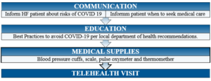
Figure 1. Heart Failure Care Suggestions During COVID-19

“The views, opinions and positions expressed within this blog are those of the author(s) alone and do not represent those of the American Heart Association. The accuracy, completeness and validity of any statements made within this article are not guaranteed. We accept no liability for any errors, omissions or representations. The copyright of this content belongs to the author and any liability with regards to infringement of intellectual property rights remains with them. The Early Career Voice blog is not intended to provide medical advice or treatment. Only your healthcare provider can provide that. The American Heart Association recommends that you consult your healthcare provider regarding your personal health matters. If you think you are having a heart attack, stroke or another emergency, please call 911 immediately.”


hidden

Peripartum Cardiomyopathy: Go Red & Recognize!

This one is dedicated to all women and their families who have been affected by Peripartum Cardiomyopathy (PPCM). Seldom detected, systolic heart failure can come as a surprise especially in young women. Developing heart failure during pregnancy, in the post-partum period or any other time throughout a subsequent pregnancy is not something any woman wants to worry about specially around the birth of their child.

The 2010 Heart Failure Association of the European Society of Cardiology Working Group defines PPCM to “an idiopathic cardiomyopathy presenting with Heart Failure (HF) secondary to Left Ventricular (LV) systolic dysfunction towards the end of pregnancy or in the months following delivery, where no other cause of heart failure is found”1, hence a diagnosis of exclusion.

The annual incidence of PPCM continues to increase. Women present with wide range of HF symptoms. Although some have complete recovery with guideline directed medical treatment, others have persistent myocardial dysfunction, advance heart failure and death which subsequently leads to devastating consequences for an entire family.  Delays in diagnosis usually occur because the symptoms and signs of PPCM can mimic the normal findings of pregnancy.  Early recognition and treatment of PPCM could lead to improvements in maternal and fetal mortality and morbidity. Easier said than done.

By the time a patient with PPCM sees a cardiovascular specialist, they often have worsening symptoms of heart failure with moderate to severe depressed left ventricular systolic function which means it was later recognized by either a primary care physician and/or an obstetrician-gynecologist prior to referral.

Awareness is key in early detection of PPCM. If you see something, say something. Think PPCM in all pregnant women. Since we mentioned that survival and recovery are both improved by early diagnosis, there is a validated self-test that can help with discerning heart failure from pregnancy related symptoms from Fett et al2. (Table 1.)


  1. Self-Test for Early Diagnosis of Peripartum Cardiomyopathy
Symptoms 0 points 1 point 2 points
Orthopnea None Need to elevate head Need to elevated 45 degrees or more
Dyspnea None Climb 8 or more steps Walking on level
Unexplained cough None At night Day and night
Excessive weight gain during last month of pregnancy None 2-4 pounds per week Over 4 pounds per week
Lower extremity edema None Below the knee Above the knee
Palpitations None When Laying down at night Day and night or any position

  The present of 4 or more points should prompt additional investigation.


Fett’s self-test can be an essential tool for the PCP and OB-GYN to aid in early detection of PPCM. Think about PPCM and use the self-test on all patients at risk to help guide further next steps in the diagnosis and management. In support of awareness of heart disease in women, think PPCM in which case the battle is half way won.


References:

  1.  Sliwa K, Hilfiker-Kleiner D, Petrie MC, et al. Current state of knowledge on aetiology, diagnosis, management, and therapy of peripartum cardiomyopathy: a position statement from the Heart Failure Association of the European Society of Cardiology Working Group on peripartum cardiomyopathy. Eur J Heart Fail 2010;12: 767–78.
  2. Fett, JD .Validation of a self-test for early diagnosis of heart failure in peripartum cardiomyopathy. Crit. Pathw. Cardiol. 10(1), 44–45 (2011).


“The views, opinions and positions expressed within this blog are those of the author(s) alone and do not represent those of the American Heart Association. The accuracy, completeness and validity of any statements made within this article are not guaranteed. We accept no liability for any errors, omissions or representations. The copyright of this content belongs to the author and any liability with regards to infringement of intellectual property rights remains with them. The Early Career Voice blog is not intended to provide medical advice or treatment. Only your healthcare provider can provide that. The American Heart Association recommends that you consult your healthcare provider regarding your personal health matters. If you think you are having a heart attack, stroke or another emergency, please call 911 immediately.”

hidden

It’s All In The Family

Our understanding of the genetic basis of adult-onset cardiomyopathy is rapidly evolving. Most of us learned to practice medicine in a paradigm that relied on detailed disease phenotyping, but now we have the ability to incorporate genetic and genomic information into routine clinical care. Even if you cannot remember the last time you thought about genes or pedigrees, here are a few modifications you can make to your practice today that will help you keep up with this exploding field of cardiovascular medicine.


1) Take a minimum three-generation family history in all patients with a primary cardiomyopathy.

  • The goals of taking a family history are to learn (1) whether the cardiomyopathy is familial, (2) about disease characteristics among family members, (3) if it is inherited in a specific pattern, and (4) to identify at-risk relatives.
  • Most adult-onset cardiomyopathies are inherited in an autosomal dominant pattern, but other inheritance patterns (e.g. dignetic, multigenic) are possible and need further study.
  • Importantly, an individual can have a genetic form of cardiovascular disease without having affected relatives. Most often, this is due to recessive inheritance, de novo mutations/variants, or reduced penetrance.


2) Use focused questions to obtain the family history.

  • The use of vague terms like “heart attack” can lead clinicians away from pursuing an inherited etiology of disease and prevent them detecting other important cardiovascular diagnoses like sudden cardiac death in family members.
  • Ask specific questions regarding heart failure symptoms (e.g. presence or absence of dyspnea at rest or on exertion, paroxysmal nocturnal dyspnea), arrhythmia symptoms (e.g. palpitations, presyncope, syncope with or without exertion), and sudden death (e.g. drowning, single-vehicle accidents) in family members.
  • Knowing about relatives’ cardiovascular procedures like arrhythmia ablation, cardiac surgery, device implantations, or heart transplantation can also be helpful.
  • If a multisystem syndrome like a laminopathy or Fabry disease is suspected, familiarize yourself with the extracardiac manifestations and include pertinent questions in your history.


3) Remember that diagnosing an individual with an inherited cardiovascular disease is just the first step in the process.

  • In cardiovascular genetics, a key concept is the transition of practice from individual patient-based care to family-based care. By incorporating the information you generate from your comprehensive multi-generational family history, you also gain insight into disease penetrance, expression, age of onset, and pleiotropy.
  • Generally, cardiomyopathies are considered “medically actionable” because evidence-based treatments to reduce morbidity and mortality exist.
  • Beyond just having effects on medical and device therapies, following this paradigm of family-based care also has implications for reproductive and family planning and lifestyle practices.


If you are interested in learning more about this topic, check out the 2018 update to the Heart Failure Society of America Practice Guideline on the Genetic Evaluation of Cardiomyopathy (PMID: 29567486).


hidden

The Unexpected Benefits of Extending Your Training

During my general cardiology fellowship, I developed a special interest in the care of patients with inherited cardiovascular disease. By virtue of the robust clinical activity of my division’s advanced heart failure and electrophysiology programs, I was exposed to clinical dilemmas like risk stratification in hypertrophic cardiomyopathy, primary prevention of sudden cardiac death in lamin A/C cardiomyopathy, and timing of heart transplantation for Danon disease early in my training. Refreshing my knowledge of clinical genetics alone was overwhelming, and I realized that while the rapid growth in genomic technologies was transforming our understanding of inherited cardiovascular disease, frontline clinicians were lagging behind in applying this knowledge to disease prevention and clinical care. To cultivate my interests further and learn to bridge this gap, I joined my institution’s new National Human Genome Research Institute (NHGRI)-supported postdoctoral training program in genomic medicine, a program created to prepare the next generation of physicians and scientists to implement genomic approaches to improve healthcare.

For M.D./D.O. trainees who have spent six consecutive years entrenched in clinical residency and fellowship programs, the idea of extending training by two years, re-entering the world of formal coursework and letter grades, and learning new skills to perform complex and unfamiliar research is more than enough to deter one from pursuing this career development track. However, participating in this program has afforded me many unexpected benefits outside the bounds of my clinical and research training:

  • Caring for patients with a new type of multidisciplinary team:
    • During my clinical training, my idea of a multidisciplinary care team was mostly grounded in my inpatient experience. While cooperating toward the same goal, physicians, nurses, advanced practice providers, therapists, nutritionists, pharmacists, social and case management workers often performed their roles asynchronously with little collaboration outside of the prescribed morning rounds. In contrast, my experience in our inherited cardiovascular disease clinic introduced me to a new paradigm essential to caring for patients and families with genetic disorders. I have been fortunate to learn about variant adjudication, pre-test and post-test counseling, cascade screening, and much more from our tremendous genetic counselors who are integral in the outpatient evaluations of our probands.
    • Though the initial years of my practice have been focused in adult medicine, I have learned about the importance of tracking variant segregation in families and of comprehensive transitions of care through our joint familial cardiomyopathy and arrhythmia programs, partnerships with our neighboring pediatric hospital.
    • Finally, I have witnessed the potential of real time bedside-to-bench-to-bedside research collaborations as shown by my mentors in their recent report of a clinical incorporation of rapid functional annotation of cardiomyopathy gene variants.1
  • Developing and sharing expertise:
    • In leading my fellowship’s didactic education curriculum as Chief Fellow, I took advantage of opportunities to share my new knowledge and skills with other fellows and residents. For our “fresh case” presentations, I often chose to present perplexing cases of cardiomyopathy to reinforce teaching points regarding the workup of genetic cardiomyopathies and the importance of taking a minimum three-generation family history.
    • After completing the Examination of Special Competence in Adult Echocardiography, I led a fellow teaching conference on echocardiography in hypertrophic cardiomyopathy. I also joined our internal medicine residents for a clinicopathologic conference as an expert discussant, a position usually reserved for faculty but generously offered to me given my interest in cardiovascular genetics and enthusiasm for teaching.
    • Pursuing these opportunities to develop and share my expertise has helped me solidify my own knowledge in the field, develop my oral and written communication skills, and grow as a peer mentor.
  • Meeting physicians and scientists outside of cardiovascular medicine:
    • The world often feels quite small while training within a medical specialty, but through my postdoctoral program, I have been exposed to physicians, scientists, and trainees in many disciplines outside of cardiovascular medicine. I heard diverse perspectives in my bioinformatics, biostatistics, and bioethics courses that have encouraged me develop my own independent opinions about my fields of interest. Multidisciplinary forums like genetics journal clubs, genetic rounds, and campus retreats have helped me contextualize the practice of genomic medicine.


My time in the postdoctoral program has shown me that these unexpected benefits of training are highly valuable to a trainee’s success. Through the genomic medicine postdoctoral program, the NHGRI “hopes to bring cross-training opportunities to individuals at different career levels and to support the training of investigators working in both basic genome science and genomic medicine” as it recognizes that this “is essential to realizing the full potential of genomics.”2


References:

  1. Lv W, Qiao L, Petrenko N, Li W, Owens AT, McDermott-Roe C, Musunuru K. Functional Annotation of TNNT2 Variants of Uncertain Significance With Genome-Edited Cardiomyocytes. Circulation. 2018;138(24):2852-2854.
  2. Green, Eric D. “NHGRI’s Research Training and Career Development: Genome Science to Genomic Medicine.” National Human Genome Research Institute. 3 Sept. 2014. https://www.genome.gov/27557674/may-5-nhgris-research-training-and-career-development-genome-science-to-genomic-medicine/


hidden

Hypertrophic Cardiomyopathy Comes in Different Shapes and Sizes

Scientific Sessions 2018 marks many firsts for me—my first time at Scientific Sessions and my inaugural blog post on the AHA Early Career Voice.  Both are tremendous opportunities.

I specifically sought out the Sunday morning session, “State of the Art in Hypertrophic Cardiomyopathy.”  As an internal medicine resident at Emory, I’ve had several experiences seeing patients with hypertrophic cardiomyopathy (HCM) in the outpatient clinic.  Unlike many other fields of cardiology, HCM is a niche dominated by young, otherwise healthy patients.  The title of this session alludes to how little we know about HCM, and how the practice of managing this complex condition truly is an “Art.”

Much of the session was an exercise in taxonomizing the umbrella term, “HCM,” splitting that pie from a number of interesting angles.  Dr. Sharlene Day divided HCM by obstructive phenotype: obstruction at rest, obstruction with provocation, and no obstruction.  Our approach to therapies has been driven by a focus on relieving obstruction, but strategies for treating symptoms in the absence of obstruction represents an open frontier.  Currently, the MAVERICK-HCM trial is studying the use of a cardiac myosin modulator in this patient population.

Dr. Jodie Ingles compared “familial” versus “non-familial” HCM.  The latter case, she argued, tends to involve men, present later, and portend a lower risk of cardiovascular events.  Discerning which cases of HCM is considered “familial” versus “non-familial,” and whether such a dichotomy truly exists, sparked much debate in the Q&A.

Drs. Elizabeth McNally, Adam Helms, and Jil Tardiff shared similarly thought-provoking insight, highlighting the heterogeneity of genotypes and phenotypes in HCM.  Multiple disparate mechanisms are responsible for producing sarcomere dysfunction, subsequent organic dysfunction, and finally clinical symptoms.  An appreciation for these finer details is necessary to guide a sophisticated approach to management.


hidden

Clinical Significance of Sigmoid Shaped Interventricular Septum

A sigmoid-shaped interventricular septum (SIS) is generally considered a normal part of the aging process and is of little clinical significance. However, certain patients with SIS may experience clinical symptoms, such as dyspnea upon effort and different types of cardiac arrhythmias. SIS is frequently observed on transthoracic echocardiography (TTE) and in cardiac magnetic resonance (MR) imaging modality in daily clinical practice. However, nothing usually occurs in subjects with SIS, and the clinical significance of the presence of SIS is unclear.

The precise mechanisms leading to isolated SIS have yet to be determined, but plausible reasons exist as to why the basal septum might be uniquely susceptible to hypertrophy. For example, Laplace’s law states that the larger a vessel’s radius, the larger the wall tension required to withstand internal fluid pressures. Because the longitudinal fibers of the basal septum have some of the largest radii in the human heart, they would be expected to experience the greatest inward component of wall stress. This is compounded by the fact that the basal septum is the last part of the ventricle to be electrically activated, so contractions from other myocardial segments further increase its wall stress (Fig. 1). Moreover, the additional load created by pressure from the right ventricle exerts additional stress on the septum. Therefore, it is conceivable that the basal septum hypertrophies earlier than other LV regions in response to increased afterload as it already operates under higher loading conditions.

Clinical Significance of Sigmoid Shaped Interventricular Septum

Prospective studies suggest that up to 20% of cardiovascular cohorts may have isolated SIS. Some researchers have reported that the cause of SIS may be aging or arteriosclerosis. This may involve a change in the spatial relationship between ascending aorta (AA) and left ventricle (LV) due to elongation or tortuosity of arteriosclerotic AA. An alternative hypothesis suggests that SIS may be a form of cardiomyopathy. However, there is no evidence to support such a hypothesis due to the limited capabilities of traditional TTE. Use of other diagnostic approaches like CMR may be needed where characteristics of LV myocardium and the spatial relationship between AA and LV and degree of arteriosclerosis of AA can be evaluated simultaneously.

It is known that LV hypertrophy with different remodeling patterns is one of the major cardiac manifestations of hypertensive heart disease, and echocardiographic LV hypertrophy could be detected in 20-40% of patients with arterial hypertension. However, there are often no specific echocardiographic features for hypertensive patients at the early stage of disease. Previous echocardiographic studies have described asymmetric septal hypertrophy with a localized septal thickening at the basal-mid portion in patients with hypertrophic cardiomyopathy or aortic valve stenosis.

Basal-septal hypertrophy may also occur in a subset of older normal subjects, with normal wall thickness (WT) elsewhere, and is considered to be an age-related anatomic variant. This morphologic echocardiographic sign is termed as septal bulge (SB), sigmoid septum, or discrete upper septal thickening or knuckle. A large community-based population study reported that SB was documented frequently in elderly individuals with higher systolic blood pressure (BP). It was shown that the overall prevalence of SB was 1.5% and was markedly higher (18%) in the eighth decades of life.

Although pathologic and echocardiographic observations have indicated that SB is a structural response in hypertensive patients, the nature and significance of the SB in subclinical arterial hypertension was never investigated. In addition, despite the fact that BP can be easily measured, AH sometimes cannot be diagnosed due to the underreported BP reading in the casual or self-measured BP measurement. BP measurement with appropriate tools is essential to diagnosing AH early as well as to guiding AH management. It has been shown that, besides resting BP measurement in the office, arterial hypertension could be clinically diagnosed by 24-hour ambulatory BP monitoring (ABPM) as well as exercise stress test in some resting normotensive individuals.

Focal hypertrophy of the basal inter-ventricular septum can be seen in up to 20% of cardiac patients without HCM, being more prevalent in the elderly and hypertensives. While it’s anatomical location plausibly renders it more susceptible to hypertrophy, evidence suggests that the basal septum enlarges mainly due to pressure overload from hypertension. This discrete upper septal hypertrophy is associated with exertional LVOT obstruction and SOB, and appears symptomatically amenable to β -blockade. While diastolic dysfunction likely also contributes to symptoms in this condition, the data to date are equivocal. Focused analyses conducted using a consensus definition of SB, in patients undergoing simultaneous assessment of myocardial systolic and diastolic performance during physiological exercise, are needed to further understand the clinical relevance of this entity.


Fawaz Alenezi Headshot

Dr. Fawaz Abdulaziz M Alenezi is a Clinical Imaging Fellow at the Duke University Health Systems. He conducts medical research on the derivation and validation of novel echocardiographic approaches to myocardial deformation and a new echocardiographic technique which assists patients with heart ventricular function.

hidden

Nursing Poster Sessions

Next to the great talks, there are also great posters at the American Heart Association. Some of these posters I would like to highlight in this blog.
 
Muna Hammash presented that interventions such as psychoeducational before and after ICD insertion may improve perceived control and, in turn, quality of life.

Muna Hammash  
Dr. Misook Chung had an interesting poster on diet quality in patients with heart failure. She found that diet quality was similar in patients with heart failure regardless of their adherence to sodium restriction diet.

Dr. Missook Chung in front of her poster at Scientific Sessions

Missook Chung poster

Solim Lee presented why patients with heart failure don’t respond to early symptoms.

Solim Lee in front of her poster at Scientific Sessions

Solim Lee conclusion

Dr. Jennifer Miller showed that living in a socio economically austere area imparts greater risk for death or hospitalization for ICD recipients.

Dr. Jennifer Miller in front of he rposter at Scientific Sessions

Hiroko Ishida presented a poster on dyad research. She found that health literacy of patients with heart failure and their caregivers was independently associated with caregivers burden. 

Hiroko Ishida in front of her poster at Scientific Sessions

Umama Gorsi had an abstract accepted about cardiac rehabilitation. She found that early outpatient cardiac rehabilitation is associated with reduced total mortality rates after myectomy for treatment of hypertrophic cardiomyopathy.

Umama Gorsi Poster

There were great presentation on self-care in patients with heart failure. Foster Osei Baah found in his research that self-care confidence moderates the relationship between marginalization and self-care maintenance in patients with heart failure.

Foster Osei Baah in front of his poster at Scientific Sessions

In another research on self-care, Miyuki Tsuchihashi-Makeya found that health literacy was associated with poor self-care. Furthermore, she found that perceived control mediated the association between health literacy and self-care behaviors in patients with heart failure.

Miyuki Tsuchihashi-Makeya Poster

I would like to thank all the researchers in this blog for their enthusiasm to discuss their work with me!

Leonie Klompstra Headshot
Leonie Klompstra is a Nurse Scientist at the Linköping University in Sweden. Her primary focus is on heart failure and rehabilitations.