hidden

Atrial Structure and Function – Methodology of Atrial Strain

“If echocardiographers are to stand still, depend on standard 2D echo imaging using equipment produced a decade ago and not upgraded since, perform “ejectionfractionograms,” focus primarily on the left ventricle and simply ‘‘eyeball’’ the other chambers, and avoid new methods such as strain imaging and contrast echo because they are perceived as ‘‘a waste of time’’, then I fear that echocardiography will be passed by. As the dinosaurs illustrated, we need to adapt and continue to evolve, or face the consequences.”   –Alan Pearlman- JASE editor, 2010

My research experience focused on heart failure and atrial function. I conducted medical research on the derivation and validation of novel echocardiographic approaches to myocardial and atrial deformations. I have been heavily involved with all projects using strain echocardiography at Duke University (approximately 50 projects over the last 7 years). I have completed over 10,000 speckle tracking strain measurements analysis on different cardiac diseases and on different cardiac chambers.

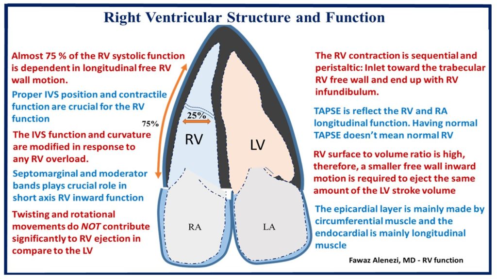
Both left and right atrium function moderate ventricular function through three components:

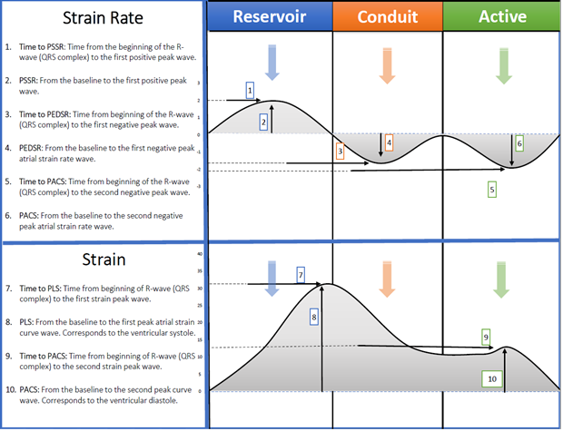
  1. Atrial diastole (Reservoir phase) or expansion during ventricular systole. This function is dependent on ventricle longitudinal function, atrial wall elastance and venous return. Reservoir function may increase during physical activity in a normal healthy heart. Increase of this phase help to increase ventricular filling.
  2. Passive atrial systolic phase (Conduit function) during ventricle early diastole. This phase is dependent on the ventricle end-diastolic pressure. Conduit function does not affect by physical activity.
  3. Active atrial systolic phase (Active contraction) (when sinus rhythm is present) during ventricle late diastole. This phase is dependent on atrial wall contractile properties and ventricular end diastolic pressure. Active contraction function is affected by physical activity in a normal heart.

Traditionally atrial function was measured indirectly by using volumes. However, this is still not resembling the actual atrial deformation and mechanics. With a high feasibility and reproducibility of speckle tracking strain echocardiography, atrial function is ready to be a part of an echo report in a daily clinical practice.

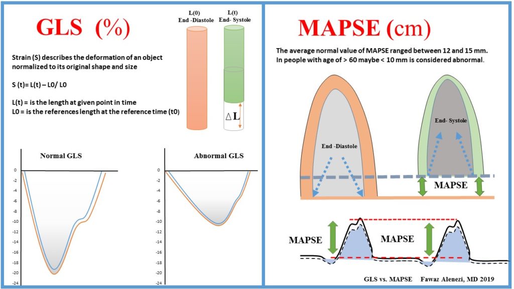
Speckle tracking strain imaging has been around for quite a while and we celebrated the first decade 3 years ago, whereas the technique described in 2004 (Leitman M-JASE) and clinical applications appeared around 2005 (Notomi Y- JACC). Since then, the interest has risen dramatically and so far, we have > 5000 publications on this topic. Left ventricular (LV) ejection fraction (LVEF), the most widely used measure of cardiac function, has important limitations including low sensitivity for incident HF, technique-related variability and does not directly assess LV contractility. Global longitudinal strain (GLS) is the most studied among strain parameters and its prognostic value has been demonstrated in several clinical scenarios.

GLS inter-Vendor variety has dramatically dropped over a short period of time. However, still if we look at the same images with different software vendors, we may have different values. This difference is due to several factors: 1) Image quality and acquisition; 2) Software used; 3) Where to measure (endocardial, myocardial, median); 4) Post-processing of data; 5) Patients age, gender variabilities and loading conditions. With regard to regional (segmental) function, we now have, for the first time, the possibility to measure regional myocardial function. However, I think this is more challenging than GLS, and there is still progress to be made. The challenges become because we have only one segment to work with, and less data to average. Tracking quality becomes more important, regional artifacts matters more, definition of sample position more relevant, and more importantly, we cannot simply measure peak values anymore. For regional analysis, the strain curve shape (not peak values, because peak values can be the same), become critical.

atrial function and structure

Technical factors that may influence atrial strain values:

  • Optimization of images quality and frame rates are vital (ideally, no less than 40 fps).
  • Start tracing at the lateral valve annulus, along the endocardial border of the atrial lateral wall, atrial roof, atrial septal wall, and ending at the septal valve annulus.
  • Difficulties tracking atrial segments relate to the thin wall, insertion of blood vessels and atrial appendages.
  • Significant age-related reductions in strain have been reported. Similarly, sex-related differences have been described, with lower deformation noted in male patients than in female patients.
  • Atrial strain increases in response to early physiological heart rate increase in the setting of exercise in normal patients. However, decreased values are found in the setting of pathological heart rate increase.
  • Atrial strain also affected by loading condition. Reporting, BP, HR and heart rhythm, IVC diameter are crucial.
  • Atrial phases function (Reservoir, conduit and atrial) is also dependent on the ventricular function. Report both atrial and ventricular functions are important.
  • Atrial strain maybe useful as an early marker of DD.

In summary, I think GLS is ready for clinical practice. Its robust, reproducible and has been shown to add unique data that can guide diagnosis and management. I recommend GLS as a valuable complement to traditional function parameters. Further studies are needed to standardize vendors, recognizing specific strain patterns and to determine if there are age, gender variabilities or loading conditions difference.


References:

Myocardial Strain Measured by Speckle-Tracking Echocardiography: Patrick Collier, MD, PHD, Dermot Phelan, MD, PHD, Allan Klein, MD VOL. 69, NO. 8, 2017 – ISSN 0735-1097

hidden

Right Ventricular Structure and Function

The effect of anticancer medication on the (LV) function and structure has been extensively investigated in comparison to the right ventricle (RV). In general, it’s known that deterioration of RV function is associated with significant morbidity and mortality. However, despite advancement in echocardiographic and other imaging techniques, the RV assessment remains challenging in comparison to the LV. In this short statement I will summarize some of the RV characteristics and distinguish

First: The right and left ventricles have distinct morphological difference. Embryologically, the RV is derived from the secondary heart field whereas the LV is derived from the primary heart field. The RV has crescentic non-ellipsoidal configuration and three anatomic divisions (an inlet region, trabeculae free wall/ apical region and an outflow tract). In addition, the RV mass and wall thickness is about 1/3 of the LV.1

Second: The RV has different cardiodynamics:

  1. The RV contraction is sequential and peristaltic: started by the RV inlet toward the trabecular free wall and end up with infundibulum. Rather than the LV contraction which is uniform, longitudinal and torsion. In case of RV volume or pressure overloads it’s become more uniform in contraction.
  2. The RV has 3 separate mechanisms of contraction:1
    1. Free wall inward movement “bellows – like effect) that is dependent on the moderator/ septomarginal band’s position and contractility.
    2. Contraction of longitudinal fibers (shortens long axis TV annulus toward apex)
    3. Traction of free wall from septal LV attachments.
  3. Shortening of RV is much greater longitudinally (75%) than radially (25%). The radius of the curvature and RV surface area do not change appreciably.
  4. The epicardial layer is mainly made with circumferential muscle and the endocardial is mainly made with longitudinal muscle loop. In comparison, the LV has helix that formed by ascending and descending obliquely oriented loops. Thus, twisting and rotational movements do NOT contribute significantly to RV ejection in compare to the LV.

Third: The RV surface/ volume ratio is high, therefore, a smaller free wall inward motion is required to eject the same amount of the LV stroke volume. Also, the RV isovolumetric contraction and relaxation are shooter than the LV, simply, because the pulmonary artery diastolic and RV filling pressure are low. In addition, about 30% of the RV systolic and stroke volume occur due to the interventricular septum (IVS) by the phenomena called “systolic ventricular interdependence.”1

Forth: Proper IVS position and contractile function are crucial for the RV function. The IVS function and curvature are modified in response to any pressure or volume overload. Under normal RV pressure, the IVS is concave toward the LV in both systole and diastole. However, in patient with pulmonary hypertension the IVS curvature become more convex shape which may help the RV to eject more blood against high pulmonary pressure.1,2

Fifth: RV failure occurs under almost two conditions, excessive RV afterload and IVS dysfunction. The most important determinant of the RV function is:1,2

  1. RV afterload (Vascular resistance and compliance)
  2. RV contractility (systolic function)
  3. The coupling of RV contractility to RV afterload
  4. Pericardial constraint/RV-LV diastole interactions

The RV function and structure assessment is challenging. An ideal index of contractility should be independent of afterload and preload, sensitive to change in inotropy, independent of heart size and mass, easy and safe to apply, and proven to be useful in the clinical setting.1,2 Invasively, the gold standard measure of RV function still the volume/ pressure relationship. The most clinically used measures of RV systolic function (RVEF, RV FAC, TAPSE, S’ tricuspid valve velocity, RV SV, strain and RVMPI) are load dependent. Just like the LV, we are slowly evolving into global longitudinal strain (GLS). Important to remember that LV GLS is a surrogate of LV ejection fraction (LVEF) and the dichotomy between the LVEF and GLS can be explained by global circumferential strain (GCS), (Figure 1).

Figure 1: Right ventricular structure and function

References:

  1. François Haddad, MD; Sharon A. Hunt, MD; David N. Rosenthal, MD; Daniel J. Murphy, MD Right Ventricular Function in Cardiovascular, Part I. Circulation 2008, 117:1436-1448
  2. François Haddad, Ramona Doyle, Daniel J. Murphy and Sharon A. Hunt. Right Ventricular Function in Cardiovascular Disease, Part II. Circulation 2008, 117:1717-1731


hidden

Cardiac Longitudinal Function: GLS vs. MAPSE

Over the history of echocardiography, there have been a multitude of research studies on the feasibility, reproducibility and the prognostic values of different echocardiographic parameters in different disease groups. Fifty years later, no single echocardiographic parameter is unique for assessment of the cardiac structure and function. Even the most widely used measurement for left ventricle ejection fraction (LVEF) has important limitations, including low sensitivity for incident heart failure (HF), specifically HF with preserved ejection fraction (HFpEF). With the advent of speckle tracking echocardiography (STE), early detection of cardiac dysfunction before signs or symptoms of HF develop (stage A or B HF) has become a target of many researchers in the last 15 years. Although, there has been some success, there is still a long way to go. In this blog, I want to discuss the differences, advantages and the limitations of mitral annular plane systolic excursion (MAPSE) vs. global longitudinal strain (GLS).

MAPSE measures LV longitudinal shortening and it has been around for over 50 years. Reduction in MAPSE was described as a marker of LV dysfunction (Figure 1). Nowadays, newer and more refined echocardiographic technologies, such as strain, are used more widely for assessment of LV function and deformation. STE imaging has been around for quite a while and we celebrated it’s first decade just 4 years ago, whereas the technique described in 2004 (Leitman M-JASE) and clinical applications appeared around 2005 (Notomi Y- JACC). Since then, the interest has risen dramatically and so far, we have around 6,000 publications on this topic. GLS is the most studied among strain parameters and its prognostic value has been demonstrated in several clinical scenarios.

Figure 1: GLS vs. MAPSE – Fawaz Alenezi -2019.


MAPSE (cm) GLS (%)

Technique

  • Real time or reconstructed M-mode
  • Measure mitral annular displacement
  • Speckle tracking
  • Measure myocardial deformation
Advantages
  • Less dependent on image quality and almost (> 95%) is feasible
  • Good correlation with LVEF
  • Simple and fast
  • Excellent reproducibility
  • High temporal resolution
  • More standardized
  • Better than LVEF in some cases
  • Vendor independent
  • Not gender or BSA dependent
 

  • Assess regional and global longitudinal/radial/circumferential function, rotation/torsion, and ventricular synchrony
  • Less angle dependent
  • Better than LVEF in some cases
  • Good correlation with LVEF


Disadvantages
  • Unable to detect regional abnormalities
  • Angle dependent
  • Age dependent
  • Pericardial effusion: The interpretation of MAPSE should be carefully applied in case of a mobile apex.
  • RV dysfunction: In patients with paradox septal motion, septal MAPSE is not only reflecting LV function but rather RV abnormalities
  • Mitral valve disease and ring calcification: Sometimes in patients with mitral valve disease, the mitral ring is extremely calcified. In these patients, the direct MAPSE measurement at the mitral ring is not possible and longitudinal functional assessment should be done slightly more above in the myocardium
  • Septal MI: Another limitation of this parameter is that small localized abnormalities (i.e. small areas of fibrosis) cannot be detected as it is only possible to assess longitudinal function of the complete wall
  • LVH: In patients with LVH due to HTN or AS will have different values
 

  • Vendor dependent
  • Although, assessment of regional function is the main advantages of strain, but studies still showing a large variability and is not ready to clinical use
  • Variable in region of interest definition
  • Highly dependent on image quality
  • Gender and BSA dependent
  • Need experience and time consuming
  • Pre and after load dependent
  • Rhythm and rate dependent
  • Frame rate dependent

Table 1: Advantages and limitations of GLS vs. MAPSE- Fawaz Alenezi – 2019


What is thought to be the main advantage of STE over MAPSE is the ability of global and regional function assessment. However, with regard to regional (segmental) function, I think this is more challenging and there is still progress to be made. The challenges exist because we have only one segment to work with and less data to average. Tracking quality becomes more important, regional artifacts matters more, definition of sample position more relevant, and more importantly, we cannot simply measure peak values anymore. For regional analysis, the strain curve shape (not peak values, because peak values can be the same) become critical. Regional strain measurements have much higher variability among vendors when compared with GLS. Recently, the HUNT study (Stoylen A et al. 2018 –Wiley echocardiography journal) showed that MAPSE and GLS measured both as MAPSEn (n= normalized for end diastolic length) and GLS have similar biological variability in adults without improvement by normalizing for length for both.

In summary, I think both GLS and MAPSE are measuring the same cardiac function. GLS is robust, reproducible and has been shown to add unique data that can guide diagnosis and management. However, it is vendor, image quality and hemodynamics dependent that need more standardization and experience. On the other hand, MAPSE is simple, easy, available in every echo machine and, more importantly, feasible even in a poor image quality. But it is not measuring the global function. Further studies are needed to standardize vendors, recognize specific strain patterns, determine if there is loading conditions difference and head-to-head comparison between both methods.


hidden

Late Breaking Trials: Scientific Sessions – Chicago 2018

Cardiovascular risk assessment is a process, not just a calculation! This year, I was very pleased to attend the 2018 guidelines for cholesterol management at the American Heart Association (AHA) Scientific Sessions – Chicago 2018. I highly recommend that you check the guidelines as there are many changes that may impact your daily practice. Here, I’m summarizing the late breaking trials that were presented at the last AHA and encouraging you to join us at the next AHA Scientific Sessions meeting – Philadelphia 2019.


Critical Questions in Cardiovascular Prevention:

The VITamin D and OmegA-3 TriaL (VITAL): Principal Results for Vitamin D and Omega-3 Fatty Acid Supplementation in the Primary Prevention of Cardiovascular Disease and Cancer.

  • RESULTS: Major CVD events and total invasive cancer not significantly reduced by Omega-3 or vitamin D3. Omega-3 significantly reduced total MI, especially in African Americans and those with lower baseline fish intake.

The Primary Results of the REDUCE-IT Trial.

  • RESULTS: High-dose icosapent ethyl vs. placebo in at-risk patients significantly reduced the composite CVD endpoint: risk of CV death, MI, stroke, coronary revascularization, and unstable angina.

Ezetimibe in Prevention of Cerebro- and Cardiovascular Events in Middle- to High-Risk, Elderly (75 Years Old or Over) Patients with Elevated LDL-Cholesterol: A Multicenter, Randomized, Controlled, Open-Label Trial.

  • RESULTS: Ezetimibe monotherapy + diet counselling vs diet counselling alone for primary prevention (elevated LDL-C; no history of CAD) in an over-75 y/o Japanese population significantly prevented cerebral and cardiovascular events.

Cost-Effectiveness of Alirocumab Based on Evidence from a Large Multinational Outcome Trial: The ODYSSEY OUTCOMES Economics Study.

  • RESULTS: For the patients in the ODYSSEY OUTCOMES trial (post-ACS and LDL-C ≥100 mg/dL), alirocumab was found to be cost effective.


Novel Approaches to CV Prevention:

The Dapagliflozin Effect on Cardiovascular Events (DECLARE)-TIMI 58 Trial.

  • RESULTS: Dapagliflozin compared to placebo in patients with T2DM was safe, reduced the composite of CV death or hospitalization for heart failure, but did not impact MACE.

The Cardiovascular Inflammation Reduction Trial (CIRT): Low Dose Methotrexate for the Prevention of Atherosclerotic Events.

  • RESULTS: Low dose methotrexate compared to placebo in patients with prior MI or multivessel CVD and either type 2 diabetes or metabolic syndrome, did not reduce inflammatory markers, and CV events weren’t lower than placebo.

Safety and Efficacy of AKCEA-APO(a)-LRx to Lower Lipoprotein(a) Levels in Patients with Established Cardiovascular Disease: A Phase 2 Dose-Ranging Trial.

  • RESULTS: Dose-dependent lowering of Lp(a) levels by AKCEA-APO(a)-LRx were seen in patients with pre-existing CV disease or PAD in this phase 2 trial


Harnessing Technology and Improving Systems for Global Health:

Effects of a Multifaceted Intervention to Narrow the Evidence-Based Gap in the Treatment of High Cardiovascular Risk Patients: The BRIDGE CV Prevention Cluster Randomized Trial.

  • RESULTS: Adherence to evidence-based therapies (antiplatelets, statins and ACE inhibitors) for high CV risk Brazilian patients was improved with use of a multifaceted quality improvement educational intervention vs routine practice.

Alert-Based Computerized Decision Support to Increase Anticoagulation Prescription Prevents Stroke and Myocardial Infarction in High-Risk Hospitalized Patients with Atrial Fibrillation: A Randomized, Controlled Trial.

  • RESULTS: Alert-based computerized decision support in high-risk hospitalized AF patients increased prescribing of anticoagulation for stroke prevention and reduced major adverse cardiovascular events, MI and stroke.

Efficacy of Electronic Clinical Decision Support in Atrial Fibrillation: Results of the Integrated Management Program Advancing Community Treatment of Atrial Fibrillation (IMPACT-AF).

  • RESULTS: An online, evidence-based computer decision support system did not significantly affect the number of patients experiencing the unplanned cardiovascular hospitalization and/or AF-related emergency room visit at 12 months.


Preserving Brain & Heart in Acute Care Cardiology:

Pre-Hospital Resuscitation Intra-Arrest Cooling Effectiveness Survival Study – The Princess Trial.

  • RESULTS: Survival with good neurologic outcome 90 days after cardiac arrest was not statistically different for pre-hospital trans-nasal evaporative intra-arrest cooling compared to standard care.

Early Goal-Directed Hemodynamic Optimization in Comatose Survivors After Out-of-Hospital Cardiac Arrest: The Neuroprotect Trial.

  • RESULTS: A higher mean arterial pressure (MAP) in the ICU during the first 36 hours after cardiac arrest improved cerebral perfusion and oxygenation but did not decrease anoxic brain damage or improve functional outcome compared to the lower MAP target of 65mmHg.

A Randomized, Double Blind, Placebo-Controlled, Parallel Group, Multicenter Clinical Trial of Low-Dose Adjunctive Alteplase During Primary PCI (T-TIME).

  • RESULTS: Compared to placebo, microvascular obstruction was not different for low-dose alteplase during primary PCI for acute STEMI.

Optimal Timing of Intervention in Non St-Elevation Acute Coronary Syndromes Without Pretreatment.

  • RESULTS: In not pre-treated intermediate and high-risk NSTEACS patients, a very early (<2 hours) invasive intervention strategy compared to a delayed one (≥12-72 hours) was highly significant for reduction in CV death or recurrent ischemic events @ 30 days.

Mechanically Unloading the Left Ventricular and Delaying Reperfusion in Patients with Anterior ST-Segment Elevation Myocardial Infarction: The Door to Unload Pilot Trial.

  • RESULTS: In patients with anterior STEMI, mechanical unloading of the left ventricle followed by either immediate vs delayed reperfusion found no difference in MACCE between the two groups and no increase in 30-day infarct size in the delayed group.


Hot News in HF:

Angiotensin Receptor-Neprilysin Inhibition in Patients Hospitalized with Acute Decompensated Heart Failure: Primary Results of the PIONEER-HF Randomized Controlled Trial.

  • RESULTS: This comparison of sacubitril/valsartan to enalapril in patients hospitalized with HFrEF found that sacubitril/valsartan resulted in significantly more reduction in NT-proBNP levels.

Withdrawal of Pharmacological Heart Failure Therapy in Recovered Dilated Cardiomyopathy – A Randomised Controlled Trial (TRED-HF).

  • RESULTS: Withdrawal of therapy in ‘recovered’ dilated cardiomyopathy patients resulted in relapse for 40% compared to 0% relapse for those who remained on therapy. “Recovered’ patents are in remission.

Effects of Rivaroxaban on Thrombotic Events in Heart Failure Patients with Coronary Disease and Sinus Rhythm.

  • RESULTS: Low-dose rivaroxaban use in HF significantly reduced the risk for thromboembolic events in the COMMANDER HF trial population.

EMPA-HEART Cardiolink-6 Trial: A Randomized Trial Evaluating the Effect of Empagliflozin on Left Ventricular Structure, Function and Biomarkers in People with Type 2 Diabetes (T2D) and Coronary Heart Disease.

  • RESULTS: In patients with T2DM and stable CAD, LV mass was significantly reduced by empagliflozin compared to placebo.


Coronary Revascularization:

Endoscopic Vein Harvest for Coronary Bypass Surgery in a Randomized Multicenter Trial with Long-Term Follow-Up.

  • RESULTS: Late findings for endoscopic vs open SVG harvesting for CABG found MACE rates were similar @ 2.7 years’ follow-up.

Long-Term Survival Following Multivessel Revascularization in Patients with Diabetes: The FREEDOM Follow-On Study.

  • RESULTS: At the 8-year FREEDOM trial follow-up, CABG showed lower rate of all-cause mortality over 8-year follow-up compared to PCI in patients with diabetes and multivessel coronary artery disease.

TiCAB: A Randomized, Double-Blind Study of Ticagrelor versus Aspirin in Patients Undergoing Coronary Bypass Surgery.

  • RESULTS: In patients having CABG, major CV events and bleeding rates were not significantly reduced with ticagrelor monotherapy compared to aspirin.

Ten-Year Clinical Outcomes After Coronary Drug-Eluting Stents with Biodegradable or Permanent Polymer Coating: Results from the Randomized ISAR-TEST 4 Trial.

  • RESULTS: 10-year CV outcomes were superior for three limus-eluting stents with different polymer coatings compared to early-generation stents.

Intramyocardial Injection of Mesenchymal Precursor Cells in Left Ventricular Assist Device Recipients: Impact on Myocardial Recovery and Morbidity.

  • RESULTS: Successful temporary LVAD weans over 6 months and 1-year mortality were similar for intramyocardial mesenchymal precursor cell injection vs sham injection.


hidden

Clinical Significance of Sigmoid Shaped Interventricular Septum

A sigmoid-shaped interventricular septum (SIS) is generally considered a normal part of the aging process and is of little clinical significance. However, certain patients with SIS may experience clinical symptoms, such as dyspnea upon effort and different types of cardiac arrhythmias. SIS is frequently observed on transthoracic echocardiography (TTE) and in cardiac magnetic resonance (MR) imaging modality in daily clinical practice. However, nothing usually occurs in subjects with SIS, and the clinical significance of the presence of SIS is unclear.

The precise mechanisms leading to isolated SIS have yet to be determined, but plausible reasons exist as to why the basal septum might be uniquely susceptible to hypertrophy. For example, Laplace’s law states that the larger a vessel’s radius, the larger the wall tension required to withstand internal fluid pressures. Because the longitudinal fibers of the basal septum have some of the largest radii in the human heart, they would be expected to experience the greatest inward component of wall stress. This is compounded by the fact that the basal septum is the last part of the ventricle to be electrically activated, so contractions from other myocardial segments further increase its wall stress (Fig. 1). Moreover, the additional load created by pressure from the right ventricle exerts additional stress on the septum. Therefore, it is conceivable that the basal septum hypertrophies earlier than other LV regions in response to increased afterload as it already operates under higher loading conditions.

Clinical Significance of Sigmoid Shaped Interventricular Septum

Prospective studies suggest that up to 20% of cardiovascular cohorts may have isolated SIS. Some researchers have reported that the cause of SIS may be aging or arteriosclerosis. This may involve a change in the spatial relationship between ascending aorta (AA) and left ventricle (LV) due to elongation or tortuosity of arteriosclerotic AA. An alternative hypothesis suggests that SIS may be a form of cardiomyopathy. However, there is no evidence to support such a hypothesis due to the limited capabilities of traditional TTE. Use of other diagnostic approaches like CMR may be needed where characteristics of LV myocardium and the spatial relationship between AA and LV and degree of arteriosclerosis of AA can be evaluated simultaneously.

It is known that LV hypertrophy with different remodeling patterns is one of the major cardiac manifestations of hypertensive heart disease, and echocardiographic LV hypertrophy could be detected in 20-40% of patients with arterial hypertension. However, there are often no specific echocardiographic features for hypertensive patients at the early stage of disease. Previous echocardiographic studies have described asymmetric septal hypertrophy with a localized septal thickening at the basal-mid portion in patients with hypertrophic cardiomyopathy or aortic valve stenosis.

Basal-septal hypertrophy may also occur in a subset of older normal subjects, with normal wall thickness (WT) elsewhere, and is considered to be an age-related anatomic variant. This morphologic echocardiographic sign is termed as septal bulge (SB), sigmoid septum, or discrete upper septal thickening or knuckle. A large community-based population study reported that SB was documented frequently in elderly individuals with higher systolic blood pressure (BP). It was shown that the overall prevalence of SB was 1.5% and was markedly higher (18%) in the eighth decades of life.

Although pathologic and echocardiographic observations have indicated that SB is a structural response in hypertensive patients, the nature and significance of the SB in subclinical arterial hypertension was never investigated. In addition, despite the fact that BP can be easily measured, AH sometimes cannot be diagnosed due to the underreported BP reading in the casual or self-measured BP measurement. BP measurement with appropriate tools is essential to diagnosing AH early as well as to guiding AH management. It has been shown that, besides resting BP measurement in the office, arterial hypertension could be clinically diagnosed by 24-hour ambulatory BP monitoring (ABPM) as well as exercise stress test in some resting normotensive individuals.

Focal hypertrophy of the basal inter-ventricular septum can be seen in up to 20% of cardiac patients without HCM, being more prevalent in the elderly and hypertensives. While it’s anatomical location plausibly renders it more susceptible to hypertrophy, evidence suggests that the basal septum enlarges mainly due to pressure overload from hypertension. This discrete upper septal hypertrophy is associated with exertional LVOT obstruction and SOB, and appears symptomatically amenable to β -blockade. While diastolic dysfunction likely also contributes to symptoms in this condition, the data to date are equivocal. Focused analyses conducted using a consensus definition of SB, in patients undergoing simultaneous assessment of myocardial systolic and diastolic performance during physiological exercise, are needed to further understand the clinical relevance of this entity.


Fawaz Alenezi Headshot

Dr. Fawaz Abdulaziz M Alenezi is a Clinical Imaging Fellow at the Duke University Health Systems. He conducts medical research on the derivation and validation of novel echocardiographic approaches to myocardial deformation and a new echocardiographic technique which assists patients with heart ventricular function.

hidden Mitral Valve Prolapse: The Complexity and Heterogeneity

Mitral Valve Prolapse: The Complexity and Heterogeneity

The concept of myxomatous mitral valve disease (MMVD) emerged in the mid-20th century, in a world dominated by rheumatic valve diseases, after the observations of astute surgeons attempting the first repairs of mitral valves (MV), of pathologists describing these atypical MV observed at autopsy, and of master clinicians intrigued by the peculiarities of systolic clicks and late systolic murmurs. The observation of the myxomatous degeneration histologically involving mitral tissue led to a “unification” of the condition under 1 central functional feature, the mitral valve prolapse (MVP).

The mitral annulus is a component of the MV apparatus and consists of a cord-like ring of collagen and elastic fibers distributed along the atrioventricular junction and giving support to the MV leaflets. It is conventionally divided into anterior and posterior portions, although the real annulus is the one that serves as hinge point for the mural posterior leaflet of the MV. The motion of the mitral annulus is passive and determined by the contraction and relaxation of adjacent atrial and ventricular musculature. As consequence, in normal condition, the posterior mitral ring and its adjacent myocardium move downward and anteriorly in systole, in synchrony with the remainder of the LV. (Figure: Annular Disjunction, Fawaz Alenezi 2018)

Figure: Annular Disjunction, Fawaz Alenezi 2018

Mitral annular disjunction (MAD) is characterized by detachment of the roots of the annulus from the ventricular myocardium to which it would normally be attached. Because the fibrous trigones are densely fibrotic, the base of the anterior leaflet is unaffected and only the area under the posterior leaflet, specifically under P1 and P2 scallops, is affected. There is no detachment of the annulus from leaflets or from atrial wall. This ventriculoannular detachment (disjunction) is difficult to diagnose and requires a keen eye to be noticed, which explains the “forgotten” nature of this component of MMVD to which it is quite specific. Indeed, in diastole the diagnosis cannot be made because the ventricular myocardium is appropriately situated under the annulus. Over systole, as the posterolateral myocardium contracts, the annulus “slides” and becomes detached from the ventricular myocardium by a variable distance, few millimeters to sometimes more than 1 cm. Often, few fibers are visible between posterolateral ventricular myocardium and annulus. A frame-by-frame analysis of high-resolution and high-frequency 2-dimensional imaging is best to visualize the defect but not the optimum technique.

The dynamic nature of MAD explains the paucity of pathological reports using flaccid hearts and of surgical reports whereby the annulus is observed from the left atrium and its disjunction from the ventricular myocardium is unnoticeable unless the posterior leaflet is separated from its implantation. The disjunction of the mitral annulus fibrous could play a role in the development of the pathological features of myxomatous valve disease through the mechanical stress incited by the excessive mobility of the mitral apparatus and the source of multiple arrhythmias.

In fact, using only echocardiography we know very little about MAD, its prevalence and consequences, with disagreements on prevalence, significance and association with MVP. Lists studies that have compared cardiac magnetic resonance (CMR) and echocardiography using quantitative methods. It details the agreement between echocardiography and CMR as well as the agreement between echocardiography and CMR among studies that were considered to have severe mitral regurgitation. In general, there is a significant degree of discordance between CMR and echocardiography when quantifying mitral regurgitation. Based on the available data, there appears to be significant discordance between 2D echocardiography and CMR. This discordance is highlighted among patients who are considered to have severe mitral regurgitation and who are likely to be referred for mitral valve surgery.

CMR has become an established noninvasive imaging modality to assess mitral regurgitation severity. Quantification of mitral regurgitant volume by CMR does not rely upon the characteristics of the regurgitant jet. Instead, the assessment of mitral regurgitation by CMR relies upon the difference between the left ventricular stroke volume and forward stroke volume, both of which are quantified using already established accurate and reproducible techniques.

In summary, the clinical and physiological understanding of MAD and MVP is a work in progress. MVP heterogeneity has been demonstrated with regard to clinical outcomes, but the analysis of MMVD morphological and physiological heterogeneity, particularly incorporating MAD description, is crucial to appropriate phenotyping of MVP and MMVD, with the hope of linking this heterogeneity to the genotyping, molecular mechanism and progression of the disease.

Fawaz Alenezi Headshot

Dr. Fawaz Abdulaziz M Alenezi is a Clinical Imaging Fellow at the Duke University Health Systems. He conducts medical research on the derivation and validation of novel echocardiographic approaches to myocardial deformation and a new echocardiographic technique which assists patients with heart ventricular function.

hidden

A Shift in Focus: From a Multi to Single Modality Approach

Since the first A-mode echocardiogram, there have been great revolutional changes and the use of noninvasive cardiac imaging has grown substantially during the past decade. Echocardiography and nuclear modality have been the primary imaging modalities for management in patients with different cardiovascular diseases. The introduction of cardiac magnetic resonance (CMR), computer tomography (CT), and three-dimensional (3D) printing, makes things quite different. Multi-modality imaging plays a role in all cardiovascular diseases that includes ventricular function, coronary artery disease, valvular disease, congenital heart disease, guidance for interventions, and vascular diseases. However, the use of each modality requires knowledge, expertise and forethought about cardiac diagnoses to avoid excess coast, inappropriate testing and to improve outcomes. Unnecessary duplication of investigations must be avoided for cost effective healthcare delivery.

For more than a decade, organizations such as the American College of Radiology and the American College of Cardiology Foundation have published criteria delineating the appropriate use of cardiac imaging. These specialty society guidance documents are designed to provide clinicians with recommendations regarding the use of imaging and are focused on reducing unnecessary and inappropriate testing. In response to rapid and unsustainable growth in the use of cardiac imaging procedures, various professional medical organizations have developed appropriate use criteria (AUC) to guide physicians and payers on effective use of these procedures. The AUC serves as a guide for physicians to incorporate symptoms, risk factors, and clinical history in selecting the patients for whom testing is most appropriate, based on the best available evidence or expert consensus; Furthermore, the AUC can help physicians steer other patients to alternative tests or no testing at all.

The AUC classify the appropriateness of testing across a range of clinical scenarios in 3 categories: “appropriate” (established value), “may be appropriate” (uncertain value), and “rarely appropriate” (no clear value). The AUC are intended to be used as part of a comprehensive disease management plan to maximize the value of care by minimizing unnecessary imaging and resultant downstream invasive procedures. (1). Appropriate use helps to avoid excess costs; enhances the value of imaging in risk stratification and decision making; and reduces radiation risk, particularly in women (2). Moreover, the principle of applying expert consensus criteria to choose the best testing strategy for each patient is widely accepted by payers, physicians, and patient groups.

  • The goal of this statement is not to showcase CMR as the exclusive modality for cardiac diagnosis but to highlight this tool as the corner stone of noninvasive cardiac imaging. By using a static and a gradient magnetic field in combination with a radiofrequency field, CMR imaging has evolved from a less commonly used technique into a tool with the potential to find an important clinical role in the near future for the noninvasive assessment of morphological and functional aspects of the heart. Consequently, CMR belongs to the fastest growing new fields of broad MR application. At the same time, CMR uses some of the strongest and fastest switching electromagnetic gradients available in MR imaging. CMR has become the primary imaging tool in many centers in characterizing disease severity and planning of patient management.
  • Volumes and Function: CMR measures ventricular volumes and mass using a simple acquisition of a 3D stack of contiguous short-axis cines with full biventricular coverage. CMR is significantly more accurate and reproducible than other techniques which makes it the technique of choice for longitudinal study of patients over time and for reducing sample size for drug studies.
  • Great Vessels: Three-dimensional angiography with gadolinium enhancement may show the lesion more clearly in high resolution, but it also shows any associated pathology (collaterals). CMR is fast and accurate for the diagnosis of aortic dissection although local issues relative to availability will determine whether CMR, CT, or echocardiography is used.
  • Valvular Heart Disease: CMR has a significant role in valve disease, but it usually acts as a second-line technique to assist when echocardiography with Doppler has proved problematic because of limited acoustic access, highly eccentric jets, or the need for quantification. The quantification of valve regurgitation is a strength of CMR because of its capability of measuring accurate ventricular stroke volumes from multislice ventricular planimetry and comparing this with the measurement of great vessel flow from velocity mapping.
  • Congenital Heart Disease: CMR is widely used to assess congenital heart disease, and when used in concert with echocardiography, the need for invasive assessment has been significantly reduced. CMR is particularly useful for the safe, accurate, and reproducible quantification of the left-to-right shunting of blood that occurs frequently in congenital heart defects such as atrial or ventricular septal defects, patent ductus arteriosus, aortopulmonary window, and partial or total anomalous pulmonary venous return.
  • Myocardial Ischemia: CMR is a safe and proven modality for detecting ischemic heart disease. Within a single study, CMR is able to assess rest and stress regional contraction, and localize and evaluate regions of myocardial ischemia and viability, of importance given the prognostic impact for the patient. With performance that is comparable to PET/SPECT and at times favorable compared to stress echocardiography, it is now an essential tool for diagnosing ischemia and for determining the likelihood of success following myocardial revascularization.
  • Non ischemic cardiomyopathy: CMR with its higher spatial resolution is considered the gold standard for evaluating ventricular mass, volumes, and ejection fraction. CMR can be used for accurate diagnosis of several conditions, especially cardiomyopathies. CMR is an invaluable tool, not only in differentiating nonischemic from ischemic cardiomyopathy, but also in aiding the accurate diagnosis of the subtype of nonischemic cardiomyopathy. CMR should routinely be integrated in the diagnostic workup of various cardiomyopathies.

CMR is a valuable tool for the evaluation of patients with, or at risk for, heart failure and has a growing impact on diagnosis, clinical management, and decision making. Through its ability to characterize the myocardium by using multiple different imaging parameters, it provides insight into the etiology of the underlying heart failure and its prognosis. CMR continues to develop across a broad range of clinical applications, and much can be expected of this technology in the future.

Fawaz Alenezi Headshot

Dr. Fawaz Abdulaziz M Alenezi is a Clinical Imaging Fellow at the Duke University Health Systems. He conducts medical research on the derivation and validation of novel echocardiographic approaches to myocardial deformation and a new echocardiographic technique which assists patients with heart ventricular function.

hidden

Methodology of Biatrial Functional Analysis

Both left and right atrium modulates ventricular filling through three components: a phase of reservoir or expansion during ventricular systole, a conduit phase during diastole, and an active contractile component (when sinus rhythm is present) during late diastole. During exercise atrial reservoir and booster functions are augmented, whereas conduit function is not; increased reservoir function may play an important role in accelerating ventricular filling by helping to maintain an enhanced atrioventricular pressure gradient during diastole and also by increasing atrial booster function through an increase in preload.

The resurgence of interest in atrial size and function has enhanced our understanding of the atrial contributions to cardiovascular performance in health and disease. Considering the limitations of conventional echocardiographic techniques for atrial functional assessment, the high feasibility and reproducibility of bi-atrial strain, and its predictive value in several clinical conditions, biatrial measured by 2D speckle tracking echocardiography could be considered to assess atrial function in the clinical routine.

Methodology of biatrial strain and strain rate: Key measurements and guidance


Methodology of biatrial measurements 2013
Figure 1: Fawaz Alenezi – Methodology of biatrial measurements 2013: 1. Time to Peak systolic strain rate [PSSR], 2. Peak systolic strain rate [PSSR], 3. Time to peak diastolic strain rate [PEDSR], 4. Peak diastolic strain rate [PEDSR], 5. Time to peak contraction strain rate [PCSR], 6. Peak contraction strain rate [PCSR], 7. Peak longitudinal strain [PLS], 8. Time to peak longitudinal strain [PLS], 9. Time to peak active contraction strain [PACS], 10.  Peak active contraction strain [PACS].

Interpretation of atrial functional indices is complicated because of the interplay between atrial and ventricular functions. Atrial dysfunction may result from an intrinsic atrial abnormality, altered load, or in an effort to compensate. Atrial dysfunction may also have different expressions at different stages of the disease process under study. The methods used to measure atrial function all have important limitations and indices from one method (e.g. volumetric) that reflect a specific atrial function often correlate poorly with other methods (e.g. tissue tracking) obtained during the same phase of the cardiac cycle. Finally, the hemodynamic and biophysical properties that are responsible for the functional changes are often assumed, not known.
Technical factors that may influence biatrial strain values:

  • Image quality: Optimization of images quality and frame rates are vital (ideally, no less than 40 fps) determinant of accurate edge detection, tracking, and strain assessment.
  • Technical differences: A number of different vendors offer strain platforms with technical differences among proprietary post-processing algorithms. There are vendor-specific differences among how overall reported atrial strain values are calculated.
  • Technical issues and trackability: Difficulties tracking atrial segments relate to the thin wall, insertion of pulmonary veins, or caval veins, the aorto-atrial or pulmono-atrial curtain (in the three-chamber views), and the lack of dedicated tracking algorithms for atrial strain assessment. Indeed, until very recently, the approach to atrial strain involved ‘‘tricking’’ the LV strain computation algorithm which have different values that a dedicated atrial software’s.
    Operator experience: The results may dependent on the individual studies that comprise the analysis and level of expertise are rarely documented.
  • Zero reference point: If the ventricular cycle is used, ventricular end-diastole (the QRS complex) is the zero reference, and the peak positive longitudinal strain corresponds to atrial reservoir function and the strain during early and late diastole correspond to conduit and atrial booster function. If the atrial cycle is used, atrial end-diastole (onset of P wave) is the zero reference, and the first negative peak strain represents the atrial booster pump function, the positive peak strain corresponds to conduit function, and their sum represents reservoir function.
  • Regional and methodological differences in atrial strain: Previous studies have described differences in mean strain ranges between two-chamber (37.6%–44.3%) and four-chamber (33.8%– 40.1%) strain. However, and again this all depend on (image quality and acquisition; software used; where to measure (endocardial, myocardial, median); and post-processing of data.
  • Age and sex differences: Significant age-related reductions in deformation have been reported. Similarly, sex-related differences have been described, with lower deformation noted in male patients than in female patients across all age groups studied.
  • Hemodynamics factors: Atrial strain increases in response to early physiological heart rate increase in the setting of exercise in normal patients. However, decreased values are found in the setting of pathological heart rate increase, most notably in sepsis.

There is now a growing body of literature supporting its use in different clinical settings. I believe biatrial strain is ready for clinical practice. Its reproducible and has been shown to add unique data that can guide diagnosis and management. I recommend atrial strain as a valuable complement to traditional function parameters. Further studies are needed to standardize vendors, recognizing specific strain patterns and to determine if there are age, gender variabilities or loading conditions difference.

Fawaz Alenezi Headshot

Dr. Fawaz Abdulaziz M Alenezi is a Clinical Imaging Fellow at the Duke University Health Systems. He conducts medical research on the derivation and validation of novel echocardiographic approaches to myocardial deformation and a new echocardiographic technique which assists patients with heart ventricular function.


hidden

Multi-Disciplinary Approach And Decision Sharing In Geri-Cardio-Oncology

Incorporating a geriatric assessment tool into the care of the geriatric cardio-oncology patient is crucial. The use of comprehensive geriatric assessments has been shown to improve overall survival, quality of life, and physical function, while decreasing hospitalizations and nursing home placement in the geriatric population. While cancer is the number one cause of mortality in patients between 60 and 79 years, heart disease is most common in people aged 80 and older. More than 40% of these cancer survivors above the age of 50 will develop cardiovascular (CV) disease. In general, older patients are affected by a number of factors, including concomitant comorbidities as well as other physiologic and functional changes that can affect prognosis, treatment, and outcomes of cancer.

Cancer therapeutics including traditional chemotherapy, targeted therapy, radiotherapy, and hormonal therapy all have short- and long-term systemic effects, often involving multiple organs. However, CV toxicities have been most concerning and can result in irreversible CV damage or reversible cardiac dysfunction. These cardiotoxic manifestations can include left-ventricular (LV) dysfunction and heart failure, myocardial ischemia and infarction, hypertension (HTN), and arrhythmias such as QT prolongation. Less frequently, these complications consist of myocarditis or pericarditis.

Anthracycline-induced cardiotoxic effects are dose dependent, and those who develop late cardiotoxicity have a high mortality. Risk factors include cumulative dose, bolus administration, high single dose, prior radiotherapy, simultaneous use of other cardiotoxic agents, female gender, bimodal age distribution, existing CV disease, elevation of cardiac biomarker during and after cancer treatment, as well as time since completion of cancer therapy. A second class of frequently used cardiotoxic agents are targeted therapies including monoclonal antibody-based tyrosine kinases (bevacizumab, trastuzumab) and small molecule tyrosine kinase inhibitors (sorafenib, sunitinib, lapatinib). HTN is a common adverse event whose mechanism is not well understood but has been attributed to the inhibition of vascular endothelial growth factor.  Progression of or acceleration of ischemic disease is more common with radiation therapy Agents such as bleomycin, etoposide, cisplatin, 5-fluorouracil have been implicated in the development of myocardial ischemia including myocardial infarction.

Risk factors for chemotherapy-related cardiac complications should be assessed in all patients diagnosed with cancer who are being considered for cancer therapy, whether it be the administration of biologics, chemotherapy, or radiation therapy. Given that advancing age has been associated with cardiac complications from chemotherapy using anthracyclines or trastuzumab-based treatments, it is recommended that all elderly patients prescribed these medications should be educated about risk stratification and risk modification. Risk stratification remains difficult in elderly patients at risk for cardiotoxicity. A comprehensive evaluation of CV comorbidities such as HTN, diabetes, dyslipidemia, and smoking needs to be evaluated prior to start of therapy.

Those patients receiving high-dose anthracyclines, high-dose radiation, and history of prior cardiac disease are at greatest risk for cardiac dysfunction are at highest risk for cardiotoxicity. Cancer-specific mortality is often higher in older patients, likely due to the impact of age-related factors. Pre-treatment evaluation of LV systolic function is a standard part of many treatment protocols. However, the utility of this approach has been debated due to a low prevalence of asymptomatic LV systolic dysfunction, with this strategy missing most patients that will ultimately have cardiotoxicity. Serum biomarkers may be useful in predicting cardiotoxicity and the role of baseline assessment of serum biomarkers prior to cancer treatment in predicting cardiotoxicity is being evaluated. An echocardiogram is the most important tool for serial evaluation of the heart during cancer therapy. Ejection fraction should be determined using biplane method of discs, according to the American Society of Echocardiography guideline. Current recommendations for imaging surveillance include monitoring of LV systolic function during treatment with both anthracyclines and trastuzumab. Current National Comprehensive Cancer Network guidelines suggest cardiac monitoring at baseline, 3, 6, and 9 months after initiating therapy for trastuzumab therapy, upon completion of treatment, and every six months for 2 years following completion of treatment.

Collaborative assessment by oncologists, cardiologists and geriatricians before the start of chemotherapy can lead to early identification of patients at risk as well as discussions about the utility and benefits of cardiotoxic medications as opposed to potential alternative therapies. In some situations, alternative noncardiotoxic chemotherapy regimens may be considered. A lower-intensity chemotherapy regimen, however, should not be prescribed based simply on a patient’s risk factors or concern for potential cardiac complications, as this has been shown to potentially worsen clinical cancer outcomes. Cardiologists and oncologists may be less familiar with and often have limited to no training in routinely performing geriatric assessments and systematically evaluating for frailty. Within the gericardio-onc collaborative framework, the geriatrician is well positioned to take an active leadership role in advocating for the patient, assisting with decision-making, and facilitating screening and long-term monitoring of CV complications.

Cardiac dysfunction developing during or after the completion of cancer therapy is a growing heath concern that should be addressed in a multidisciplinary setting. There is a need for research on early biomarkers of toxicity as well as monitoring, surveillance, and treatment of older patients with cancer receiving potentially cardiotoxic therapy. The results of several studies are imperative in determining how best to risk-stratify and treat elderly patients with cancer while preserving their quality of life and functional outcomes.


Fawaz Alenezi Headshot
Dr. Fawaz Abdulaziz M Alenezi is a post-doctorate associate at the Duke University Health Systems. He conducts medical research on the derivation and validation of novel echocardiographic approaches to myocardial deformation and a new echocardiographic technique which assists patients with heart ventricular function.


hidden

Relevance Of Imaging In The Setting Of Advanced Age Related Complexities

The population older than 65 years of age is expected to peak at 20 percent of the population in 2030. By that year, more than 19.5 million U.S. adults will be older than 80 years of age. Due to this large increase in the geriatric population, major efforts are needed to ensure adequate and appropriate health care for the elderly. The prevalence of systolic and diastolic heart failure, atrial fibrillation, aortic stenosis, and electrical conduction defects increases with age, resulting in a significant burden of cardiovascular disease (CVD). Aging itself creates distinctive dimensions to CVD management as both absolute risk reduction, and the potential for harm from treatment, increase with advancing age.

Cardiovascular imaging (CVI) is used for screening (detecting asymptomatic cardiac disease), diagnosing the cause of symptoms, defining the extent of cardiac disorder in a patient with known disease (including risk stratification), monitoring the progression (or regression) of disease, and guiding therapeutic management, including decisions about aggressive therapy. There is a paucity of evidence to guide the rational use of many CVI modalities in patients of advanced age. There are senescent effects in cardiac morphology and physiology as well as broader organ systems changes in elderly patients. Older patients used to be clinically heterogeneous with different age related arterial compliance and left ventricular (LV) diastolic dysfunction. Although, the Appropriate Use Criteria (AUC) do not integrate age as a factor in determining the appropriateness of an imaging test, in part due to a lack of data. Nonetheless, there are a number of other important factors associated with aging which impact the use of cardiac imaging in the senior population. Older patients, regardless of underlying disease severity or comorbidities, undergo fewer invasive cardiovascular evaluations than younger patients.

A key aspect of advanced age, is that the concept of high risk burden must be considered in the context of age-related management efficacy. In contrast to younger populations, the high risk, high prevalence and high burden of disease in the elderly population leads to a situation in which CVI is likely to detect disease for which the implications of therapy are uncertain. Thus, if structural or functional abnormalities are detected in an asymptomatic patient, management decisions may still remain ambiguous. Detection of non-obstructive atherosclerotic coronary artery disease (CAD) in an elderly diabetic with peripheral vascular disease will, for example, not likely lead to any intensification of medical therapy beyond that which is already indicated. In contrast, in other scenarios imaging has substantial clinical impact, including rationale for selecting patients mostly likely to benefit from interventions that may prolong and improve quality of life.

The elderly group had a lower prevalence of CAD risk factors but high degrees of ischemic events and cardiac mortality. Given the high pretest probability of CAD in relation to advanced age, the prognostic utility of a stress study may be particularly useful. CVI adds additional prognostic power and is necessary as part of pharmacological stress, for the majority of the elderly who cannot complete a treadmill exercise protocol. With the growing focus on transcatheter aortic valve replacement (TAVR) the particular value of imaging for common valvular decisions in older adults has advanced as a clinical priority. Transthoracic echocardiography (TTE) is the central imaging modality to detect and characterize the degree of aortic stenosis. In addition to TTE, contrast-enhanced computer tomography imaging in necessary to ensure that vasculature will be able to tolerate the large bore catheters that are required for the procedure. Newer percutaneous mitral valve repair techniques are also options for some elderly patients who are poor surgical candidates and TEE is necessary to demonstrate leaflet anatomy amenable to novel percutaneous interventions.

Approximately 1%–2% of the adult population in developed countries has heart failure (HF), with the prevalence rising to ≥10% among persons 70 years of age or older. HF is underdiagnosed in elderly patients who often lack specific symptoms and usually present with comorbidities that can confound diagnostic assessments based primarily on symptoms. Therefore, TTE plays an important complementary role in establishing a diagnosis for patients with HF symptoms. In addition to guiding management, cardiac imaging plays a central role in prognostication in HF. Currently, LV ejection fraction (LVEF) is the primary imaging criteria to determine which patients are candidates for implantation with a biventricular pacemaker for cardiac resynchronization therapy. While such devices have demonstrated to improve survival and quality of life, they also entail procedural risk, particularly for older candidates.

CVI is essential in the treatment of CVD in the elderly, but more data are needed on the optimal use of imaging in this population. Better delineation of clinical indications for imaging relevant for older adults in the context of broader age related complexity is a key need. While decisions should be individualized for each patient, investigations into imaging in subpopulations of the elderly may provide clinicians (and, perhaps, future AUC task forces) necessary data to consider patient-level factors such as comorbidities, frailty, functional limitations and cognitive decline in determining the appropriateness of imaging modalities for certain indications.

Fawaz Alenezi Headshot
Dr. Fawaz Abdulaziz M Alenezi is a Clinical Imaging Fellow at the Duke University Health Systems. He conducts medical research on the derivation and validation of novel echocardiographic approaches to myocardial deformation and a new echocardiographic technique which assists patients with heart ventricular function.