hidden

MINOCA – The New Unique Type of Myocardial Infarction

Myocardial infarctions have been claiming lives since ancient times, yet we are still understanding the condition itself. With the emergence of acute coronary angiography in the 80s, it became evident nearly 90% of myocardial infarctions are associated with occluded coronary arteries. This led to the advances in clinical approaches to reduce the myocardial damage by reopening the obstructed coronary arteries as quickly as possible with the aid of mechanical and pharmacological interventions. Among all this, a distinctive form of myocardial infarction have been silently increasing in prevalence over the years without drawing much attention. This subtype of myocardial infarction has recently been named “myocardial infarction with non-obstructive coronary arteries’ or ‘MINOCA’. As the name implies, it refers to patients presenting with myocardial infarct symptoms without obstructive coronary artery disease. The lack of obstructive coronary artery disease in this group often leads clinicians to disregard them as “false-positive presentations” and patients are discharged with “it doesn’t seem like there is anything wrong with you and there is not much I can do about it at this stage.” Despite a myocardial infarct presentation, patients are discharged to home with minimal to no medical management and no explanation. With the widespread use of coronary angiography and the advance of more sensitive cardiac biomarkers, the MINOCA presentations have started to gain attention among cardiologists and researchers in recent years.

This year at Scientific Sessions 2018, Dr Jacqueline E. Tamis-Holland and Dr Harmony Reynolds addressed MINOCA and the existing knowledge gap. Here is a summary of the key points discussed at the meeting:
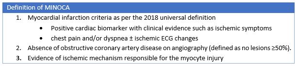
 

What is MINOCA?

Approximately 5-10% of myocardial infarct presentations are suspected as MINOCA and available data suggests that they are likely be younger, females and have lower cardiovascular risk factors than myocardial infarct patients with obstructed arteries. The recent 4th universal definition of myocardial infarction published in 2018 highlighted that the diagnosis of MINOCA indicates that there is an ischemic mechanism responsible for the myocyte injury. Therefore, the MINOCA diagnosis is not applied to patients with clinical evidence of aberrant troponin changes as a result of non-ischemic or non-cardiac causes such as myocarditis or pulmonary embolism.


What causes MINOCA and what are the additional recommended tests?

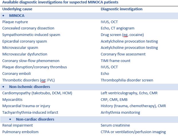
The dilemma with treating MINOCA is delineating MINOCA presentations from those with troponin rise and/or fall due to non-ischemic and non-cardiac causes as this not feasible based on the presentation itself. When a patient is suspected as MINOCA following coronary angiography, the patient should be clinically re-evaluated with multiple potential causes in mind. The following are the key underlying causes and corresponding diagnostic investigations.


Prognosis of MINOCA

The available literature demonstrates that overall suspected MINOCA patients have a favorable prognosis compared to those with the classic myocardial infarction (associated with obstructive CAD). However, careful examination of literature shows suspected MINOCA patients have the equivalent 12-month all-cause mortality to those with myocardial infarction associated with single- or double-vessel coronary artery disease. However, the prognosis associated with MINOCA with only ischemic mechanisms in mind is yet to be studied.


Treatment for MINOCA

There are no randomized trials addressing this question. However, a recent publication by Lindahl and colleagues stemming from the SWEDEHEART (Swedish Web-System for Enhancement and Development of Evidence-Based Care in Heart Disease Evaluated According to Recommended Therapy) registry provides the first insights into potential long-term prognostic benefit of medical therapy in the management of MINOCA. The authors have showed benefits of statins, angiotensin-converting enzyme inhibitors/angiotensin receptor blockers and beta-blocker therapy in MINOCA cohort.

The MINOCA BAT Trial (Randomized Evaluation of β‐Blocker and Angiotensin‐Converting Enzyme Inhibitor/Angiotensin Receptor Blocker Treatment in MINOCA Patients) is the first randomized clinical trial initiative in MINOCA patients and expected to begin enrollment in Australia and Europe in 2018 and also plans to expand enrollment to the United States and Canada in the next year. This will be a pragmatic prospective, randomized, multicentre, open-label clinical trial, with 2×2 factorial design. All outcomes will be analyzed using the intention-to-treat principle. The study aims to determine whether oral beta-blockade and/or ACEI/ARB impacts on MACE in patients discharged with MINOCA, where MACE is defined as the 4-year composite endpoint of all-cause mortality or hospital admission for AMI, ischemic stroke or heart failure.

As evident from Scientific Sessions 2018, the current available MINOCA literature demonstrates the importance of diagnosing and treating patients with MINOCA, although substantial knowledge gaps exist that require future research to identify optimal management.


Key points about MINOCA

  • MINOCA is not uncommon occurring in approximately 5-10% of patients.
  • MINOCA indicates that there is an ischemic mechanism responsible for the myocyte injury.
  • MINOCA diagnosis is not applied to patients with clinical evidence of aberrant troponin changes
  • There are various etiologies for MINOCA and it is important to perform a careful evaluation to identify the cause
  • Treatment will vary depending on the underlying cause but there may be some role for cardioprotective therapies in MINOCA
  • The prognosis of MINOCA is not benign, once again emphasizing proper diagnosis and aggressive treatment for this condition


“The good physician treats the disease; the great physician treats the patient who has the disease” – William Osler


Do you have any thoughts on MINOCA?


hidden

A Shift in Focus: From a Multi to Single Modality Approach

Since the first A-mode echocardiogram, there have been great revolutional changes and the use of noninvasive cardiac imaging has grown substantially during the past decade. Echocardiography and nuclear modality have been the primary imaging modalities for management in patients with different cardiovascular diseases. The introduction of cardiac magnetic resonance (CMR), computer tomography (CT), and three-dimensional (3D) printing, makes things quite different. Multi-modality imaging plays a role in all cardiovascular diseases that includes ventricular function, coronary artery disease, valvular disease, congenital heart disease, guidance for interventions, and vascular diseases. However, the use of each modality requires knowledge, expertise and forethought about cardiac diagnoses to avoid excess coast, inappropriate testing and to improve outcomes. Unnecessary duplication of investigations must be avoided for cost effective healthcare delivery.

For more than a decade, organizations such as the American College of Radiology and the American College of Cardiology Foundation have published criteria delineating the appropriate use of cardiac imaging. These specialty society guidance documents are designed to provide clinicians with recommendations regarding the use of imaging and are focused on reducing unnecessary and inappropriate testing. In response to rapid and unsustainable growth in the use of cardiac imaging procedures, various professional medical organizations have developed appropriate use criteria (AUC) to guide physicians and payers on effective use of these procedures. The AUC serves as a guide for physicians to incorporate symptoms, risk factors, and clinical history in selecting the patients for whom testing is most appropriate, based on the best available evidence or expert consensus; Furthermore, the AUC can help physicians steer other patients to alternative tests or no testing at all.

The AUC classify the appropriateness of testing across a range of clinical scenarios in 3 categories: “appropriate” (established value), “may be appropriate” (uncertain value), and “rarely appropriate” (no clear value). The AUC are intended to be used as part of a comprehensive disease management plan to maximize the value of care by minimizing unnecessary imaging and resultant downstream invasive procedures. (1). Appropriate use helps to avoid excess costs; enhances the value of imaging in risk stratification and decision making; and reduces radiation risk, particularly in women (2). Moreover, the principle of applying expert consensus criteria to choose the best testing strategy for each patient is widely accepted by payers, physicians, and patient groups.

  • The goal of this statement is not to showcase CMR as the exclusive modality for cardiac diagnosis but to highlight this tool as the corner stone of noninvasive cardiac imaging. By using a static and a gradient magnetic field in combination with a radiofrequency field, CMR imaging has evolved from a less commonly used technique into a tool with the potential to find an important clinical role in the near future for the noninvasive assessment of morphological and functional aspects of the heart. Consequently, CMR belongs to the fastest growing new fields of broad MR application. At the same time, CMR uses some of the strongest and fastest switching electromagnetic gradients available in MR imaging. CMR has become the primary imaging tool in many centers in characterizing disease severity and planning of patient management.
  • Volumes and Function: CMR measures ventricular volumes and mass using a simple acquisition of a 3D stack of contiguous short-axis cines with full biventricular coverage. CMR is significantly more accurate and reproducible than other techniques which makes it the technique of choice for longitudinal study of patients over time and for reducing sample size for drug studies.
  • Great Vessels: Three-dimensional angiography with gadolinium enhancement may show the lesion more clearly in high resolution, but it also shows any associated pathology (collaterals). CMR is fast and accurate for the diagnosis of aortic dissection although local issues relative to availability will determine whether CMR, CT, or echocardiography is used.
  • Valvular Heart Disease: CMR has a significant role in valve disease, but it usually acts as a second-line technique to assist when echocardiography with Doppler has proved problematic because of limited acoustic access, highly eccentric jets, or the need for quantification. The quantification of valve regurgitation is a strength of CMR because of its capability of measuring accurate ventricular stroke volumes from multislice ventricular planimetry and comparing this with the measurement of great vessel flow from velocity mapping.
  • Congenital Heart Disease: CMR is widely used to assess congenital heart disease, and when used in concert with echocardiography, the need for invasive assessment has been significantly reduced. CMR is particularly useful for the safe, accurate, and reproducible quantification of the left-to-right shunting of blood that occurs frequently in congenital heart defects such as atrial or ventricular septal defects, patent ductus arteriosus, aortopulmonary window, and partial or total anomalous pulmonary venous return.
  • Myocardial Ischemia: CMR is a safe and proven modality for detecting ischemic heart disease. Within a single study, CMR is able to assess rest and stress regional contraction, and localize and evaluate regions of myocardial ischemia and viability, of importance given the prognostic impact for the patient. With performance that is comparable to PET/SPECT and at times favorable compared to stress echocardiography, it is now an essential tool for diagnosing ischemia and for determining the likelihood of success following myocardial revascularization.
  • Non ischemic cardiomyopathy: CMR with its higher spatial resolution is considered the gold standard for evaluating ventricular mass, volumes, and ejection fraction. CMR can be used for accurate diagnosis of several conditions, especially cardiomyopathies. CMR is an invaluable tool, not only in differentiating nonischemic from ischemic cardiomyopathy, but also in aiding the accurate diagnosis of the subtype of nonischemic cardiomyopathy. CMR should routinely be integrated in the diagnostic workup of various cardiomyopathies.

CMR is a valuable tool for the evaluation of patients with, or at risk for, heart failure and has a growing impact on diagnosis, clinical management, and decision making. Through its ability to characterize the myocardium by using multiple different imaging parameters, it provides insight into the etiology of the underlying heart failure and its prognosis. CMR continues to develop across a broad range of clinical applications, and much can be expected of this technology in the future.

Fawaz Alenezi Headshot

Dr. Fawaz Abdulaziz M Alenezi is a Clinical Imaging Fellow at the Duke University Health Systems. He conducts medical research on the derivation and validation of novel echocardiographic approaches to myocardial deformation and a new echocardiographic technique which assists patients with heart ventricular function.