hidden

The Needle Moves Slowly on MINOCA

I remember being a medical student and listening to a podcast where I first heard the term MINOCA (myocardial infarction with nonobstructive coronary arteries) in 2019. I was deciding between internal medicine and OBGYN at this time, and learning about heart disease specific to and common in women naturally grasped my attention. Dr. Bairey Merz from Cedars-Sinai provides a fantastic overview of the disease process and evaluation. I kept thinking about all the ways we’ve made incredible strides in heart disease over the years. Now, I was thinking they were unequal.

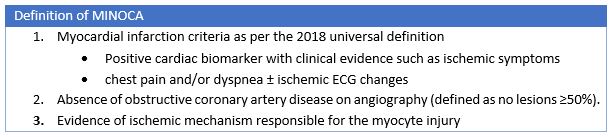
Dr. Brent Gudenkauf, a PGY-2 at the Johns Hopkins Hospital, et al. recently published a review in the Journal of the American Heart Association entitled “Role of Multimodality Imaging in the Assessment of Myocardial Infarction with Nonobstructive Coronary Arteries: Beyond Conventional Coronary Angiography1. They do a wonderful job of taking us through diagnostic criteria, preferred imaging modalities, and guideline recommendations regarding MINOCA. This term is still not commonplace outside of cardiology, and in the early days some thought these patients were having “false positive MIs” as they outline in the paper. This led to mostly women missing out on necessary diagnostic work up and targeted therapies. Today we have specific diagnostic criteria from the AHA and ESC including positive serum myocardial biomarkers and clinical evidence of MI (which can include ischemic symptoms, new ST segment changes, new LBBB, new pathologic Q waves among others) and no epicardial coronary lesions >50% stenosis on angiography.2,3

What I find disheartening is how slowly our needle has moved in terms of therapies. As they highlight in this paper, even after diagnosis of MINOCA 25% of patients continue to experience angina5 and experience worse quality of life compared with MI-CAD (MI associated with obstructive coronary artery disease) due to persistent anginal symptoms and inadequate treatment with existing antianginal therapies. They were less often treated with beta blockers and less often referred to cardiac rehab5. It is clear that this patient population of majority young women is faring worse than its traditional myocardial infarction counterpart in terms of therapies and quality of life.

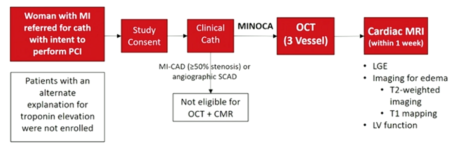
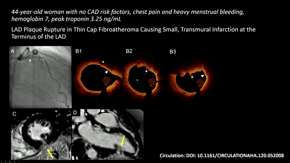
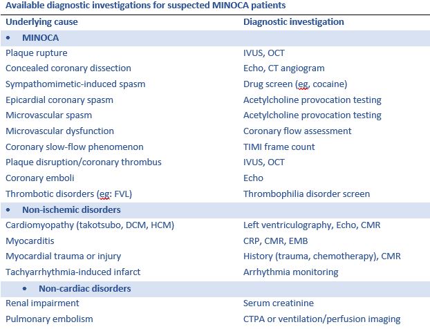
For these reasons, we should all develop a good understanding of diagnostic pathways and targeted treatments. The recommended imaging modality is IVUS (intravascular ultrasound) or OCT (optical coherence tomography) 2,4. As a young trainee myself, I am not familiar with either of these modalities and was introduced to these concepts via #AHA21. OCT is an optical analogue of IVUS and can “differentiate tissue characteristics such as fibrous, calcified, or lipid-rich plaque and identify thin-cap fibroatheroma”6. During PCI, OCT can also provide information about dissection, tissue prolapse, and thrombi6; this is significant given SCAD (spontaneous coronary artery dissection), in situ thrombosis, and epicardial and microvascular spasms are all causes that can lead to MINOCA1. Cardiac MR is also useful when MINOCA is suspected as it will show late gadolinium enhancement and can also uncover mimics like myocarditis and Takotsubo cardiomyopathy. Additionally, if embolism to coronary arteries is suspected then thrombophilia workup is recommended. They do a wonderful job outlining this algorithm in Figure 2 in the paper by Gudenkauf et al.1 We should all be working to familiarize ourselves with this figure and its recommendations and integrating this into our evaluation for chest pain.

Although the advancements in diagnosis and evaluation are exciting and important, there are no randomized clinical trials evaluating treatments for patients with MINOCA. The MINCOA-BAT trial is an upcoming randomized multi-center study which will hopefully help to move the needle forward in evidence-based targeted therapies (clinicaltrials.gov, NCT 03686696). This excellent review by Gudenkauf et al should be shared widely as this is an important and still too often underdiagnosed and undertreated condition among our patients.


References

  1. Gudenkauf, B., Hays, A. G., Tamis‐Holland, J., Trost, J., Ambinder, D. I., Wu, K. C., Arbab‐Zadeh, A., Blumenthal, R. S., & Sharma, G. (2021). Role of multimodality imaging in the assessment of myocardial infarction with nonobstructive coronary arteries: Beyond conventional coronary angiography. Journal of the American Heart Association. https://doi.org/10.1161/jaha.121.022787
  2. Tamis‐Holland JE, Jneid H, Reynolds HR, Agewall S, Brilakis ES, Brown TM, Lerman A, Cushman M, Kumbhani DJ, Arslanian‐Engoren C, et al. Contemporary diagnosis and management of patients with myocardial infarction in the absence of obstructive coronary artery disease: a scientific statement from the American Heart Association. Circulation. 2019; 139:e891–e908. doi: 10.1161/CIR.0000000000000670
  3. Ibanez B, James S, Agewall S, Antunes MJ, Bucciarelli‐Ducci C, Bueno H, Caforio ALP, Crea F, Goudevenos JA, Halvorsen S, et al. 2017 ESC guidelines for the management of acute myocardial infarction in patients presenting with ST‐segment elevation: the Task Force for the management of acute myocardial infarction in patients presenting with ST‐segment elevation of the European Society of Cardiology (ESC). Eur Heart J. 2018; 39:119–177. doi: 10.1093/eurheartj/ehx393
  4. Agewall S, Beltrame JF, Reynolds HR, Niessner A, Rosano G, Caforio AL, De Caterina R, Zimarino M, Roffi M, Kjeldsen K, et al. ESC working group position paper on myocardial infarction with non‐obstructive coronary arteries. Eur Heart J. 2017; 38:143–153. doi: 10.1093/eurheartj/ehw149
  5. Grodzinsky A, Arnold SV, Gosch K, Spertus JA, Foody JM, Beltrame J, Maddox TM, Parashar S, Kosiborod M. Angina frequency after acute myocardial infarction in patients without obstructive coronary artery disease. Eur Heart J Qual Care Clin Outcomes. 2015; 1:92–99. doi: 10.1093/ehjqcco/qcv014
  6. Terashima, M., Kaneda, H., & Suzuki, T. (2012). The role of optical coherence tomography in coronary intervention. The Korean journal of internal medicine, 27(1), 1–12. https://doi.org/10.3904/kjim.2012.27.1.1.

“The views, opinions and positions expressed within this blog are those of the author(s) alone and do not represent those of the American Heart Association. The accuracy, completeness and validity of any statements made within this article are not guaranteed. We accept no liability for any errors, omissions or representations. The copyright of this content belongs to the author and any liability with regards to infringement of intellectual property rights remains with them. The Early Career Voice blog is not intended to provide medical advice or treatment. Only your healthcare provider can provide that. The American Heart Association recommends that you consult your healthcare provider regarding your personal health matters. If you think you are having a heart attack, stroke or another emergency, please call 911 immediately.”

hidden

An ALL-Woman Trial on MINOCA Takes a Seat in the Main Area as Late Breaking Science Addressing Challenges in Coronary Care

As an interventional cardiologist who’s passionate about reducing the disparities in diagnosis and management in women with cardiovascular disease, I was captivated by the late-breaking science that took the main arena for Current Challenges in Coronary and Valve Disease at Scientific Sessions 2020. The Coronary OCT and Cardiac MRI (CMR) to Determine Underlying Cause of MINOCA (Myocardial Infarction with Nonobstructive Coronary Arteries) in Women Trial from the HARP (Women’s Heart Attack Research Program) investigators led by Dr. Reynolds and colleagues is truly groundbreaking for women patients with MINOCA (1).  HARP-MINOCA was a multimodality imaging trial that included all women. Yes—that’s correct. An ALL Woman Trial. Why would a study enroll all women?  Because MINOCA is a condition that disproportionately affects women (3). In women presenting with MI, the presence of MINOCA is 10.5% compared to 3.4% in men (4).  MINOCA is a disease process that has largely been met with controversy such that clinicians have challenged the presence of a “true” myocardial infarction in the absence of coronary artery disease. Furthermore, the lack of an obstructive lesion on a coronary angiogram has misled women to believe that they’re “fine” just because their coronary arteries don’t require stenting.  This is completely false. Patients with MINOCA have clinical outcomes similar to patients with obstructive CAD at the time of myocardial infarction (MI) (3).  Our patients have suffered in this variability of treatment and assessment which ultimately impacts their care. The findings of this study area additive to the American Heart Association’s scientific statement on the contemporary diagnosis and management of MINOCA which sought to standardize the definition of MINOCA and create a clinically useful diagnostic framework and treatment algorithm (2). Thus, the HARP-MINOCA multimodality imaging findings deepen the roots of MINOCA as a true disease process that requires a proper diagnosis with multimodality imaging to optimize management for improved patient outcomes.

This prospective multicenter study enrolled women a total of 301 women with a clinical diagnosis of MI across 16 different sites of which 170 had MINOCA. Once invasive coronary angiography was performed and revealed <50% stenosis in all major arteries, multi-vessel optical coherence tomography (OCT) was performed, followed by CMR (cine imaging, late gadolinium enhancement, and T2-weighted imaging and/or T1 mapping). [Figure 1]

Figure 1

Dual imaging allowed for a thorough investigation of the following: 1) Vascular causes of MINOCA by OCT, 2) Myocardial abnormalities on CMR and 3) integration of the various underlying etiologies on OCT and CMR.  The findings from OCT investigation revealed that a culprit lesion in 46% of cases included plaque rupture, thrombus without plaque rupture, intra-plaque cavity, layered plaque, dissection, or spasm.  The CMR findings revealed infarction with late gadolinium enhancement 33% of cases, regional pattern of ischemic injury in 21% and 21% had non-ischemic pattern giving them an alternate diagnosis. The study did have a few limitations included a low rate of STEMI enrollment, regional myocarditis cannot be excluded from the CMR definition of ischemic injury (defined as a single coronary territory with myocardial edema) that was used, the contribution of coronary vasospasm to the presentation was not evaluated, and not all women underwent 3-vessel OCT and CMR leaving some diagnoses that may have been missed.

The integration of the dual-imaging findings revealed a specific cause for MINOCA in 85% of cases.  When an OCT culprit lesion, there was CMR evidence of infarction or regional ischemic injury in 75% of cases.  Multi-modality imaging was better than either imaging modality alone leading to an identified cause of MINOCA in 85% of cases. One of the cases shared during the trial presentation was eye-opening.  Despite no evidence of an obstructive lesion in the LAD vessel on a coronary angiogram, OCT performed in the LAD revealed plaque rupture and subsequent CMR demonstrated a small, transmural infarction in the terminal segment of the LAD. [Figure 2]. The importance of establishing the diagnosis with additional multimodality imaging is the key findings here. MINOCA patients have pathophysiology hidden deep beyond the limitations of coronary angiography and without additional imaging, they could be subject to a missed diagnosis and ultimately poor long-term care and management.

Figure 2

The 2020 European Society of Cardiology for non-ST elevation MI gave the use of CMR a Class IB recommendation for MINOCA evaluation.  Unfortunately, there are no such recommendations for the use of intracoronary imaging. As a community-based interventionalist who performs emergent percutaneous coronary interventions at ST-Elevation MI (STEMI) receiving centers without cardiothoracic surgical support, we have restrictions as operators when performing interventions such an intravascular imaging when no intervention is planned.  The implications of this OCT relevant data would be practice-changing for interventional cardiologists practicing in my clinical setting.  The feasibility of OCT may bring its own challenges with operator-experience, staff support, and contrast use.  However, education regarding the important data noted from OCT in MINOCA patients is a very important first step to implement change in one’s cath lab.

This study presented here at sessions continues to advance the growing field of MINOCA science. Late-breaking science advancing the understanding of this heterogeneous population of MINOCA patients is incredibly exciting and I’m looking forward to the continuum of knowledge to transform the algorithm for diagnostic assessment and framework for MINOCA treatment.


References:

  1. Reynolds et al. Coronary Optical Coherence Tomography and Cardiac Magnetic Resonance Imaging to Determine Underlying Causes of MINOCA in Women. Circulation 2020; Epub. 10.1161/CIRCULATIONAHA.120.052008
  2. Tamis-Holland JE et al. Contemporary diagnosis and management of patients with myocardial Infarction in the absence of obstructive coronary artery disease: a scientific statement from the American Heart Association. Circulation. 2019;139(18):e891–908.
  3. Safdar B et al. Presentation, clinical profile, and prognosis of young patients with myocardial infarction with nonobstructive coronary arteries (MINOCA): results from the VIRGO study. J Am Heart Assoc. 2018;7(13)
  4. Smilowitz NR et al. Mortality of myocardial infarction by sex, age, and obstructive coronary artery disease status in the ACTION registry-GWTG (acute coronary treatment and intervention outcomes network registry-get with the guidelines). Circ Cardiovasc Qual Outcomes. 2017;10(12):e003443.

“The views, opinions and positions expressed within this blog are those of the author(s) alone and do not represent those of the American Heart Association. The accuracy, completeness and validity of any statements made within this article are not guaranteed. We accept no liability for any errors, omissions or representations. The copyright of this content belongs to the author and any liability with regards to infringement of intellectual property rights remains with them. The Early Career Voice blog is not intended to provide medical advice or treatment. Only your healthcare provider can provide that. The American Heart Association recommends that you consult your healthcare provider regarding your personal health matters. If you think you are having a heart attack, stroke or another emergency, please call 911 immediately.”

hidden

AHA Scientific Statement on Diagnosis and Management of Myocardial Infarction with Non-Obstructive Coronary Arteries (MINOCA)

Pathophysiology of a classic acute MI is attributed to the concept of coronary atherothrombosis leading to myocardial ischaemia and ultimately infarction. The overall prognostic benefit with coronary revascularization has been established in these patients. Recently, there is a significant research and clinical interest in acute MI presentations without evidence of significant atherothromotic lesions, so that revascularization therapies are considered inappropriate. These presentations are referred as Myocardial Infarction with Non-Obstructive Coronary Arteries (MINOCA) and is now an established clinical entity. The AHA just released the first scientific statement on diagnosis and management of MINOCA and is an important read1. The document provides the first formal updated definition of MINOCA and clinically useful framework and algorithms for the diagnostic evaluation and management of these patients.

The key points from the statement are:


The diagnosis of MINOCA is made in patients with acute MI due to myocardial ischemia.

  1. Acute myocardial infarction as per the “Fourth Universal definition of MI” Criteria
  2. Nonobstructive coronary arteries on angiography: the absence of obstructive disease on angiography (ie, no coronary artery stenosis ≥50%) in any major epicardial vessel
  3. No specific alternate diagnosis for the clinical presentation: Alternate diagnoses include but are not limited to non-ischemic causes such as sepsis, pulmonary embolism, and myocarditis


The “Traffic Light” Sequence for the Diagnosis of MINOCA.

Involving a clever adaptation of traffic light sequence, a very detailed diagnostic algorithm was provided for the diagnosis of MINOCA.

Red:  to exclude myocardial injury causes without ischemic context (Eg: Sepsis, Pulmonary Embolism)

Yellow: to exclude clinically subtle non-ischemic mechanisms of myocardial injury (Eg: Clinically overlooked CAD, Takotsubo, Myocardits)

Green: the final diagnosis of MINOCA is made upon a clear evidence of an ischemic context.


Specific causes of MINOCA Presentations: Atherosclerotic vs Nonatherosclerotic Causes of Myocardial Necrosis

Plaque disruption:

  • Reported in approximately 1/3 of MINOCA undergoing IVUS.
  • Authors recommend invasive imaging studies (IVUS or OCT) if available

Coronary Spasm:

  • Reported in approximately 50% of MINOCA undergoing provocative spasm testing.
  • Predilection for spasm in Asians compared with Caucasians.
  • Spasm testing appears to be safe in MINOCA cohort.

Microvascular Dysfunction:

  • Need to be studied in MINOCA population

Coronary embolism/Thrombosis:

  • Consider the inherited hypercoagulable states in patients with MINOCA, especially in younger women

Spontaneous Coronary Artery Dissection

  • Rare
  • Should be suspected mainly in young women


Management strategies for MINOCA

Given that there is currently no randomized clinical trials or guidelines on treating MINOCA, the statement suggests careful considerations in managing patients. Overall, a ‘working diagnosis’ approach should be adopted, with cardioprotective therapies and treatments targeting the underlying cause considered.


The full AHA statement on MINOCA can be found here.


Reference:

  1. Tamis-Holland Jacqueline E, Jneid H, Reynolds Harmony R, Agewall S, Brilakis Emmanouil S, Brown Todd M, Lerman A, Cushman M, Kumbhani Dharam J, Arslanian-Engoren C, Bolger Ann F, Beltrame John F and null n. Contemporary Diagnosis and Management of Patients With Myocardial Infarction in the Absence of Obstructive Coronary Artery Disease: A Scientific Statement From the American Heart Association. Circulation. 0:CIR.0000000000000670.
hidden

MINOCA – The New Unique Type of Myocardial Infarction

Myocardial infarctions have been claiming lives since ancient times, yet we are still understanding the condition itself. With the emergence of acute coronary angiography in the 80s, it became evident nearly 90% of myocardial infarctions are associated with occluded coronary arteries. This led to the advances in clinical approaches to reduce the myocardial damage by reopening the obstructed coronary arteries as quickly as possible with the aid of mechanical and pharmacological interventions. Among all this, a distinctive form of myocardial infarction have been silently increasing in prevalence over the years without drawing much attention. This subtype of myocardial infarction has recently been named “myocardial infarction with non-obstructive coronary arteries’ or ‘MINOCA’. As the name implies, it refers to patients presenting with myocardial infarct symptoms without obstructive coronary artery disease. The lack of obstructive coronary artery disease in this group often leads clinicians to disregard them as “false-positive presentations” and patients are discharged with “it doesn’t seem like there is anything wrong with you and there is not much I can do about it at this stage.” Despite a myocardial infarct presentation, patients are discharged to home with minimal to no medical management and no explanation. With the widespread use of coronary angiography and the advance of more sensitive cardiac biomarkers, the MINOCA presentations have started to gain attention among cardiologists and researchers in recent years.

This year at Scientific Sessions 2018, Dr Jacqueline E. Tamis-Holland and Dr Harmony Reynolds addressed MINOCA and the existing knowledge gap. Here is a summary of the key points discussed at the meeting:


What is MINOCA?

Approximately 5-10% of myocardial infarct presentations are suspected as MINOCA and available data suggests that they are likely be younger, females and have lower cardiovascular risk factors than myocardial infarct patients with obstructed arteries. The recent 4th universal definition of myocardial infarction published in 2018 highlighted that the diagnosis of MINOCA indicates that there is an ischemic mechanism responsible for the myocyte injury. Therefore, the MINOCA diagnosis is not applied to patients with clinical evidence of aberrant troponin changes as a result of non-ischemic or non-cardiac causes such as myocarditis or pulmonary embolism.


What causes MINOCA and what are the additional recommended tests?

The dilemma with treating MINOCA is delineating MINOCA presentations from those with troponin rise and/or fall due to non-ischemic and non-cardiac causes as this not feasible based on the presentation itself. When a patient is suspected as MINOCA following coronary angiography, the patient should be clinically re-evaluated with multiple potential causes in mind. The following are the key underlying causes and corresponding diagnostic investigations.


Prognosis of MINOCA

The available literature demonstrates that overall suspected MINOCA patients have a favorable prognosis compared to those with the classic myocardial infarction (associated with obstructive CAD). However, careful examination of literature shows suspected MINOCA patients have the equivalent 12-month all-cause mortality to those with myocardial infarction associated with single- or double-vessel coronary artery disease. However, the prognosis associated with MINOCA with only ischemic mechanisms in mind is yet to be studied.


Treatment for MINOCA

There are no randomized trials addressing this question. However, a recent publication by Lindahl and colleagues stemming from the SWEDEHEART (Swedish Web-System for Enhancement and Development of Evidence-Based Care in Heart Disease Evaluated According to Recommended Therapy) registry provides the first insights into potential long-term prognostic benefit of medical therapy in the management of MINOCA. The authors have showed benefits of statins, angiotensin-converting enzyme inhibitors/angiotensin receptor blockers and beta-blocker therapy in MINOCA cohort.

The MINOCA BAT Trial (Randomized Evaluation of β‐Blocker and Angiotensin‐Converting Enzyme Inhibitor/Angiotensin Receptor Blocker Treatment in MINOCA Patients) is the first randomized clinical trial initiative in MINOCA patients and expected to begin enrollment in Australia and Europe in 2018 and also plans to expand enrollment to the United States and Canada in the next year. This will be a pragmatic prospective, randomized, multicentre, open-label clinical trial, with 2×2 factorial design. All outcomes will be analyzed using the intention-to-treat principle. The study aims to determine whether oral beta-blockade and/or ACEI/ARB impacts on MACE in patients discharged with MINOCA, where MACE is defined as the 4-year composite endpoint of all-cause mortality or hospital admission for AMI, ischemic stroke or heart failure.

As evident from Scientific Sessions 2018, the current available MINOCA literature demonstrates the importance of diagnosing and treating patients with MINOCA, although substantial knowledge gaps exist that require future research to identify optimal management.


Key points about MINOCA

  • MINOCA is not uncommon occurring in approximately 5-10% of patients.
  • MINOCA indicates that there is an ischemic mechanism responsible for the myocyte injury.
  • MINOCA diagnosis is not applied to patients with clinical evidence of aberrant troponin changes
  • There are various etiologies for MINOCA and it is important to perform a careful evaluation to identify the cause
  • Treatment will vary depending on the underlying cause but there may be some role for cardioprotective therapies in MINOCA
  • The prognosis of MINOCA is not benign, once again emphasizing proper diagnosis and aggressive treatment for this condition


“The good physician treats the disease; the great physician treats the patient who has the disease” – William Osler


Do you have any thoughts on MINOCA?


hidden

Highlights of the 1st Annual Sex and Gender Conference at AHA18

Walking into the Palmer House Hotel, the longest continuously operating hotel in the United States, you can’t help but pause in awe at the intricate décor and take in the most photographed ceiling in the world. I make my way to the Honoré Ballroom, named after Bertha Honoré Palmer, the wife of Palmer and an astute businesswoman and well-known Chicago socialite of her time, not knowing what to expect for the 1st annual Sex and Gender Influence on Cardiovascular Disease (CVD) conference.

Annabelle Volgman, medical director of the Rush Heart Center for Women, kicks off the evening by thanking the speakers and planning members, and encouraging photography and social media sharing. The many photos of the evening include Bertha Honoré’s portrait adjacent to the colorful and modern logo that, I think, will become a recognized image at future AHA Scientific Session meetings.

Dr. Annabelle Volgman welcomes attendees to the 1st Annual Sex and Gender Influences on Cardiovascular Disease at the Palmer Hotel in Chicago, IL (November 11, 2018).


Dr. Nanette Wenger of the Emory Women’s Heart Center starts the conversation with her presentation titled “Why is Mortality from Cardiovascular Disease Rising in Men and Women?” She flashes a graph of CVD mortality on the screen, highlighting the steep decline in the past decades, but the leveling off and reversal in recent years, particularly in women under the age of 55 years. The parallel rise in obesity and diabetes, as well as “non-traditional” CVD risk factors such as depression and perceived stress disproportionally affect women, she explains, and may be responsible for this reversal in CVD death rates. Summarizing the recent paper, “Defining the New Normal in Cardiovascular Risk Factors” by Dr. Donald Lloyd-Jones and Dr. Philip Greenland she points to a combination of health behaviors and ideal levels of total cholesterol, blood pressure, and fasting blood glucose, as key factors in achieving cardiovascular health.

“Behavior change,” she says, “is the ‘Holy Grail’ of heart health” and as “health professionals take back the role [of health educator] and address lifestyle behaviors” we will see favorable trends in biomarker targets we’re so interested in.

Later during the Q+A panel, when asked about the best way to approach behavior change with patients, she advises to first, “Give information – if your patient does not have the information, they can’t make a change. Then, let them start with what they would like to start with. Don’t give them 8-10 [health behaviors] to change – they will tune you out.” Dr. Gina Lundberg, co-director of the Emory Women’s Heart Center, chimes in that the clinician’s “approach to weight loss is similar to smoking cessation. Identify the obstacles in the patient’s way – money, time, desire – and often just identifying those hurdles will lead to improvement.”

Dr. Laxmi Mehta, director of the Women’s Cardiovascular Health Program at the Ohio State University Wexner Medical Center, adds that she includes an emotional appeal – “Where is the patient going and what do they want?” Seeing a child’s wedding or playing with their grandkids, developing rapport with patients and fitting your recommendations to their goals can start the health behavior change process, even in a 5 minute clinician-patient discussion.