hidden

Happy July 1: Cardiology Fellowship Begins

Anyone in the medical field knows the significance of July 1.  Don’t get sick in July, they say, because the hospital is full of brand-new residents and fellows.  For me, cardiology fellowship begins right where internal medicine residency left off—at Emory.  At least I know where to park and how to find the bathrooms.

This year we have a tight-knit group of six clinical fellows.  At orientation, we practiced performing echocardiograms on each other, taking turns squinting at gray speckles on a dark screen.  That night, we raised our drinks to say a toast—and to wash away our nerves.  And soon enough, I’m strolling into the hospital sporting my new white coat, which drapes over my shoulders like an oversized tent.  The coat fits awkwardly, in both a physical and figurative sense.

I’m told you don’t even feel like a real cardiologist until you’ve learned to perform heart catheterizations and read echocardiograms.  Ask me again in six months.  For now, I start with general consults, where I’m the cardiology consultant for other physicians in the hospital.  The hardest part about inexperience is the decision-making fatigue—even trivial decisions require excessive mental effort.  To overcome this, the goal is to see as many bread-and-butter cases as possible, to build a sort of muscle memory.

It’s been a wonderful year on this blog, reminiscing the end of residency, chronicling the start of fellowship, and pondering the milestones yet to come.  What gives me comfort at this moment is the supportive culture of my program, where I can always lean on co-fellows and attendings.  I’ll keep this mind as I tackle the next major hurdle—my first overnight call.  Just thinking about it gives me palpitations.

hidden

Heart Attack and Stroke: Same Disease, Different Organs?

I’m spending the last month of internal medicine residency on a neurology rotation.  I suppose that’s fair; my wife, a neurology resident, had to do a whole year of medicine.  To me, the most interesting part of neurology is the parallel between stroke and acute myocardial infarction (AMI).  Conceptually these are two manifestations of a common underlying disease process.  Yet, there are glaring differences in their management, and I can’t help but wonder why.

For instance, neurologists and cardiologists use different protocols for anticoagulation and thrombolysis.  Tissue plasminogen activator (tPA) is a first line therapy for ischemic stroke unless there are contraindications, including recent use of anticoagulation.  Thrombolytic therapy is also used to treat STEMI when percutaneous coronary intervention (PCI) is not immediately available.  In contrast to stroke, STEMI thrombolysis calls for higher doses of tPA as well as concurrent infusion of heparin to prevent recurrent thrombosis.1  Perhaps thrombolysis after stroke is a more cautious affair due to the risk of reperfusion injury and hemorrhagic conversion.

For decades STEMI PCI has largely replaced tPA, yet endovascular therapy for stroke is a relatively recent innovation and its utility is limited to proximal large vessel occlusions.  While PCI relies on balloon expandable stents designed to prevent restenosis, stenting is perhaps a less attractive option in stroke due to the tortuous anatomy of intracranial vessels and the bleeding risk associated with dual antiplatelet therapy.2 Instead, neurologists perform mechanical thrombectomy using stent retrievers and aspiration catheters.  While routine thrombectomy during STEMI PCI is generally not beneficial,3 aspiration and rheolytic catheters can be used selectively in the event of large thrombus burden.

Finally, evidence does not support facilitated PCI (i.e. pretreatment with tPA prior to PCI).4-5  Interestingly, it is common practice among neurologists to pretreat with tPA prior to mechanical thrombectomy.  Theoretically pretreatment may facilitate clot extraction, but does this strategy outweigh the additional bleeding risk?6

Heart attack and stroke are similar diseases occurring in different organs.  With widespread adoption of mechanical thrombectomy for acute stroke, the fields of neurology and cardiology increasingly share similar practices.  Still, there are striking differences in stroke and AMI management—no doubt a constant source of cognitive dissonance as I complete my neurology rotation and start cardiology fellowship.


References:

  1. Kijpaisalratana N, Chutinet A, Suwanwela N. Hyperacute simultaneous cardiocerebral infarction: Rescuing the brain or the heart first? Frontiers in Neurology 2017;8:664.
  2. Gralla J, Brekenfeld C, Mordasini P, Schroth G. Mechanical thrombolysis and stenting in acute ischemic stroke. Stroke 2012;43:280-285.
  3. Jolly SS, James S, Dzavik V, et al. Thrombus aspiration in ST-segment elevation myocardial infarction. An Individual Patient Meta-Analysis: Thrombectomy Trialists Collaboration. Circulation. 2016;135:143–152.
  4. The ASSENT-4 PCI Investigators. Primary versus tenecteplase-facilitated percutaneous coronary intervention in patients with ST-segment elevation acute myocardial infarction (ASSENT-4 PCI). Lancet. 2006;367:569–578.
  5. Ellis SG, Tendera M, de Belder MA, et al. Facilitated PCI in patients with ST-elevation myocardial infarction. N Engl J Med. 2008;358:2205–17.
  6. Kasemacher J, Mordasini P, Arnold M, et al. Direct mechanical thrombectomy in tPA-ineligible and -eligible patients versus the bridging approach: a meta-analysis. J Neurointerv Surg. 2019;11:20-27.
hidden

Small Conferences Are More Worthwhile

I ditched the hospital early last Friday and sped over to the Emory Conference Center.  There, gray-haired luminaries and neatly combed trainees filled the lobbies with their crisp suits and smart banter.  This is the scene of the Emory Practical Intervention Course, with all the pomp of a proper nerd ball.  The annual meeting attracts interventional cardiologists from across the Southeast region with live case demonstrations and simulations.  But more importantly, it’s an opportunity for old colleagues to reunite, and for young trainees like me to rub shoulders with them.  There were academic faculty and community interventionists, and they all seemed to know each other.

The EPIC meeting reminded me of another smaller gathering last November—AHA Resuscitation Science Symposium (ReSS).  Tucked away from the massive Scientific Sessions next door, ReSS served a tight circle of investigators studying survival after cardiac arrest.  As I showed off my poster on bystander AED use, I was approached by a man with particularly insightful input.  A glance at his name tag revealed him to be the author of a manuscript I had cited in my work.  Small, intimate meetings foster this type of serendipity.  It’s easy for young trainee to feel intimidated or lost in the crowd at a national meeting.  That’s why smaller conferences (such as the upcoming AHA Basic Cardiovascular Sciences (BCVS) meeting) can have a bigger payoff.

hidden

Cardiac CT: The Future of Diagnostic Cardiology?

As a medical student eyeing the field of radiology, the science of imaging of was all too seductive.  Ultimately, a love for cardiac physiology won me over, but an interest in imaging lingered.  As it turns out, cardiologists are part-time radiologists with expertise in a number of cardiac imaging modalities.

CT has become the latest frontier in cardiac imaging with a number of useful applications.

By now, coronary calcium scoring is a well-established tool for risk stratification in subclinical coronary artery disease.  Cross-sectional imaging is also useful for evaluating pericardial thickening in constrictive pericarditis.  Beyond these traditional applications, newer techniques are poised to change the way we use CT to evaluate heart disease.


Coronary CT Angiography

Using fast, EKG-gated scanners, coronary CT angiography (CCTA) is a noninvasive means to detect coronary anomalies and obstructive plaque.  CCTA is a sensitive tool for excluding coronary disease, with a nearly perfect negative predictive value in the ACCURACY trial1.  However, specificity is poor and the presence of stents or calcium degrades image quality.

The specificity of CCTA is improved with FFR-CT (HeartFlow), a noninvasive method that mimics invasive fractional flow reserve measurements.  Computational fluid dynamics are applied to a 3D model of coronary anatomy in order to simulate the hemodynamic effects of stenotic lesions.  The PLATFORM trial2 showed how these technologies can safely reduce unnecessary catheterizations with no detriment to outcomes.


CT Myocardial Perfusion Imaging

CT myocardial perfusion imaging is also possible.  Indeed, a key advantage of CT is the ability to combine anatomic and physiologic evaluation in a single study.  However, exposure to radiation and iodinated contrast is an important consideration when comparing this to SPECT imaging.


As our diagnostic tools multiply, cardiac testing will become less invasive yet choosing the right study will become more complicated.  Cardiology is a fortunate field that controls much of its own imaging, but with the emergence of cardiac CT, we will need to collaborate with our radiology colleagues to push our fields forward in tandem.


References:

1Budoff MJ, Dowe D, Jollis JG, et al. Diagnostic performance of 64-multidetector row coronary computed tomographic angiography for evaluation of coronary artery stenosis in individuals without known coronary artery disease: results from the prospective multicenter ACCURACY (Assessment by Coronary Computed Tomographic Angiography of Individuals Undergoing Invasive Coronary Angiography) trial. J Am Coll Cardiol 2008;52:1724-32.

2Douglas PS, Pontone G, Hlatky MA, et al. Clinical outcomes of fractional flow reserve by computed tomographic angiography-guided diagnostic strategies vs. usual care in patients with suspected coronary artery disease: the prospective longitudinal trial of FFR(CT): outcome and resource impacts study. Eur Heart J. 2015;36:3359–3367. doi: 10.1093/eurheartj/ehv444.

hidden

Tech in Cardiology

Tech in Cardiology

On a recent flight from San Francisco, I found myself sitting in a dreaded middle seat.  To my left was a programmer typing way in Python, and to my right was an oncologist flipping through a slide set on chemotherapy trials.  While this may sound like the beginning of a bad joke, I remember this moment because it got me thinking about the influence of tech on medicine.  The purpose of my trip, by the way, was to interview for a fellowship position in cardiology, a specialty with arguably some of the most impressive tech.


Wearables

Not to discount advances in medical devices (e.g. leadless pacemakers, bioprosthetic valves), the emergence of consumer-facing wearable devices is as trendy as ever.  Google recently collaborated with AHA to build its fitness app (Google Fit), which uses algorithms to quantify physical activity in terms of “heart points.”1  The Apple Health app now incorporates EKG capabilities, allowing patients to record episodes of arrhythmias—something I have certainly witnessed in cardiology clinic.2


Big data

Big data is an increasingly prominent component of clinical research, and a number of joint ventures with medical and tech leaders have emerged.  One Brave Idea3 is a research collaboration between AHA and Verily (Alphabet’s life sciences division) which uses genomics to study coronary artery disease.  Meanwhile, Verily’s Project Baseline4 is a massive longitudinal observational study—a modern version of the Framingham Heart Study.


Artificial intelligence

AI could eventually play a prominent role in medical diagnosis and decision-making.  The Stanford Machine Learning Group5 has developed a neural network that outperforms cardiologists in diagnosing arrhythmias on EKG—a significant improvement on existing algorithms which are often unreliable.  AI also carries vast potential in radiologic interpretation.  Already, Veril is using machine learning to interpret retinal images not only to detect diabetic retinopathy and macular edema but also to extrapolate information about cardiovascular risk.6


EMR

Electronic medical records represent an obvious space for tech innovation.  Fast Healthcare Interoperability Resources (FHIR) are making it easier to share health information across our disjointed EMR systems.  Providers are now able to push health data directly to patients’ iPhones using Apple Health Records.7  One can only speculate whether we will see a legacy software giant compete directly in the EMR space.


Cardiology and the rest of medicine has long excelled at basic science and translational research, but digital tech is increasingly creeping in.  We are in a tech zeitgeist, and this is good for both patients and providers.


References:

  1. https://www.heart.org/en/news/2018/08/21/google-just-launched-heart-points-here-are-5-things-you-need-to-know
  2. https://www.apple.com/healthcare/site/docs/Apple_Watch_Arrhythmia_Detection.pdf
  3. https://www.onebraveidea.org/
  4. https://verily.com/projects/precision-medicine/baseline-study/
  5. https://stanfordmlgroup.github.io/projects/ecg/
  6. https://blog.verily.com/2018/02/eyes-window-into-heart-health.htm
  7. https://www.apple.com/healthcare/health-records/
hidden

A Trainee’s Notes on the MitraClip: MITRA-FR versus COAPT

As an internal medicine resident, I’ve been peripherally involved in managing patients undergoing percutaneous mitral valve repair and conducting research on procedural outcomes.  Developed as an alternative to conventional mitral valve surgery, the MitraClip device provides a percutaneous means to treat mitral regurgitation (MR).  The device was originally approved for degenerative MR based on data from the EVEREST trials.  As recent headlines show, the role for MitraClip in functional MR is the subject of ongoing debate.

Marking a first stab at answering this question, the MITRA-FR trial gave us a neutral result.  In spite of symptomatic improvement, there was no reduction in the primary composite endpoint of all-cause mortality and cardiac rehospitalization.  Shortly thereafter, the COAPT trial offered new hope, demonstrating a marked reduction in cardiac rehospitalization (NNT=3.1) and mortality (NNT=5.9).

There are a few ways to reconcile these differences.  MITRA-FR enrolled half as many patients and followed them for half as long compared to COAPT; therefore, the study may have lacked statistical power.  Others have questioned operator experience, pointing to a high proportion of patients in MITRA-FR with significant residual MR.  Finally, patients in the COAPT study had more severe MR and less dilated ventricles, making them better candidates for valve repair.  In MITRA-FR patients with very low ejection fraction, valve repair may have had negligible effect in the face of severe underlying cardiomyopathy.

This last point underscores the importance of patient selection and the need to identify prognosticators of clinical outcomes.  To this end, my mentors and I conducted a retrospective review of patients undergoing percutaneous mitral valve repair at Emory University Hospital.  We found that intraprocedural improvement in pulmonary venous doppler waveforms predicted 1-year survival and freedom from cardiac rehospitalization in both degenerative MR and functional MR subgroups.

It’s exciting to “grow up” and enter the field of cardiology in an era of advanced structural interventions.  On the horizon, RESHAPE-HF is yet another study examining the role for MitraClip in functional MR.  The results are highly anticipated, and I hope it brings clarity to this contentious topic.


References:

  • Obadia J-F, Messika-Zeitoun D, Leurent G, et al. Percutaneous repair or medical treatment for secondary mitral regurgitation. N Engl J Med 2018;379(24):2297-2306.
  • Stone GW, Lindenfeld JA, Abraham WT, et al., on behalf of the COAPT Investigators. Transcatheter Mitral-Valve Repair in Patients With Heart Failure. N Engl J Med 2018;379:2307-18.
  • Corrigan FE III, Chen JH, Maini A, et al. Pulmonary venous waveforms predict rehospitalization and mortality after percutaneous mitral valve repair. J Am Coll Cardiol Img 2018;11:xxx–x.
hidden

The Art of Medicine

The Senior Capstone presentation is considered a milestone for third year internal medicine residents at Emory.  Residents have wide latitude in choosing a topic and are encouraged to delve into the literature in an area related to their career interests.  Some of my co-residents pursuing cardiology have discussed advancements in LVAD technology, reviewed seminal trials for PCI, and explored data behind the DASH diet.  I wanted to stray from the beaten path and wade into the medical humanities.

Here’s how my presentation begins:


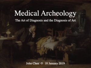
Slide 1. My talk today is called medical archaeology: the art of diagnosis and the diagnosis of art.  My goal is to spend some time at the intersection of medicine, art, and history, and see what lessons we can learn about making observations and deciphering a diagnosis.


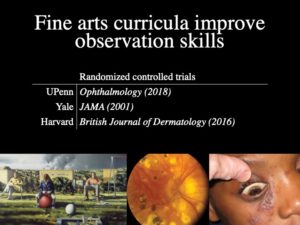
Slide 2. A couple years ago, the University of Pennsylvania started teaching their medical students how to study paintings.  The goal was to practice observing and describing the human form, skills that are fundamental to any astute clinician.  Last year, they published a randomized controlled trial. Here’s how it worked:

During their ophthalmology block, half the students went to the Philadelphia Museum of Art to get taught by professional art instructors.  Afterwards, they were tested on their ability to describe three types of images: paintings, retinal images, and clinical photos.  Students in the intervention arm scored significantly better in each of these categories compared to the control arm.

It turns out they’re not the only ones doing this.  Yale published a similar randomized controlled trial in JAMA back in 2001. Meanwhile, Harvard has been sending its dermatology residents to the Boston Museum of Fine Arts. We don’t have time today for a field trip to the High Museum, but what we can do is take a virtual tour of art and history as it relates to medicine.

Midway through my talk is an interesting example about an ancient drug we still use in modern cardiology.


Slide 3. Over the centuries, often by accident, paintings have captured clues that might be relevant to trained medical eye.  Sometimes, the striking thing about a painting is not what’s portrayed, but how it’s portrayed–the style and technique.  Here, we’re going to use art to give us clues about its creator.

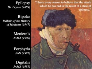
Our next artist was a Dutch painter who was known around town as the “redheaded madman.” In letters to his brother, Van Gogh complained of chronic abdominal pain, hallucinations, and insomnia.  He was hospitalized multiple times for episodes of insanity.  During one of these spells, he famously cut off his ear.  Doctors at the time diagnosed him with epilepsy.

At the age of 37, Van Gogh died of suicide–shot himself in the chest. Since his death, the scientific literature on Van Gogh has become as prolific as the artist himself. And while doctors back then called it epilepsy, doctors of the modern era have expanded the differential. A 1947 paper was the first to propose bipolar disorder, pointing out descriptions of manic and depressive episodes. A JAMA manuscript from 1990 argued for Meniere’s disease, suggesting tinnitus drove Van Gogh to perform surgery on his own ear. According to a 1991 article in the BMJ, Van Gogh may have had acute intermittent porphyria, a disorder involving heme synthesis which causes attacks of abdominal pain and psychiatric symptoms.


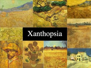
Slide 4. But the most well-known theory comes from examining Van Gogh’s artwork itself.  In fact, many of you probably use Van Gogh’s artistic style as a mnemonic to help you remember signs of digoxin toxicity.  In his later years, as his health got worse, Van Gogh’s color palate turned intensely yellow, suggesting he may have developed xanthopsia or yellow vision.


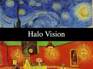
Slide 5. The swirling halos in pieces like Starry Night or Night Café are also big clues.  Digoxin is known to cause blurry outlines, a type of halo vision.  Recall that GI side effects are also common, consistent with his chronic nausea and abdominal pain.


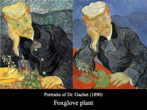
Slide 6. If you’re a real trivia buff, you might know that digitalis was originally extract from the Fox Glove plant.  This plant was widely used during the 19th century not just for cardiac problems, but also for various neurologic and psychiatric ailments.  So it’s likely that Van Gogh’s doctors gave him Fox Glove to treat his epilepsy.  In fact, on two separate occasions, Van Gogh painted his personal doctor posing with Fox Glove.  Once again, the proof is in the painting.


References:

  1. Gurwin J, et al. A randomized controlled study of art observation training to improve medical student ophthalmology skills. Ophthalmology. 2018;125:8–14.
  2. Huang JT, et al. Fine arts training improves observational skills of dermatology trainees: a pilot study. Br J Dermatol. 2016;175:815–817.
  3. Dolev JC, et al. Use of fine art to enhance visual diagnostic skills. JAMA. 2001;286:1020–1021.
  4. Perry I. Vincent van Gogh’s illness: a case record. Bulletin of the History of Medicine. 1947;21:146-72.
  5. Blumer D. The Illness of Vincent van Gogh. Am J Psychiatry. 2002;159(4):519–526.
  6. Arenberg IK, et al. Van Gogh had Ménière’s disease and not epilepsy. JAMA. 1990;264(4):491-493.
  7. Loftus LS, et al. Vincent van Gogh’s illness: acute intermittent porphyria. BMJ. 1991;303:1589–1591.
  8. Lee TC. Van Gogh’s vision digitalis intoxication. JAMA. 1981;245:728–9.


hidden

Notes from November: Medical Training is a Journey

November has been a whirlwind of angst and excitement for me.  It began with cramming for my final USMLE board exam.  After sitting for the two-day test, I flew directly to Chicago, where I attended my first AHA Scientific Sessions and presented a poster on bystander AED use at the Resuscitation Science Symposium.  Upon returning home to Atlanta, I pored over the list of fellowship interviews I had attended in the last two months and agonized over last-minute adjustments to my rank list.  Such is the life of a third-year internal medicine resident.

This Wednesday, I stumbled home after a 24-hour hospital shift and opened my email account.  That’s when I found out I had not only passed my board exam but also matched at Emory, my home institution and top fellowship choice.  In spite of my exhaustion, I was so elated that it was hard to fall asleep.

Reflecting back on the month, I’m reminded of Dr. Ivor Benjamin’s address at the AHA Presidential Session.  He waxed poetic about his circuitous journey–growing up in Guyana, traveling to the U.S., and training at several premiere academic medical centers.  He spoke about fortuitous relationships with key mentors that propelled his career as a basic science researcher.

Listening to Dr. Benjamin’s narrative was a delight and an inspiration.  His account echoed the stories of many impressive residents and fellows I met at AHA.  It was also a reminder of my own humble roots—born in Shanghai, growing up in the rural Midwest, and studying at Vanderbilt and Emory.  For each of us, medical training is long, and it is transformative.  I look forward to the next stage, and I hope to return for Scientific Sessions in 2019.  By then I will be a cardiologist in the making.


hidden

Hypertrophic Cardiomyopathy Comes in Different Shapes and Sizes

Scientific Sessions 2018 marks many firsts for me—my first time at Scientific Sessions and my inaugural blog post on the AHA Early Career Voice.  Both are tremendous opportunities.

I specifically sought out the Sunday morning session, “State of the Art in Hypertrophic Cardiomyopathy.”  As an internal medicine resident at Emory, I’ve had several experiences seeing patients with hypertrophic cardiomyopathy (HCM) in the outpatient clinic.  Unlike many other fields of cardiology, HCM is a niche dominated by young, otherwise healthy patients.  The title of this session alludes to how little we know about HCM, and how the practice of managing this complex condition truly is an “Art.”

Much of the session was an exercise in taxonomizing the umbrella term, “HCM,” splitting that pie from a number of interesting angles.  Dr. Sharlene Day divided HCM by obstructive phenotype: obstruction at rest, obstruction with provocation, and no obstruction.  Our approach to therapies has been driven by a focus on relieving obstruction, but strategies for treating symptoms in the absence of obstruction represents an open frontier.  Currently, the MAVERICK-HCM trial is studying the use of a cardiac myosin modulator in this patient population.

Dr. Jodie Ingles compared “familial” versus “non-familial” HCM.  The latter case, she argued, tends to involve men, present later, and portend a lower risk of cardiovascular events.  Discerning which cases of HCM is considered “familial” versus “non-familial,” and whether such a dichotomy truly exists, sparked much debate in the Q&A.

Drs. Elizabeth McNally, Adam Helms, and Jil Tardiff shared similarly thought-provoking insight, highlighting the heterogeneity of genotypes and phenotypes in HCM.  Multiple disparate mechanisms are responsible for producing sarcomere dysfunction, subsequent organic dysfunction, and finally clinical symptoms.  An appreciation for these finer details is necessary to guide a sophisticated approach to management.