hidden

Women in Electrophysiology

While I was chatting with a few fellows in our hospital hallway, I met one of the fellows who was very interested in electrophysiology (EP). We had a very interesting chat about her application and future career forward. In this blog, I summarize my chat with Jasneet Devgun, an aspiring electrophysiologist.

Question: Hi Jasneet, great to have you here! Let’s start with this question: When and how did you know you love EP?

  • Answer: EP is something I never really thought of pursuing initially. In fact, I was interested in interventional cardiology since my second year of medical school. It was not until I met an electrophysiologist at the University of Chicago during my third year of medical school that I thought of EP as a possible future career. He was so excited to show me the world of EP and frequently took me to the lab to see EP in action. I still remember the day he said, “we need more women in EP…you should consider it.” From then on, my curiosity grew. I found myself drawn to the lab, scrubbing in on cases in residency and fellowship. The unique therapeutics, cutting-edge procedures and technology, intellectual and logical nature of EP, alongside very memorable and rewarding encounters with patients and wonderful attendings, made me realized that EP was the right field for me.

Question: This is great!! What are your thoughts about women in EP?

  • Answer: Last year, Dr. Kamala Tamirisa wrote a very thoughtful piece for EP Lab Digest on the EP fellow shortage.1 At the time, the National Resident Matching Program (NRMP) demonstrated that approximately 40% of 130 EP fellowship positions in the US were unfilled.2 In 2021, that number drastically declined to 4%. Despite clear rising interest in EP, there remains a paucity of women in the field. The American Board of Internal Medicine (ABIM) reported that women comprised only 10% of first year EP fellows, while remaining steady at this rate for the past 10 years.3

The paucity of women pursing EP is a multi-faceted issue. A recent survey of cardiology fellows-in-training published in the Journal of American College of Cardiology showed that the most significant reasons women did not choose EP were greater interest in another field, radiation concerns, lack of female role models, a perceived “old boys’ club” culture, and discrimination/harassment concerns.4 Another reason was length of training. Reasons why women did choose EP were positive mentorship, unique features about the specialty, expertise, and the presence of a female role model, the latter being the major influencer.

These results are not surprising, but there are ways we can tackle the question at hand.

Question: Absolutely!! And this brings up the importance of mentorship, can you share your experience with that?

  • Answer: I cannot stress enough the value of a good mentor. A good mentor inspires and cultivates the foundations of turning one’s future into reality. This was personally a huge factor for me; I did not know anything about, let alone consider, EP until I met electrophysiologists who had a genuine interest in my career development. Interestingly, none of them were female. Our male colleagues can be some of our biggest advocates. I certainly see how a female role model is uniquely relatable and valuable. However, the gap will remain until more females in EP exist. That said, networking with female electrophysiologists through existing organizations, as well as creating outreach/interest groups in-person and on social media to involve residents, medical students, and even undergraduate students, would be very effective.

Question: What advice do you have for fellows who do not know much about EP or are not sure if they would want to pursue it? What are some possible barriers to developing interest?

  • Answer: Exposure is key! Many trainees do not have much exposure to EP, and therefore may not know enough to develop an interest for it. Fellows should be aware of the distinctive benefits and exciting features unique to EP, which can only be achieved by increasing their time with electrophysiologists and the EP lab. This can also dispel concerns about radiation safety. For example, female cardiology fellows concerned about radiation in the survey may not know about the use of 3D mapping systems and multi-modality imaging, affording “low-fluoro” or “fluoro-less” cases and reduction in procedure times. Moreover, the development of robotically assisted procedures has provided an avenue to reduce occupational hazards.
  • I was fortunate that my residency program offered an EP elective, where I met electrophysiologists who were excited to show me their world. A structured elective early in residency and more electives for medical students, with some exposure to the lab, may help bridge the gap and channel early interest. The earlier it is, the better, since interests develop quickly (as did mine!).

Question: What are your thoughts on the length of training and how this may be impacting fellows’ wellness and career decisions?

  • Answer: Length of training is an important issue.1,4 This time overlaps with childbearing years and critical family development. Fellows, both male and female, should not have to feel like they must choose one over the other. There must be a genuine culture of promoting work-life balance during the long years of training. Fellows are also consequently faced with prolonged financial strains from student loan debt. These are things that fellows consider when deciding to pursue another fellowship. As medicine progresses towards a milieu of sub-specialties requiring ever-more training, the training structure must be modernized to optimize the workforce. Unfortunately, many people, including female fellows, may be missing out on great sub-specialties like EP because of these issues. Some have proposed modifying the last 6 months of cardiology fellowship as the beginning of CCEP, which is a great short-term goal.1 A “fast-track” program in fellowship that may even extend into residency may be a proposition for much later in the future. These changes can make a major difference in fellows’ career decisions, health, and well-being.  

Despite the paucity of women in EP, I am positive that the great strengths of this field will surpass any barriers to recruiting them. Building exposure early, having more visible role models and mentors, modification of the training structure, and many other solutions previously stated will allow for tremendous progress. Even simple interventions can make leaps and bounds in bringing more women into the wonderful world of electrophysiology.

I would like to thank Jasneet Devgun, DO, who is currently a general cardiology fellow at Henry Ford Hospital, and an aspiring electrophysiologist, for sharing her experience and thoughts with us. A special thank you goes to Dr. Judith Mackall and Dr. Cristina Tita who helped in writing this blog.

References:

  1. Tamarisa, K. The Importance of Choosing Cardiac Electrophysiology as a Career: Thoughts on the EP Fellow Shortage. EP Lab Digest. Available at https://www.hmpgloballearningnetwork.com/site/eplab/importance-choosing-cardiac-electrophysiology-career-thoughts-ep-fellow-shortage. Accessed October 9, 2021.
  1. Fellowship Match Data and Reports. National Resident Matching Program. Available at http://www.nrmp.org/fellowship-match-data/. Accessed October 9, 2021.
  2. Percentage of First-Year Fellows by Gender and Type of Medical School Attended. Available at https://www.abim.org/about/statistics-data/resident-fellow-workforce-data/first-year-fellows-by-gender-type-of-medical-school-attended.aspx. Accessed October 9, 2021.
  1. Abdulsalam N, Gillis AM, Rzeszut AK, Yong CM, Duvernoy CS, Langan MN, West K, Velagapudi P, Killic S, O’Leary EL. Gender Differences in the Pursuit of Cardiac Electrophysiology Training in North America. J Am Coll Cardiol. 2021 Aug 31;78(9):898-909. doi: 10.1016/j.jacc.2021.06.033. PMID: 34446162.

“The views, opinions, and positions expressed within this blog are those of the author(s) alone and do not represent those of the American Heart Association. The accuracy, completeness, and validity of any statements made within this article are not guaranteed. We accept no liability for any errors, omissions, or representations. The copyright of this content belongs to the author and any liability with regards to infringement of intellectual property rights remains with them. The Early Career Voice blog is not intended to provide medical advice or treatment. Only your healthcare provider can provide that. The American Heart Association recommends that you consult your healthcare provider regarding your health matters. If you think you are having a heart attack, stroke, or another emergency, please call 911 immediately.”

hidden

Space traveling and CVD risk: RACE to MACE

Picture Reference: https://twitter.com/glitchandgrace/status/711782004497326081 @glitchandgrace

Lately, the space race has been the talk of the town and while it might seem that we are on the verge between science-fiction and reality, there is a long way to go before humans can overcome the health hazards associated with space travel and adapt to their new environment. Humans will likely be exposed to many health hazards emerging from the “space exposome” during their journey into space. Many of these health hazards can affect the cardiovascular (CV) system.

Astronauts are required to undergo an extensive training program including engineering and paramedic courses, intensive health check and various assessments to see how they can cope under stressful conditions. From the start, there is a specific selection process to identify the most ‘fit’ of people who can qualify for space travel. However, even the most ‘fit’ of people can’t escape the health hazards that are awaiting us when we leave mother Earth.

One of the most prominent health hazards associated with long distance space travel affecting the CV system is space radiation. Space radiation usually consists of solar radiation and, the more dangerous, cosmic radiation. Among the components of cosmic radiation, the HZE ions are the most hazardous to the human body because they are highly penetrating and can even generate secondary particles when they interact with shielding materials such as spacecraft or spacesuit. Space radiation damages DNA either directly by energy absorption leading to clustered DNA damage, mutations, chromosome exchanges, carcinogenesis and apoptotic cell death, or indirectly via the production of reactive oxygen species (ROS) from the radiolysis of water molecules.

Exposure to various types of radiation can lead to radiation-induced cardiovascular disease (RICVD) which is a known complication in patients undergoing radiation therapy to treat thoracic cancers and in Japanese atomic bomb survivors. RICVD following space radiation exposure can develop acutely in the form of pericarditis or chronically leading to myocardial remodeling and fibrosis, accelerated development of atherosclerosis, cardiomyopathies, valve abnormalities, arrhythmias and conduction disorders. RICVD can also develop 10-15 years following exposure.

The number of astronauts that have surpassed the low Earth Orbit, where the space radiation amount becomes significantly higher, were only from the Apollo mission which limits the amount of data available. Therefore, several studies to investigate RICVD have been done in animal models using different types of radiations to mimic, to a certain degree, RICVD in humans. These studies have shown that 56Fe ions, the most prominent heavy ion found in cosmic radiation, lead to cardiac hypertrophy and myocardial remodeling. In addition, people that have been exposed to higher levels of radiation compared to the general population had increased risk of myocardial infarction due to atherosclerosis. Mouse models further showed that the effects of irradiation are rather local than systemic where atherosclerosis developed in the areas which were specifically subject to irradiation using 56Fe ions. Plaques in these mice had a thickened intima (carotid), indicating a damage to the arterial wall, and a large necrotic core (aortic root) which increases the risk of instability and thrombogenic complications. The biological processes that have been suggested to induce RICVD include endothelial dysfunction leading to a pro-inflammatory and a pro-fibrogenic environment, apoptotic cell death of various cardiovascular cell types and alterations in DNA methylation.

To date, extensive research is going into improving the shielding methods to protect astronauts from the effects of space radiation. The administration of anti-oxidants such as N-acetyl cysteine (NAC), ascorbic acid (vitamin C), vitamin B, coenzyme Q10 and vitamin E can complement the vitamin deficiencies that humans are subject to during space travel and also remove the generated ROS limiting DNA damage and protecting from space radiation exposure. Some drugs such as angiotensin converting enzyme (ACE) inhibitors and statins have also shown to reduce radiation-induced cardiopulmonary complications and radiation induced atherosclerosis in animal models respectively but need further exploration. Of note, altered DNA methylation can serve as an early biomarker for space radiation exposure and might lead to personalized treatment based on the level of altered DNA methylation after exposure. Space radiation can also increase the risk of cancer and diseases of the central nervous system such as impaired motor function, neurobehavioral changes, Alzheimer’s disease or accelerated aging which can complicate an existing CVD.

Apart from space radiation, other health hazards are also associated with space traveling. Prolonged exposure to microgravity induces bone and muscle atrophy as well as cardiovascular deconditioning. This requires humans to exercise constantly to remain healthy which could be beneficial for CV outcomes. Being confined in a closed environment, disrupted circadian rhythms and the stress for being away from mother Earth also add another layer of psychological and mental challenges to be overcome when venturing into space.


References:

  1. Meerman M, Bracco Gartner TCL, Buikema JW, Wu SM, Siddiqi S, Bouten CVC, et al. Myocardial Disease and Long-Distance Space Travel: Solving the Radiation Problem. Front Cardiovasc Med. 2021;8:27.
  2. Patel ZS, Brunstetter TJ, Tarver WJ, Whitmire AM, Zwart SR, Smith SM, et al. Red risks for a journey to the red planet: The highest priority human health risks for a mission to Mars. Npj Microgravity. 2020 Nov 5;6(1):1–13.
  3. Patel S. The effects of microgravity and space radiation on cardiovascular health: From low-Earth orbit and beyond. IJC Heart Vasc. 2020 Oct 1;30:100595.
  4. Delp MD, Charvat JM, Limoli CL, Globus RK, Ghosh P. Apollo Lunar Astronauts Show Higher Cardiovascular Disease Mortality: Possible Deep Space Radiation Effects on the Vascular Endothelium. Sci Rep. 2016 Jul 28;6(1):29901.
  5. Glitch And Grace Art. Love #glitchandgrace #watercolour #watercolor #space #anatomical-heart #lovewins https://t.co/RIugIHw13W [Internet]. @glitchandgrace. 2016 [cited 2022 Jan 11]. Available from: https://twitter.com/glitchandgrace/status/711782004497326081
hidden

Sleep and Ideal Cardiovascular Health

During the AHA’s Scientific Session 2021, heart health, defined by the Life’s Simple 7 metric, was often woven into health equity conversations.

Empirical evidence consistently supports AHA’s recognition of these seven risk factors that people can avoid or improve on through lifestyle changes to help achieve ideal cardiovascular health. Regardless of how challenging this goal is for the average American, Life’s Simple 7 is an essential benchmark for staying heart-healthy.

More attention is now being directed at the role of sleep in maintaining heart-healthy lifestyle practices. Sleep plays an important role in overall health and well-being. In-kind, there exists a reciprocal relationship between the quality of one’s diet, physical activity, and stress on the quality of sleep achieved. Ideally, sleep needs to be deep and restorative to support good cardiovascular health. Specifically, the Centers for Disease Control and Prevention recommend that adults between 18-65 years aim for at least seven hours of quality sleep per night. However, sleeping well is not common. 4 in 10 adults report consistently good sleep at night, and 50 million to 70 million American adults suffer from chronic sleep problems or sleep disorders.

As an early career epidemiologist, who was not too long ago a pre-doctoral candidate, I am familiar with several factors that contribute to trouble with sleep. These have included staying up late to work on an analysis or drafting a manuscript whose internal deadline was already past due; following up on emails while binging a popular streaming series and munching on some snacks; juggling a busy household with two young children that always find a reason to wake up sometime after midnight. Perhaps these experiences are relatable. Often lifestyle choices, poor sleep habits, stress, and medical conditions can play a role in why you can’t sleep.

Alcohol

  • A glass of wine before bed might not interfere with your ability to drift off but indulging in more servings of alcohol before bedtime may impair your sleep by interfering with your sleep cycle, especially REM sleep. This leads to fragmented, unrefreshing rest.

Poor Sleep Habits

  • Habits that make it harder to fall and stay asleep may include (1) staying up late, (2) watching television in bed, (3) playing or browsing on your phone in bed, (4) having an irregular sleep schedule. Simple lifestyle changes to your nightly routines could help to remedy these issues.

Bed Sharing

  • Whether with a partner, child, or pet, reduced sleep quality can be caused by sharing your bed. Anything that can make you uncomfortable (i.e., snores, crowding, pulled covers, or mismatched sleep condition preferences like temperature, light, or noise level) will disrupt your sleep.

Poor Sleep Environment

  • Sleeping environments that are too hot or too cool will disrupt your sleep. Sleep experts recommend a bedroom temperature at a moderate climate between 65 to 72 degrees Fahrenheit at night. The body needs to cool at night for the most refreshing sleep, but a too-cold room will cause you to wake up. Don’t forget about light exposure. Whether it’s from a reading lamp, television, streetlight, or even the glow from a device, this could be enough to signal your brain to wake up.

Caffeine

  • Some folks may argue that coffee has no effect on their ability to sleep at night and will enjoy a cup before bed. However, caffeine has a half-life of three to five hours, so even a late afternoon caffeinated beverage can disrupt your sleep later that night.

Stress

  • Often the events of the day that creep back into our minds at night are not the positive experiences but the ones that fill our minds with worry and stress. Stress is one of the most cited symptoms of sleep problems.

Exercise

  • Exercise, like an evening walk, is excellent for fostering better sleep. But intense, heart-pumping, and sweat-inducing cardio workouts within three hours of your bedtime may be too much. Both your body temperature and heart rate naturally drop as you fall asleep. Exercise stimulates your entire nervous system and raises these two body functions making it difficult to sleep.

Snack Choices

  • Snacks or meals high in fat or protein consumed right before bedtime can overstimulate your digestive system, cause heartburn and make it difficult to sleep. A late-night sugar rush can also lead to hunger pangs and drops in blood sugar, causing you to wake up.

Sleep Disorders and Mental Health

  • Importantly other factors like sleep disorders and mental health problems can make it difficult to sleep. You should talk with your doctor if you suspect that a medical or mental health condition may be contributing to your poor sleep.

Insufficient sleep and poor sleep quality, in addition to sleep disorders, are linked to a wide range of adverse health effects. Major physical and mental health consequences include anxiety, bipolar disorder, hormone imbalances, weakened immune system, cardiovascular disease, and major heart disease risk factors like obesity, inflammation, Type 2 diabetes, high blood pressure. Additionally, poor sleep is linked to overall decreased quality of life and increased mortality risk.

If good sleep habits are not currently part of your daily routine, consider some useful strategies to alleviate some of the factors that have interfered with your quality and quantity of sleep. Here in this infographic are a few tips and techniques developed by AHA to help those who do not have a sleep disorder make small daily changes to establish healthier sleep habits.

Working to alleviate factors that contribute to insufficient sleep and poor sleep quality may also be another critical metric for cardiovascular health. Preliminary findings presented at the AHA’s 2021 Epidemiology and Prevention/Lifestyle and Cardiometabolic Health Scientific Sessions recommended revising the AHA’s Life’s Simple 7 to include sleep as a metric creating a new “Simple 8 or Essential 8” metric measuring cardiovascular health. The study’s lead investigator, Nour Makarem, Ph.D., explained that while sleep is a health behavior that people engage in every day, like diet and exercise, it has received far less attention. However, increasing evidence links sleep to heart disease and risk factors for cardiovascular disease.

Along with her team of investigators, Dr. Makarem assessed whether a cardiovascular health score that includes the Life’s Simple 7 combined with sleep metrics would be more strongly associated with cardiovascular disease than the Life’s Simple 7 score. The study found that when at least one measure of sleep was added with the Life’s Simple 7 measures, the new heart health score was more strongly associated with cardiovascular disease than the traditional Life’s Simple 7. The results were compelling and showed, for example, that study participants who received seven to eight hours of sleep a night in addition to meeting Life’s Simple 7 guidelines had up to 61% lower odds of having heart disease. Those who got less than six hours of sleep scored lower for overall cardiovascular health and had a higher prevalence of overweight and obesity, Type 2 diabetes, and high blood pressure. Sleep duration and the other sleep metrics included in the study made the cardiovascular health scores more predictive of cardiovascular disease risk than the seven metrics alone.


Like several current Life’s Simple 7 measures, clocking 7-9 hours of sleep per day can be challenging. However, the traditional cardiovascular health metric may need to be revisited for a potential upgrade in providing yet another vital benchmark for predicting and promoting ideal cardiovascular health.

References:

  1. Lloyd-Jones, Donald M., et al. “Defining and setting national goals for cardiovascular health promotion and disease reduction: the American Heart Association’s strategic Impact Goal through 2020 and beyond.”Circulation 4 (2010): 586-613.
  2. https://www.heart.org/en/health-topics/sleep-disorders/sleep-and-heart-health
  3. https://www.heart.org/en/news/2020/03/06/sleep-should-be-added-as-measure-of-heart-health-study-says
  4. https://www.verywellmind.com/reasons-for-not-sleeping-well-and-how-to-fix-350760
  5. https://www.heart.org/en/healthy-living/healthy-lifestyle/sleep/sleep-well-infographic
  6. https://www.ahajournals.org/doi/10.1161/circ.141.suppl_1.36

“The views, opinions and positions expressed within this blog are those of the author(s) alone and do not represent those of the American Heart Association. The accuracy, completeness and validity of any statements made within this article are not guaranteed. We accept no liability for any errors, omissions or representations. The copyright of this content belongs to the author and any liability with regards to infringement of intellectual property rights remains with them. The Early Career Voice blog is not intended to provide medical advice or treatment. Only your healthcare provider can provide that. The American Heart Association recommends that you consult your healthcare provider regarding your personal health matters. If you think you are having a heart attack, stroke or another emergency, please call 911 immediately.”


hidden

The tension between generalizability and inclusivity in population research

As I have continued my journey in epidemiologic research, one of my primary aims is to illuminate health disparities within my projects. Currently located in New York City, identifying diverse populations is hardly an issue. However, I have thought more about populations around the United States as I apply to PhD programs and consider the projects of potential mentors. One project in particular was out of a prestigious university in the Midwest in which the investigators planned to include equal proportions of individuals of different racial groups(e.g. 25% non-Hispanic Black, 25% White, 25% Asian, 25% Hispanic) in an effort to not exclude demographics.

This raises the following important question: How do we choose the best sampling method and accommodate for disparate demographics in research in a way that preserves data validity?

Diversity in health research populations is crucial because data is needed not only on dominant populations but also on populations which are potentially affected disproportionately by the illnesses examined in the studies. When including individuals in studies, random sampling is beneficial because it reduces bias and improves validity. However, random sampling may under-represent individuals in affected demographics when sampling is done among racially disparate populations. Essentially, investigators may need to choose between representativeness of diverse populations and confidence in their ability to make inferences about a general population.

Let us review the most common methods of sampling and recruitment for studies. There are about six main sampling methods which can generally be considered probability sampling or non-probability sampling. Probability sampling consists of random sampling in which there are about equal chances of being selected into a sample; and stratified sampling in which subsets of the population have low incidence relative to other subsets, and individuals are chosen based on and interval selections method (e.g. each nth individual is selected.1 In non-probability sampling, individuals are selected by non-random methods.1 One example is quota sampling, which is a non-probability equivalent of stratified sampling. Convenience sampling is another method in which accessible individuals in the population of interest are chosen.1 In judgment sampling, a population utilized in the study is considered to be representative of a larger population (e.g. a town in the US is thought to be representative of the US population). Finally, In snowball sampling, participants may recruit other individuals into the study via their own networks.1 Biases may occur with each sampling method, however minimal bias occurs in random sampling methods.

When it comes to ensuring that individuals of various groups or characteristics are included in the study population, some researchers turn to oversampling. In essence, the number of individuals of a particular group may be included disproportionately to the distribution to accommodate for an imbalance in the data.

The issue of racial diversity in study populations is subtle but needs to be addressed according to the specifics of each project. When necessary, choosing between generalizability and inclusion of disparate populations is a decision which requires careful consideration and may implicate possible biases in study results and findings.

References

  1. Tyrer, S., & Heyman, B. (2016). Sampling in epidemiological research: issues, hazards and pitfalls. BJPsych Bulletin, 40(2), 57–60. https://doi.org/10.1192/pb.bp.114.050203

“The views, opinions and positions expressed within this blog are those of the author(s) alone and do not represent those of the American Heart Association. The accuracy, completeness and validity of any statements made within this article are not guaranteed. We accept no liability for any errors, omissions or representations. The copyright of this content belongs to the author and any liability with regards to infringement of intellectual property rights remains with them. The Early Career Voice blog is not intended to provide medical advice or treatment. Only your healthcare provider can provide that. The American Heart Association recommends that you consult your healthcare provider regarding your personal health matters. If you think you are having a heart attack, stroke or another emergency, please call 911 immediately.”


hidden

Electronic Cigarette Customization Matters.

Electronic nicotine delivery systems (ENDS) are increasingly popular as an alternative to standard ‘combustion’ cigarettes.  ENDS come in a large variety of forms and offer adulterant choices that enhance the user’s experience, such as flavors, humectants, and nicotine in different concentrations. There is a common perception that vaping is a safer alternative to traditional tobacco cigarettes as the ‘smoke’ lacks tars and other toxicants found in cigarette smoke(1). That may be true to some extent, however the ability of users to modify ENDS, like adjusting the power wattage, changing the type of heating element, and use of e-liquids with different flavor and nicotine concentrations, will influence the amounts of toxic chemicals in the inhaled aerosol. Under normal circumstances, the vapor contains, contrary to popular opinion, some of the same toxic compounds (formaldehyde, acetaldehyde, acrolein…) that are found in CCs (2). The ability to add custom adulterants to the vape fluid can add to the complexity of assessing potential risks. How modifications, or customizations might contribute to health effects and the generation of harmful chemicals is a topic that deserves more attention.

Devices are usually composed of a heating element, often a disposable metal heating coil, and atomizer tanks which directly produce the vapor. The most common heating coils and atomizer units can be comprised of different metals, such as stainless teel, nickel-chromium or nichrome, Kanthal nickel, or titanium. At usual setting these all work well, however they degrade with extended use. In some devices, users can set temperatures sufficiently high that degradation products of the device hardware such as metals are aerosolized and inhaled along with the vaporized e-liquid containing nicotine, flavoring, and solvents (3). Environmental metal contaminants are known risk factors for cardiovascular disease. Users of eC devices could inadvertently select vaping profiles that promote chemical reactions with the normally low-toxicity e-liquid to generate harmful chemicals in the aerosol they are inhaling at levels even exceeding traditional cigarettes.

Many studies have already brought insight into how device settings can generate levels of metals in inhaled aerosol that are unique to the modifiable aspects of eC devices. One study found increased concentrations of metals such as arsenic (As), chromium (Cr), copper (Cu), iron (Fe), manganese (Mn), nickel (Ni), lead (Pb), antimony (Sb), tin (Sn), and zinc (Zn) concentrations in eC aerosols when the device power is increased from low (20 Watts) to intermediate (40 Watts) settings (4). These exposure levels to metals like Ni from ENDS could exceed those of traditional reference cigarettes. It is interesting to note that ENDS from different manufactures emit different concentrations of metals, suggesting that heating and cooling patterns of individual devices could influence the amount of metals released. A similar study also found higher metal levels in aerosol and e-liquid after it comes in contact with the metal heating coil, compared to the original e-liquid in the dispenser vial (5). Elevated levels of metal biomarkers such as Cu, Cr, Sn, and Pb were found in the urine of electronic cigarette  users. These studies highlight the need to further study how the unique customizable aspects of ENDS technology contribute to the formation of varying levels of contaminants in the inhaled aerosol.  There is no question that smoking is a risk factor for cardiovascular disease and can increase blood pressure, heart rate, among other health outcomes. Educating  the public about the health burden that ENDS pose and informing users about the potential dangers of vaping at elevated temperatures or using degraded heating components can be a step towards reducing use of new tobacco products.

References:

  1. Benowitz, N. L., & Burbank, A. D. (2016). Cardiovascular toxicity of nicotine: Implications for electronic cigarette use. Trends in cardiovascular medicine26(6), 515–523. https://doi.org/10.1016/j.tcm.2016.03.001
  2. Perraud, V., M.J. Lawler, K.T. Malecha, R.M. Johnson, D.A. Herman, N. Staimer et al.: Chemical characterization of nanoparticles and volatiles present in mainstream hookah smoke. Aerosol Science and Technology 53(9): 1023-1039 (2019)
  3. Wylie, B. J., Hauptman, M., Hacker, M. R., & Hawkins, S. S. (2021). Understanding Rising Electronic Cigarette Use. Obstetrics and gynecology137(3), 521–527. https://doi.org/10.1097/AOG.0000000000004282
  4. Zhao, D., Navas-Acien, A., Ilievski, V., Slavkovich, V., Olmedo, P., Adria-Mora, B., Domingo-Relloso, A., Aherrera, A., Kleiman, N. J., Rule, A. M., & Hilpert, M. (2019). Metal concentrations in electronic cigarette aerosol: Effect of open-system and closed-system devices and power settings. Environmental research174, 125–134. https://doi.org/10.1016/j.envres.2019.04.003
  5. Olmedo, P., Rodrigo, L., Grau-Pérez, M., Hilpert, M., Navas-Acién, A., Téllez-Plaza, M., Pla, A., & Gil, F. (2021). Metal exposure and biomarker levels among e-cigarette users in Spain. Environmental research202, 111667. Advance online publication. https://doi.org/10.1016/j.envres.2021.111667
  6. Bhatnagar A, Whitsel LP, Blaha MJ, Huffman MD, Krishan-Sarin S, Maa J, Rigotti N, Robertson RM, Warner JJ; on behalf of the American Heart Association. New and emerging tobacco products and the nicotine endgame: the role of robust regulation and comprehensive tobacco control and prevention: a presidential advisory from the American Heart Association. 2019;139:e937–e958. doi: 10.1161/CIR.0000000000000669.

“The views, opinions and positions expressed within this blog are those of the author(s) alone and do not represent those of the American Heart Association. The accuracy, completeness and validity of any statements made within this article are not guaranteed. We accept no liability for any errors, omissions or representations. The copyright of this content belongs to the author and any liability with regards to infringement of intellectual property rights remains with them. The Early Career Voice blog is not intended to provide medical advice or treatment. Only your healthcare provider can provide that. The American Heart Association recommends that you consult your healthcare provider regarding your personal health matters. If you think you are having a heart attack, stroke or another emergency, please call 911 immediately.”

hidden

Zooming In: The Impact of the COVID-19 Pandemic on PhD Interviews

The COVID-19 pandemic has brought significant changes to multiple layers of academia, including the biological and biomedical sciences PhD admissions process in the United States. Typically, prospective applicants are selected to interview in-person at the destination campus not only as a part of the evaluation process, but also as an opportunity for the applicants to gauge program fit. The travel disruptions caused by the pandemic put this opportunity on hold in 2020, and for many programs, lasted into the 2021 admissions cycle. As such, many programs opted for virtual interviews instead.


Rama Alhariri, a PhD student in the Human Genetics and Molecular Biology program at Johns Hopkins University, was unable to visit the universities she applied to when she was choosing PhD programs in 2020. All her interview processes took place virtually. Although there was slight variation in the format, she had an opportunity to talk to current students and faculty members about their research interests and the PhD program in general. In addition to the interviews, her virtual programs included sessions introduce applicants to the university and the city itself, such as a panel session about things to do in the area and a live virtual tour of the program. However, this experience did not quite resemble the in-person visits.

“I would’ve liked a higher quality of the tour of facilities as some programs lacked that altogether or it was a little unclear. Additionally, I would’ve liked a greater interaction with other interviewees without the presence of upper-class students or faculty so that we can get to know one another better, the way we might if we were in an in-person interview. It is unfortunate because it’s these interactions that also shape how well you might integrate with other students,” she added.

When asked about how she gauged the fit of the PhD program, Alhariri said that she tried to focus on her interactions with the students, faculty, and other interviewees. From these conversations, she was able to get a glimpse into the campus culture and the overall level of formality and professionalism among the faculty members and students.

“Ultimately the program I chose, while it was also the highest ranking one, was the one in which I felt most comfortable with the upper-class students as well as those that could potentially be within my cohort. I sought an environment with a good balance between professional and somewhat relaxed, which would be the best fit for me.”

Although virtual interviews have become more common in the PhD admissions process in the past two year, they are not new. International students who reside in other countries, for example, typically have limited opportunities to travel due to visa issues and a lack of financial reimbursement for long-distance travel. When I was choosing a PhD program to commit to in 2018, I was finishing up my undergraduate studies outside of the United States. Because of that, my campus visits were limited to places that I could afford flying to. Additionally, my virtual interviews were limited to conversations with faculty members, which was not enough to give me a comprehensive picture.

Alhariri shared a few tips for current applicants who are unable to visit campuses in-person, “It’s hard to make a decision on what program you want to choose based off limited virtual interactions. I say trust your gut and try to support your intuition with evidence. I obtained information about living in the city through online searching. Because faith is important to me, I also checked social media sites of the Muslim student faith group – I wanted to know that there was a large enough Muslim community within the city and even within the university at large, not limited to just my program.”

Ultimately, completing a PhD is a long-term commitment, and the decision making to commit to a program looks different from person to person. Self-introspection can help a lot in this case: what is important for you? Will the environment nurture your academic curiosity? Do you like the idea of living in the area for several years? Based on my own experience of interviewing virtually, I would suggest doing the following things. First, try to talk to as many people in the PhD program as you can. Do not hesitate to ask to be connected to faculty members you are interested in working with and current students to get more information about the culture of the university. Second, try to connect with previous applicants who ended up choosing a different program to understand their perspectives and add information to your decision making. Lastly, if you are moving to a new environment, do some research about the area itself – including cost of living and things to do beyond academic work – to ensure that you will adapt well.

“The views, opinions and positions expressed within this blog are those of the author(s) alone and do not represent those of the American Heart Association. The accuracy, completeness and validity of any statements made within this article are not guaranteed. We accept no liability for any errors, omissions or representations. The copyright of this content belongs to the author and any liability with regards to infringement of intellectual property rights remains with them. The Early Career Voice blog is not intended to provide medical advice or treatment. Only your healthcare provider can provide that. The American Heart Association recommends that you consult your healthcare provider regarding your personal health matters. If you think you are having a heart attack, stroke or another emergency, please call 911 immediately.”


hidden

A Scientific Statement from the American Heart Association on the Management of Cardiovascular Risk Factors for Adults with Type 2 Diabetes

The prevalence of diabetes is increasing at an alarming rate, with more than 34 million Americans suffering from diabetes1. Patients with type 2 diabetes make up 90% to 95% of total diabetes cases1. Cardiovascular diseases (CVD) are the principal cause of death and disability in type 1 and type 2 diabetes patients2. American Heart Association (AHA) recommends a comprehensive and patient-centered approach involving lifestyle management and pharmacological interventions to manage cardiovascular risk factors, such as smoking, obesity, glycemia, blood pressure (BP), and lipid abnormalities, in type 2 diabetes patients3.

A healthy lifestyle can substantially lower the risk of CVD events in type 2 diabetic patients. Lifestyle management involves physical activity, nutrition, psychological and emotional well-being, and smoking cessation3. The lifestyle interventions using meal replacement products and at least 175 minutes of moderate-intensity physical activity /week, with a calorie goal of 1200 to 1800 kcal per day (with <30% from fat and >15% from protein), resulted in weight loss and hemoglobin A1c. Individuals with either ≥10% reduction in their body weight or >2 metabolic equivalent increase in fitness experienced reductions in cardiovascular outcomes; however, the rate of a major adverse cardiovascular event (MACE) was not reduced4. Mediterranean, vegetarian, low-carbohydrate, and diets rich in protein and nuts can lower blood glucose and body weight in type 2 diabetes patients3. Mediterranean diet over 4.8 years exhibited the highest benefits in blood glucose regulation and 29% of CVD events3,5. Further, increased exercise and physical activity can improve blood glucose, blood pressure, insulin sensitivity, lipid profile, and inflammation in type 2 diabetes3. American Diabetes Association (ADA) has recommended more than recommends ≥150 minutes/week of moderate-to-vigorous intensity aerobic activity with no more than 2 days of inactivity in diabetes patients3,6. Including 2-3 sessions/week each of resistance and balance training is also recommended. Further, vigorous activity for a short duration (>75 minutes/week) or interval training is beneficial3,7. Patients with BMI ≥27 kg/m2 can use US Food and Drug Administration (FDA) approved weight loss medication. Orlistat, lorcaserin, liraglutide, naltrexone/bupropion are some of the weight management drugs approved by FDA have demonstrated ability to lower A1c3. Liraglutide at a lower dose can reduce cardiovascular risk in high-risk patients8. However, medicines should be immediately stopped if weight loss after 3 months is less than 5% or any safety concerns arise. Patients with BMI ≥40 kg/m2 or BMI 35.0 to 39.9 kg/m2 with no benefit with nonsurgical methods can consider metabolic surgery3. The long-term effects of weight-loss drugs and metabolic surgery on reducing cardiovascular events are yet to be studied.

Smoking is linked with abnormal lipid profile, worsening of glycemic measures, and increased pro-inflammatory marker in type 2 diabetes. Therefore, cessation of smoking is recommended3. Interestingly, light to moderate alcohol consumption compared to no drinking, particularly wine, has been associated with fewer heart attacks, whereas heavy alcohol consumption increases CVD risk3. Despite the benefits of moderate alcohol intake, non-drinks should not be advised to drink, and adults with diabetes should be mindful of the risk of hypoglycemia, weight gain, and hypertension. No more than 1 drink/day for women and 2 drinks/day for men are recommended3. In America, 12-ounce beer or 5-ounce wine, or 1.5 ounces of distilled spirits are considered as one drink.

In addition to lifestyle management, intensive glycemic control can be valuable to prevent cardiovascular disease events in diabetes patients. Randomized trials involving intensive glucose control using insulin reported a 17% reduction in myocardial infarction (MI), 15% reduction in coronary heart disease, 16% reduction in nonfatal MI, but no effect on stroke or all-cause mortality3. However, tight glucose control increases the two-fold risk of severe hypoglycemia and 47% risk of heart failure3. The research involving intensive glucose control using new anti-hyperglycemic agents is undergoing. Some newer agents are Dipeptidyl peptidase-4 (DPP4) inhibitors, GLP-1 receptor agonists, and sodium-glucose cotransporter-2 (SGLT-2) inhibitors. DPP4 inhibitors inhibit the DPP4 enzyme, thereby prolonging the action of incretin hormone GLP-1 and insulinotropic polypeptide, which ultimately results in increased insulin secretion and lower glucose. DPP4 inhibitors agents successfully lowered A1C but showed no reduction in MACE, and one of the agents was associated with an increased risk of heart failure3. GLP-1 receptor agonist stimulates insulin release and slows down gastric emptying to decrease glucose absorption. The intervention with GLP-1 receptor agonists resulted in a significant reduction in MACE, heart attack, stroke, and cardiovascular death; however, it had no beneficial effect on heart failure. The use of GLP-1 receptor agonists is associated with increased heart rate, pancreatitis, pancreatic cancer, thyroid cancer, and retinopathy. SGLT-2 inhibitors limit glucose reabsorption in the renal tubules3. SGLT-2 inhibitors lower the risk of hypertensive heart failure by 27-35%, MACE BY 11%, heart attacks by 11%, and cardiovascular death by 16%. SGLT-2 inhibitors are associated with genital and urinary infections, polyuria, acute kidney injury (with a higher dose), and reduction in bone mineral density3.

CVD risk increases with low and high blood pressure in patients with type 2 diabetes. When initiated at baseline (BP > 140/90 mmHg), Antihypertensive therapy resulted in CVD risk reduction but did not have a robust effect in type 2 diabetes patients compared to patients without diabetes3. ADA does not recommend a specific BP target but suggests risk classification to avoid overtreatment and polypharmacy3. In addition to BP abnormalities, an altered lipid profile is also a central risk factor for CVD in diabetes. The most common lipid abnormalities encountered in diabetes include:

  • Increased serum triglycerides.
  • Triglyceride-rich, very-low-density lipoprotein.
  • Mild increase in small dense low-density lipoprotein cholesterol (LDL-C).
  • Decreased HDL-C.

The lipid therapies include lowering LDL with statin/ non-statin, lowering triglycerides, and increasing HDL. Statin therapies reduce cardiovascular risk by 25%9, but HDL raising therapies had no effect3.

Lastly, clinical care only accounts for 10-20% of health outcomes; the rest 80-90% is contributed by social determinants, including socioeconomic factors, racism, environment, and individual behavior3. Therefore, we need a multifaced approach to address social determinants to eliminate disparities in CVD health. AHA recommends using a patient-centered approach and considering the patient’s family, community, and society while planning their cardiovascular risk management3. ADA and AHA have initiated a “Know Diabetes by Heart” program to improve CVD and outpatient care of type 2 diabetes patients. The program raises awareness about the link between diabetes and CVD, supports clinicians in patient engagement, and empowers patients3.

 Reference

  1. National Diabetes Statistics Report. Accessed January 14, 2022. https://www.cdc.gov/diabetes/data/statistics-report/index.html
  2. Cheng YJ, Imperatore G, Geiss LS, et al. Trends and Disparities in Cardiovascular Mortality Among U.S. Adults With and Without Self-Reported Diabetes, 1988-2015. Diabetes Care. 11 2018;41(11):2306-2315. doi:10.2337/dc18-0831
  3. Joseph JJ, Deedwania P, Acharya T, et al. Comprehensive Management of Cardiovascular Risk Factors for Adults With Type 2 Diabetes: A Scientific Statement From the American Heart Association. Circulation. Jan 10 2022:CIR0000000000001040. doi:10.1161/CIR.0000000000001040
  4. Fox CS, Golden SH, Anderson C, et al. Update on Prevention of Cardiovascular Disease in Adults With Type 2 Diabetes Mellitus in Light of Recent Evidence: A Scientific Statement From the American Heart Association and the American Diabetes Association. Circulation. Aug 25 2015;132(8):691-718. doi:10.1161/CIR.0000000000000230
  5. Esposito K, Maiorino MI, Bellastella G, Chiodini P, Panagiotakos D, Giugliano D. A journey into a Mediterranean diet and type 2 diabetes: a systematic review with meta-analyses. BMJ Open. Aug 10 2015;5(8):e008222. doi:10.1136/bmjopen-2015-008222
  6. Association AD. 5. Facilitating Behavior Change and Well-being to Improve Health Outcomes:. Diabetes Care. 01 2020;43(Suppl 1):S48-S65. doi:10.2337/dc20-S005
  7. Arnett DK, Blumenthal RS, Albert MA, et al. 2019 ACC/AHA Guideline on the Primary Prevention of Cardiovascular Disease: A Report of the American College of Cardiology/American Heart Association Task Force on Clinical Practice Guidelines. Circulation. 09 10 2019;140(11):e596-e646. doi:10.1161/CIR.0000000000000678
  8. Marso SP, Daniels GH, Brown-Frandsen K, et al. Liraglutide and Cardiovascular Outcomes in Type 2 Diabetes. N Engl J Med. 07 28 2016;375(4):311-22. doi:10.1056/NEJMoa1603827
  9. de Vries FM, Denig P, Pouwels KB, Postma MJ, Hak E. Primary prevention of major cardiovascular and cerebrovascular events with statins in diabetic patients: a meta-analysis. Drugs. Dec 24 2012;72(18):2365-73. doi:10.2165/11638240-000000000-00000

“The views, opinions and positions expressed within this blog are those of the author(s) alone and do not represent those of the American Heart Association. The accuracy, completeness and validity of any statements made within this article are not guaranteed. We accept no liability for any errors, omissions or representations. The copyright of this content belongs to the author and any liability with regards to infringement of intellectual property rights remains with them. The Early Career Voice blog is not intended to provide medical advice or treatment. Only your healthcare provider can provide that. The American Heart Association recommends that you consult your healthcare provider regarding your personal health matters. If you think you are having a heart attack, stroke or another emergency, please call 911 immediately.”

hidden

The darkness before the dawn–Long COVID is lurking around

As we are starting to live with the facts that COVID-19 is not leaving us any time soon, sense of danger and urgency start to fade away. More than two years have passed, we have made great strides in combating the pandemics. With advanced technologies, vaccine and antiviral drug developments provide us potential means out of this seemingly never-ending battle. Viruses constantly change through mutation. While Delta variant is phasing out, Omicron variant begins its domination globally. Current research suggest that Omicron variant is less deadly compared to the Delta variant, especially to fully vaccinated people. However, the high spread rate of Omicron variant is a major concern to the public.


Some of us may wonder, it might not be a big deal. Since most evidence suggest that the symptoms of fully vaccinated patients are rather minor, especially the chance of suffering severe symptoms if you had a booster shot is even less. After a few days discomfort, you might end up obtaining better immunity after building up antibodies through infection. It might be partially true to some people. However, some unlucky ones continue to suffer from post-COVID conditions even after four or more weeks. These post-COVID conditions are also known as long COVID, long-haul COVID, post-acute COVID, long-term effects of COVID, or chronic COVID.


What is long COVID?

According to the US Centers for Disease Control and Prevention (CDC), long COVID 
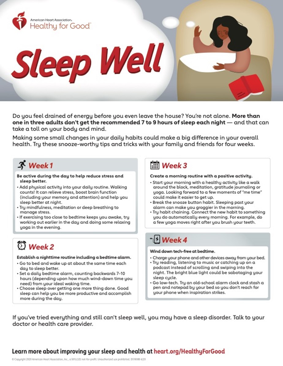
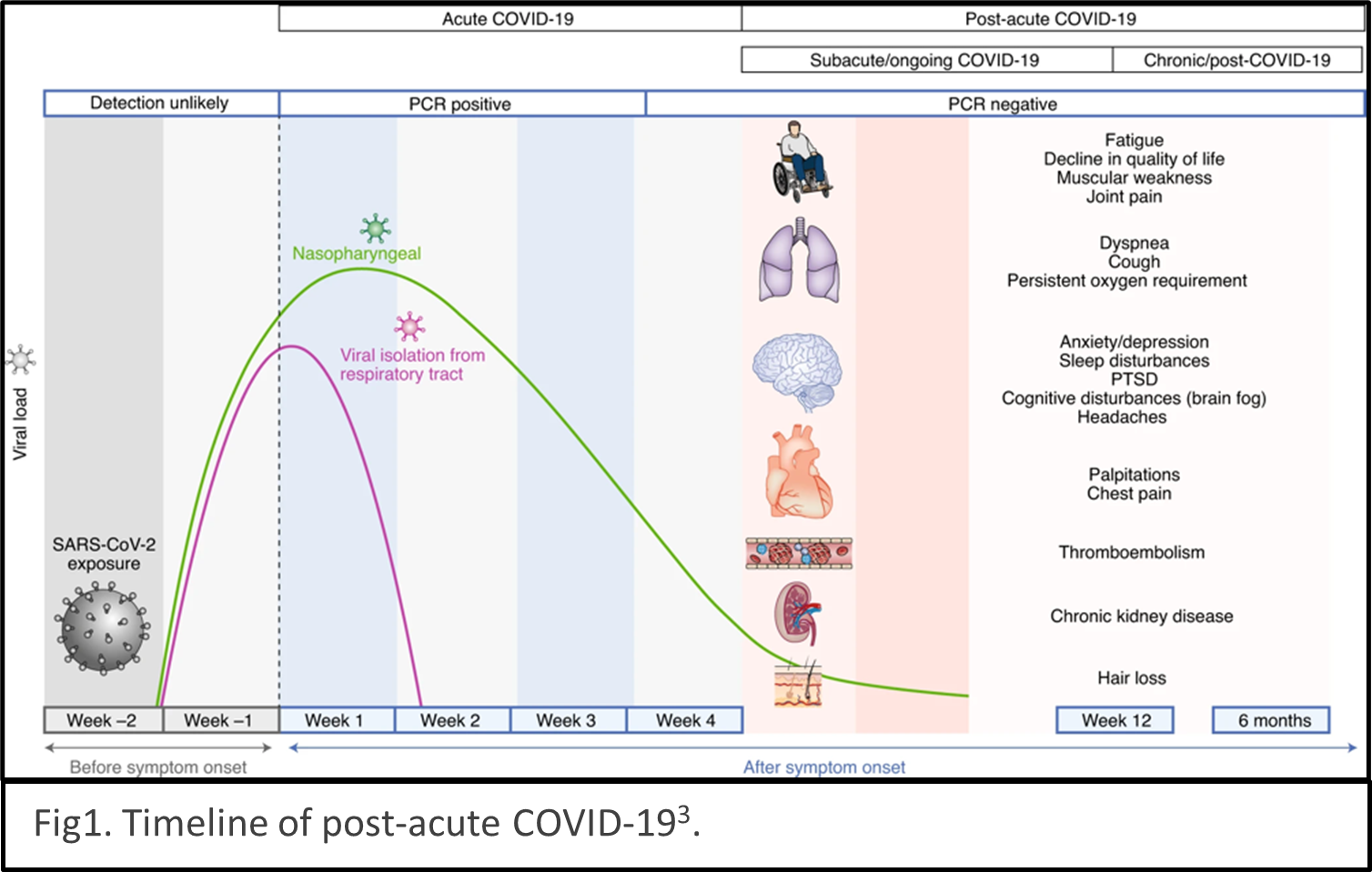
describes the condition as sequelae that extend beyond four weeks after initial infection1. The timeline of post-acute COVID-19 shows as Fig1. The list of common symptoms of post-COVID conditions is growing. Symptoms which people commonly report include difficulty breathing, fatigue, brain fog, cough, chest/stomach pain, headache, heart palpitations, muscle pain, diarrhea, sleep problems, fever, lightheadedness, rash, mood changes, changes in smell or taste, and changes in menstrual period cycles2,3. The challenges of diagnosing long COVID are multiple layers. The social isolation resulting from pandemic prevention measures can cause mental health stress such as depression, anxiety, and mood changes. Complications of pre-existing conditions may unmask after COVID infections. 
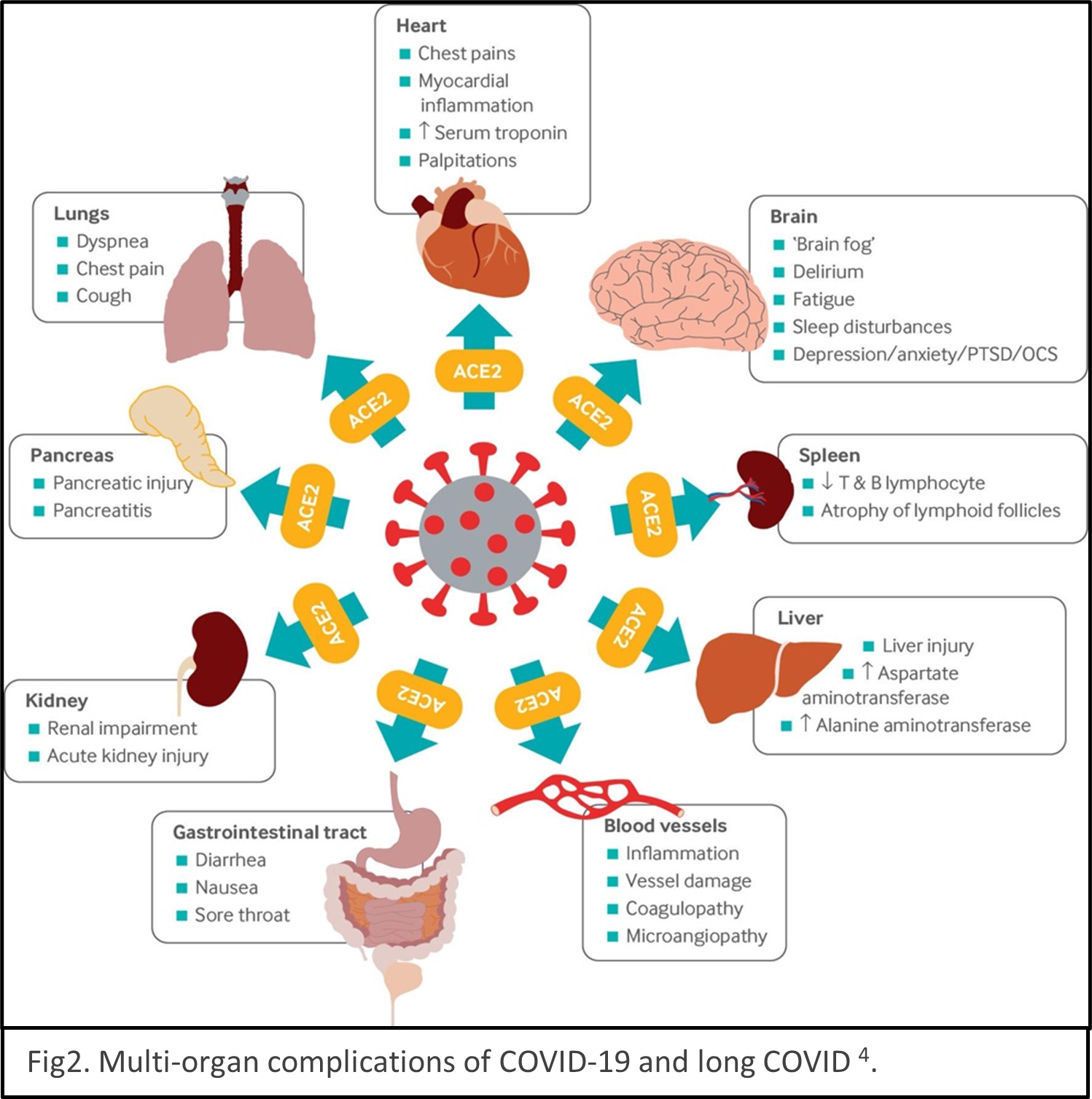
Reinfection of COVID could be mistaken as persistent symptoms. Multiple organs are reported being the victims of SARS-CoV-2 infection, for example, lungs, heart, brain, kidney, spleen, liver and the cardiovascular systems4 (Fig2). Some people have severe illness with COVID experience combinations of multiorgan effects or autoimmune conditions with symptoms lasting for weeks or months after initial infection. Long COVID is a serious concern. We just begin to understand it, and the path to be able to treat it with ease is winding.

Various guidelines have been established focus on treating and managing COVID5,6 and long COVID7. While the guidelines will undoubtedly improve as new evidence comes to light. The etiology, mechanism, and consequences of COVID and long COVID remain elusive currently. Large epidemiology studies are undergoing to help us understand mechanisms and develop targeted treatments. American Heart Association just establishes a new funding program to invite more researchers to study the mechanisms underlying cardiovascular consequences associated with COVID-19 and long COVID in January 2022. Scientific communities are racing to understand COVID and long COVID.


The COVID pandemics is a serious public crisis. Although the situation may seem be getting better, healthcare staff shortages caused by pervasive Omicron variant infections and the lack of rapid COVID tests are posing imminent danger to the public healthcare system. Moreover, the effects of long COVID vary person by person, it’s better to stay vigilant and not get infected than taking it callously and passing it to vulnerable people unintentionally.


REFERECE

  1. Datta SD, Talwar A, Lee JT. A Proposed Framework and Timeline of the Spectrum of Disease Due to SARS-CoV-2 Infection: Illness Beyond Acute Infection and Public Health Implications. JAMA. 2020;324(22):2251–2252.
  2. Al-Aly Z, Xie Y, Bowe B. High-dimensional characterization of post-acute sequelae of COVID-19. Nature. 2021;594(7862):259–264.
  3. Nalbandian A, Sehgal K, Gupta A, Madhavan M V, McGroder C, Stevens JS, Cook JR, Nordvig AS, Shalev D, Sehrawat TS, Ahluwalia N, Bikdeli B, Dietz D, Der-Nigoghossian C, Liyanage-Don N, et al. Post-acute COVID-19 syndrome. Nature Medicine. 2021;27(4):601–615.
  4. Crook H, Raza S, Nowell J, Young M, Edison P. Long covid–mechanisms, risk factors, and management. BMJ. 2021;374.
  5. World Health Organization. COVID-19 clinical management: living guidance. 2021. https://www.who.int/publications/i/item/WHO-2019-nCoV-clinical-2021-1
  6. National Institute of Health. Coronavirus disease 2019 (COVID-19) treatment guidelines. 2021. https://www.covid19treatmentguidelines.nih.gov/
  7. National Institute for Health and Care Excellence. COVID-19 rapid guideline: managing the long-term effects of COVID-19 NICE guideline; c2020. https://www.nice.org.uk/guidance/ng188


hidden

Moyamoya Disease

Moyamoya disease is a progressive cerebrovascular disorder that often begins during childhood. Patients with Moyamoya disease have injuries in their blood vessels at the base of the brain. The term ‘moyamoya’ comes from Japanese and means ‘puff of smoke’, which describes the appearance of abnormal blood vessels. Symptoms of Moyamoya disease include acute strokes, transient ischemic attacks (TIA), blurred vision, and weakness of one side of the body. Some researchers believe that Moyamoya disease is the result of inherited genetic disorders because individuals with Moyamoya often have close relatives who are also affected.

If the disease is not treated properly, patients will suffer from mental decline and recurrent strokes due to insufficient blood supply caused by moyamoya vessels in the brain. In severe cases, Moyamoya disease can cause death and life-long disabilities. The primary goal of treatment is to reduce the risk of strokes using medical and/or surgical interventions. In mild cases, medications are effective to prevent acute or recurrent strokes. In more severe cases, revascularization surgeries can restore normal blood flow bypassing affected blood vessels.

Imaging exams (such as CT or MRI) are often performed to diagnose Moyamoya disease and identify the location of the affected blood vessels. The most widely used techniques include CT perfusion, digital subtraction angiography, and MR angiography. In recent years, more advanced techniques such as arterial spin labeling have been employed to evaluate the blood flow and circulation without radiation or contrast agents.

AHA has been a pioneer in providing support for Moyamoya patients and funding research on this disease. I am honored to be supported by AHA to develop advanced imaging technologies to identify Moyamoya patients with a higher risk of stroke.

A recent story of a patient recovering from Moyamoya disease was covered by the AHA news:

https://www.heart.org/en/news/2019/03/27/after-4-strokes-rare-disease-and-brain-surgery-woman-helps-others

Image source: Moyamoya Foundation


References:

https://www.ninds.nih.gov/Disorders/All-Disorders/Moyamoya-Disease-Information-Page

https://stanfordhealthcare.org/medical-clinics/moyamoya-center.html

https://moyamoya-foundation.org/inspiration


“The views, opinions and positions expressed within this blog are those of the author(s) alone and do not represent those of the American Heart Association. The accuracy, completeness and validity of any statements made within this article are not guaranteed. We accept no liability for any errors, omissions or representations. The copyright of this content belongs to the author and any liability with regards to infringement of intellectual property rights remains with them. The Early Career Voice blog is not intended to provide medical advice or treatment. Only your healthcare provider can provide that. The American Heart Association recommends that you consult your healthcare provider regarding your personal health matters. If you think you are having a heart attack, stroke or another emergency, please call 911 immediately.”

hidden

Elevated Lipoprotein A, are we ready to intervene?

Lipid abnormalities can be classified into four clinical groups:1) elevated triglycerides, 2) low high-density lipoprotein cholesterol (HDL-C),3) elevated low-density lipoprotein cholesterol (LDL-C), and 4) elevated lipoprotein A.  Lipoprotein A disorder has been the least lipid abnormality studied from a clinical standpoint. Although many mendelian and genome-wide association studies have shown an association between elevated lipoprotein A and risk of incident atherosclerotic cardiovascular disease (ASCVD), the causality relationship still lacks the final corroboration of a randomized Lp(a) lowering intervention clinical trial1.

In 1963, for the first time, Kare Berg described the existence of Lp(a). Lp(a) consisted of apolipoprotein (a) bound to apolipoprotein B-100, an apolipoprotein also found on LDL-C particles. Distinct from other lipoproteins greatly affected by diet and genetics, Lp (a) is determined by more than 90% by individuals’ genetics. Lp(a) is synthesized in hepatocytes and is released into blood circulation. Plasma concentrations of Lp(a) are inversely associated with the size of apolipoprotein(a). It is postulated that small isoforms can be synthesized faster than large ones, and apolipoprotein(a) size can account for up to 70% influence on Lp(a) blood concentration. Other known factors include single nucleotide polymorphisms, sex hormones, inflammatory mediators, and dietary factors2.

Many medications have been tested to lower Lp(a) concentration. A large-scale meta-analysis of 5,256 patients (1,371 on placebo and 3,885 on statin) enrolled in a randomized clinical trial showed that most statins significantly increase Lp(a) concentration by 8-24%3. Niacin may lower Lp(a) concentration by 20-30%. However, studies did not show any significant reduction in risk of ASCVD event, once niacin was added to the medication list of patients already receiving a statin. Estrogen can also lower Lp(a) concentration by 20%; however, the usage is limited due to concern about increasing the risk of thrombotic events. Proprotein convertase subtilisin Kexin type 9 inhibitors (PCSK9 inhibitors) have been shown to lower Lp(a) concentrations. In 2 major PCSK-9 inhibitors clinical trials- FOURIER and ODYSSEY OUTCOME- the fourth quartile of Lp(a) in the treatment arm was associated with a 25% increase risk of major adverse cardiovascular events. This implies the presence of residual risk of ASCVD despite very low LDL-C4. In a recent sub-analysis of ODYSSEY OUTCOME trial, alirocumab reduced Lp(a) concentration. Each 5mg/dl decrease in Lp(a) by alirocumab resulted in a 2.5% reduction in risk of cardiovascular events5. It should be noted that none of the above medications have primarily been initially designed to test the hypotheses of the beneficial effect of medication on Lp(a) concentration.

Genetic studies have shed light on the role of each allele on Lp(a) concentrations and have provided a base for pharmacological intervention. Antisense oligonucleotides (ASO) have revolutionized the treatment of Lp(a). ASOs are short DNA fragments designed complementary for a target messenger RNA. LPA gene transcribes the two alleles of apolipoprotein(a) mRNA. ASO like Pelacarsen binds to the mRNA and generates the mRNA-ASO complex. Hepatocytes recognized this complex as foreign objects, and RNase H1 cleaves the sense strand. Four clinical trials have been conducted to assess the efficacy of ASOs to lower Lp(a) concentrations. All the clinical trials have shown promising results, from 40% to up to 90% reduction in Lp(a) depending on the type of ASO, dosage, and frequency of administrations6.

Currently, the pivotal phase 3 of Lp(a)HORIZON7 (ClinicalTrials.gov Identifier: NCT04023552) randomized controlled trial is enrolling up to 8280 individuals, aged >18 years with Lp(a) ≥ 70 mg/dl and with a history of myocardial infarction, ischemic stroke, or symptomatic peripheral artery disease. Participants are double blindly randomized to receive TQJ230 80 mg injected monthly subcutaneously or placebo and will be followed for the primary outcome of MACE (CV death, non-fatal MI, non-fatal stroke, and urgent coronary revascularization). The estimated primary completion date is May 29, 2025. So, we need to be patient and wait to see if the causal relationship between Lp(a) and ASCVD will be established by 2025.


References:

  1. Miksenas H, Januzzi JL, Jr. and Natarajan P. Lipoprotein(a) and Cardiovascular Diseases. JAMA. 2021;326:352-353.
  2. Tsimikas S. A Test in Context: Lipoprotein(a): Diagnosis, Prognosis, Controversies, and Emerging Therapies. J Am Coll Cardiol. 2017;69:692-711.
  3. Tsimikas S, Gordts P, Nora C, Yeang C and Witztum JL. Statin therapy increases lipoprotein(a) levels. Eur Heart J. 2020;41:2275-2284.
  4. Ruscica M, Greco MF, Ferri N and Corsini A. Lipoprotein(a) and PCSK9 inhibition: clinical evidence. Eur Heart J Suppl. 2020;22:L53-L56.
  5. Szarek M, Bittner VA, Aylward P, Baccara-Dinet M, Bhatt DL, Diaz R, Fras Z, Goodman SG, Halvorsen S, Harrington RA, Jukema JW, Moriarty PM, Pordy R, Ray KK, Sinnaeve P, Tsimikas S, Vogel R, White HD, Zahger D, Zeiher AM, Steg PG, Schwartz GG and Investigators OO. Lipoprotein(a) lowering by alirocumab reduces the total burden of cardiovascular events independent of low-density lipoprotein cholesterol lowering: ODYSSEY OUTCOMES trial. Eur Heart J. 2020;41:4245-4255.
  6. Tsimikas S, Moriarty PM and Stroes ES. Emerging RNA Therapeutics to Lower Blood Levels of Lp(a): JACC Focus Seminar 2/4. J Am Coll Cardiol. 2021;77:1576-1589.
  7. Assessing the Impact of Lipoprotein (a) Lowering With TQJ230 on Major Cardiovascular Events in Patients With CVD (Lp(a)HORIZON). https://clinicaltrialsgov/ct2/show/NCT04023552. 2019.

“The views, opinions and positions expressed within this blog are those of the author(s) alone and do not represent those of the American Heart Association. The accuracy, completeness and validity of any statements made within this article are not guaranteed. We accept no liability for any errors, omissions or representations. The copyright of this content belongs to the author and any liability with regards to infringement of intellectual property rights remains with them. The Early Career Voice blog is not intended to provide medical advice or treatment. Only your healthcare provider can provide that. The American Heart Association recommends that you consult your healthcare provider regarding your personal health matters. If you think you are having a heart attack, stroke or another emergency, please call 911 immediately.”