hidden

COVID-19 and Endothelial Cell Dysfunction

Photo by CDC on Unsplash

COVID-19, caused by severe acute respiratory syndrome coronavirus 2 (SARS-CoV-2), represents a global health crisis. Cough, fever, and shortness of breath are the most common reported symptoms; however, neurological and gastroenterological manifestations can also be present1. Angiotensin-converting enzyme 2 (ACE-2) has been shown to act as a co-receptor to facilitate coronavirus entry by efficiently binding to the S1 domain of spike protein, a surface glycoprotein of SARS-CoV2. The pathogenesis of COVID-19 depends on the localization of the coronavirus co-receptors. The epithelium of lungs and intestine is rich in ACE-2 expression, thereby providing a possible route of entry for SARS-CoV-2. Further, ACE-2 is also expressed on Type I and type II pneumocytes providing other entry sites for SARS-CoV-2. Virus entry can cause pathological changes at the alveoli-capillary interface. Additionally, copious expression of ACE-2 on the type II alveolar cells can promote rapid viral expansion resulting in further alveolar damage and hyperinflammation3,4.

ACE-2 is also present on endothelial cells, smooth muscle cells, and perivascular pericytes in all the organs, indicating that if SAR-COV-2 is transmitted to the blood circulation, the virus can quickly spread throughout the body3. The postmortem lung tissues of COVID-19 patients exhibited a higher number of ACE-2 positive endothelial cells and a higher prevalence of endothelial injury (disruption of cell membrane and presence of the intracellular virus) and microthrombi than lung tissues of patients who died from influenza-associated respiratory failure1,5. The most common comorbidities observed in COVID-19 patients are hypertension, diabetes, and obesity, all of which are associated with endothelial dysfunction. Further, COVID-19 patients are projected to be at a higher risk of deep vein thrombosis, systemic vasculitis, and acute pulmonary embolism6,7, possibly due to underlying endothelial cell injury and inflammation. Thrombi can directly affect gas exchange and cause and cause multisystem organ dysfunction in COVID-19 pneumonia8. Upon activation, platelets release polyphosphates, which accelerate the activation of factors V and XI, inhibit tissue factor pathway inhibitor and contribute to thicker fibrin strands synthesis. Further, the cytokine release can activate endothelial cells resulting in a prothrombotic environment1.

Acute respiratory distress syndrome is suggested to be caused by the dissociation between lung mechanics, loss of lung perfusion regulation and hypoxic vasoconstriction, and severe hypoxemia9. The loss of hypoxic vasoconstriction can be due to increased mitochondrial oxidative stress resulting in pulmonary endothelial cell dysfunction10. SARS-CoV-2 elements, accumulation of inflammatory cells, intracellular virus, and disrupted cell membranes are detected in the endothelial cells of COVID-19 patients5,11, further indicating endotheliitis /endothelial cell dysfunction during COVID-19 infection. Endothelial cell dysfunction can cause abnormalities in microcirculation in different vascular beds and contribute to life-threatening complications of COVID-19, including thromboembolism and multiple organ damage.

References:

  1. Huertas A, Montani D, Savale L, et al. Endothelial cell dysfunction: a major player in SARS-CoV-2 infection (COVID-19)? Eur Respir J. 07 2020;56(1)doi:10.1183/13993003.01634-2020
  2. Ziegler CGK, Allon SJ, Nyquist SK, et al. SARS-CoV-2 Receptor ACE2 Is an Interferon-Stimulated Gene in Human Airway Epithelial Cells and Is Detected in Specific Cell Subsets across Tissues. Cell. 05 28 2020;181(5):1016-1035.e19. doi:10.1016/j.cell.2020.04.035
  3. Hamming I, Timens W, Bulthuis ML, Lely AT, Navis G, van Goor H. Tissue distribution of ACE2 protein, the functional receptor for SARS coronavirus. A first step in understanding SARS pathogenesis. J Pathol. Jun 2004;203(2):631-7. doi:10.1002/path.1570
  4. Mehta P, McAuley DF, Brown M, et al. COVID-19: consider cytokine storm syndromes and immunosuppression. Lancet. 03 28 2020;395(10229):1033-1034. doi:10.1016/S0140-6736(20)30628-0
  5. Ackermann M, Verleden SE, Kuehnel M, et al. Pulmonary Vascular Endothelialitis, Thrombosis, and Angiogenesis in Covid-19. N Engl J Med. 07 09 2020;383(2):120-128. doi:10.1056/NEJMoa2015432
  6. Bompard F, Monnier H, Saab I, et al. Pulmonary embolism in patients with COVID-19 pneumonia. Eur Respir J. Jul 2020;56(1)doi:10.1183/13993003.01365-2020
  7. Criel M, Falter M, Jaeken J, et al. Venous thromboembolism in SARS-CoV-2 patients: only a problem in ventilated ICU patients, or is there more to it? Eur Respir J. Jul 2020;56(1)doi:10.1183/13993003.01201-2020
  8. Poor HD, Ventetuolo CE, Tolbert T, et al. COVID-19 critical illness pathophysiology driven by diffuse pulmonary thrombi and pulmonary endothelial dysfunction responsive to thrombolysis. Clin Transl Med. Jun 2020;10(2):e44. doi:10.1002/ctm2.44
  9. Gattinoni L, Coppola S, Cressoni M, Busana M, Rossi S, Chiumello D. COVID-19 Does Not Lead to a “Typical” Acute Respiratory Distress Syndrome. Am J Respir Crit Care Med. 05 15 2020;201(10):1299-1300. doi:10.1164/rccm.202003-0817LE
  10. Guignabert C, Phan C, Seferian A, et al. Dasatinib induces lung vascular toxicity and predisposes to pulmonary hypertension. J Clin Invest. 09 01 2016;126(9):3207-18. doi:10.1172/JCI86249
  11. Varga Z, Flammer AJ, Steiger P, et al. Endothelial cell infection and endotheliitis in COVID-19. Lancet. 05 02 2020;395(10234):1417-1418. doi:10.1016/S0140-6736(20)30937-5

“The views, opinions, and positions expressed within this blog are those of the author(s) alone and do not represent those of the American Heart Association. The accuracy, completeness, and validity of any statements made within this article are not guaranteed. We accept no liability for any errors, omissions, or representations. The copyright of this content belongs to the author and any liability with regards to infringement of intellectual property rights remains with them. The Early Career Voice blog is not intended to provide medical advice or treatment. Only your healthcare provider can provide that. The American Heart Association recommends that you consult your healthcare provider regarding your health matters. If you think you are having a heart attack, stroke, or another emergency, please call 911 immediately.”

hidden

The darkness before the dawn–Long COVID is lurking around

As we are starting to live with the facts that COVID-19 is not leaving us any time soon, sense of danger and urgency start to fade away. More than two years have passed, we have made great strides in combating the pandemics. With advanced technologies, vaccine and antiviral drug developments provide us potential means out of this seemingly never-ending battle. Viruses constantly change through mutation. While Delta variant is phasing out, Omicron variant begins its domination globally. Current research suggest that Omicron variant is less deadly compared to the Delta variant, especially to fully vaccinated people. However, the high spread rate of Omicron variant is a major concern to the public.


Some of us may wonder, it might not be a big deal. Since most evidence suggest that the symptoms of fully vaccinated patients are rather minor, especially the chance of suffering severe symptoms if you had a booster shot is even less. After a few days discomfort, you might end up obtaining better immunity after building up antibodies through infection. It might be partially true to some people. However, some unlucky ones continue to suffer from post-COVID conditions even after four or more weeks. These post-COVID conditions are also known as long COVID, long-haul COVID, post-acute COVID, long-term effects of COVID, or chronic COVID.


What is long COVID?

According to the US Centers for Disease Control and Prevention (CDC), long COVID 
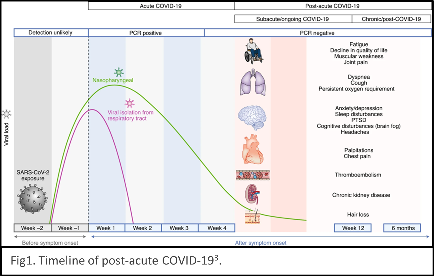
describes the condition as sequelae that extend beyond four weeks after initial infection1. The timeline of post-acute COVID-19 shows as Fig1. The list of common symptoms of post-COVID conditions is growing. Symptoms which people commonly report include difficulty breathing, fatigue, brain fog, cough, chest/stomach pain, headache, heart palpitations, muscle pain, diarrhea, sleep problems, fever, lightheadedness, rash, mood changes, changes in smell or taste, and changes in menstrual period cycles2,3. The challenges of diagnosing long COVID are multiple layers. The social isolation resulting from pandemic prevention measures can cause mental health stress such as depression, anxiety, and mood changes. Complications of pre-existing conditions may unmask after COVID infections. 
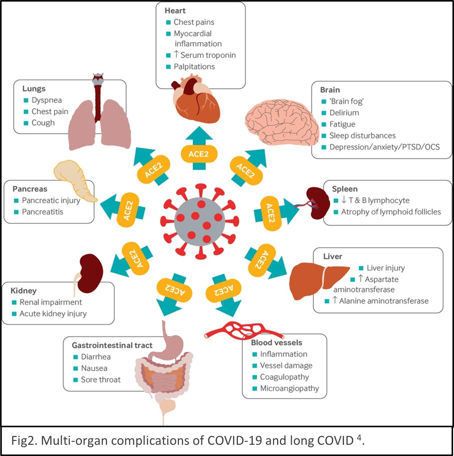
Reinfection of COVID could be mistaken as persistent symptoms. Multiple organs are reported being the victims of SARS-CoV-2 infection, for example, lungs, heart, brain, kidney, spleen, liver and the cardiovascular systems4 (Fig2). Some people have severe illness with COVID experience combinations of multiorgan effects or autoimmune conditions with symptoms lasting for weeks or months after initial infection. Long COVID is a serious concern. We just begin to understand it, and the path to be able to treat it with ease is winding.

Various guidelines have been established focus on treating and managing COVID5,6 and long COVID7. While the guidelines will undoubtedly improve as new evidence comes to light. The etiology, mechanism, and consequences of COVID and long COVID remain elusive currently. Large epidemiology studies are undergoing to help us understand mechanisms and develop targeted treatments. American Heart Association just establishes a new funding program to invite more researchers to study the mechanisms underlying cardiovascular consequences associated with COVID-19 and long COVID in January 2022. Scientific communities are racing to understand COVID and long COVID.


The COVID pandemics is a serious public crisis. Although the situation may seem be getting better, healthcare staff shortages caused by pervasive Omicron variant infections and the lack of rapid COVID tests are posing imminent danger to the public healthcare system. Moreover, the effects of long COVID vary person by person, it’s better to stay vigilant and not get infected than taking it callously and passing it to vulnerable people unintentionally.


REFERECE

  1. Datta SD, Talwar A, Lee JT. A Proposed Framework and Timeline of the Spectrum of Disease Due to SARS-CoV-2 Infection: Illness Beyond Acute Infection and Public Health Implications. JAMA. 2020;324(22):2251–2252.
  2. Al-Aly Z, Xie Y, Bowe B. High-dimensional characterization of post-acute sequelae of COVID-19. Nature. 2021;594(7862):259–264.
  3. Nalbandian A, Sehgal K, Gupta A, Madhavan M V, McGroder C, Stevens JS, Cook JR, Nordvig AS, Shalev D, Sehrawat TS, Ahluwalia N, Bikdeli B, Dietz D, Der-Nigoghossian C, Liyanage-Don N, et al. Post-acute COVID-19 syndrome. Nature Medicine. 2021;27(4):601–615.
  4. Crook H, Raza S, Nowell J, Young M, Edison P. Long covid–mechanisms, risk factors, and management. BMJ. 2021;374.
  5. World Health Organization. COVID-19 clinical management: living guidance. 2021. https://www.who.int/publications/i/item/WHO-2019-nCoV-clinical-2021-1
  6. National Institute of Health. Coronavirus disease 2019 (COVID-19) treatment guidelines. 2021. https://www.covid19treatmentguidelines.nih.gov/
  7. National Institute for Health and Care Excellence. COVID-19 rapid guideline: managing the long-term effects of COVID-19 NICE guideline; c2020. https://www.nice.org.uk/guidance/ng188


hidden

Last Day of #AHA20: COVID-19 Galore!

The last day of the amazing #AHA20 featured a series of COVID-19-related research presentations.

First, data from the AHA COVID-19 Registry, a large database collecting data about COVID-19 patients and outcomes around the country, were shared. The registry includes data from 109 hospitals and over 22,500 records of patients who were hospitalized with COVID-19. Notably, large numbers of COVID-19 patients in this registry had cardiovascular risk factors such as hypertension and diabetes. Prior cardiovascular disease was also common. The disease was additionally noted to have a high morbidity and mortality rate, with more than 20% of hospitalized COVID-19 patients requiring mechanical ventilation.

One interesting study examined racial and ethnic differences in the AHA COVID-19 Registry of patients hospitalized with COVID-19, focusing primarily on the association of these factors with in-hospital death as the primary outcome and secondary outcomes such as major adverse cardiovascular events (MACE: death, myocardial infarction, stroke, new onset heart failure or cardiogenic shock) or COVID-19 cardio-respiratory disease severity scale. Notably, Black and Hispanic patients accounted for >50% of hospitalizations in this Registry, suggesting significant over-representation of Black and Hispanic patients compared with the census demographics in their areas. Cardiovascular risk factors such as obesity and hypertension were also more common in Black and Hispanic patients. Mechanical ventilation and need for renal replacement therapy were more likely in Black patients. Overall in-hospital mortality was high at 18.4%, and particularly high for those older than 70 years old.

In fully adjusted models taking into account age, medical history and sociodemographic features, there was no statistically significant difference in mortality and MACE among different racial or ethnic groups, though Asian patients had a higher COVID-10 disease severity on presentation. These findings suggest that though race and ethnicity are not independently associated with worse in-hospital outcomes in COVID-19 patients, Black and Hispanic patients bear a greater burden of morbidity associated with COVID due to their disproportionate representation among patients hospitalized with CVOID-19. This study was simultaneously published online in Circulation.

One additional study examined the association between body mass index (BMI) with a composite of in-hospital death and/or mechanical ventilation (primary outcome), as well as with MACE (a composite of in-hospital all-cause death, stroke, heart failure, myocardial infarction), deep vein thrombosis and renal replacement therapy (secondary outcomes). Patients with a higher BMI were more likely to be admitted to the hospital with COVID-19. In analyses adjusting for age, sex, ethnicity, comorbidities, cardiovascular disease and chronic kidney disease, higher class obesity was associated with higher likelihood of in-hospital mortality or mechanical ventilation. MACE was not associated with obesity class. Deep venous thrombosis or pulmonary embolism were not associated with obesity class. Class I, II and III obesity, however, were noted to have a higher likelihood of need for mechanical ventilation, regardless of age. Moreover, when stratified by age, BMI >40 kg/m2 was associated with a higher risk of in-hospital death only in lower age groups (<50 years old). These findings suggest that better public health messaging may be required for younger obese individuals who may underestimate their own risk related to COVID-19. This study was also simultaneously published in Circulation.


“The views, opinions and positions expressed within this blog are those of the author(s) alone and do not represent those of the American Heart Association. The accuracy, completeness and validity of any statements made within this article are not guaranteed. We accept no liability for any errors, omissions or representations. The copyright of this content belongs to the author and any liability with regards to infringement of intellectual property rights remains with them. The Early Career Voice blog is not intended to provide medical advice or treatment. Only your healthcare provider can provide that. The American Heart Association recommends that you consult your healthcare provider regarding your personal health matters. If you think you are having a heart attack, stroke or another emergency, please call 911 immediately.”

hidden

How will COVID-19 Affect Return to High School Sports and ECG Screening?

August is traditionally very busy month in the pediatric cardiology office; visits for “sports clearance” flood the schedule due to something picked up on a high school sports physical such as chest pain. Most of these will be non-cardiac and receive reassurance; however, the cardiac causes in the pediatric population can be quite distressing. With the current COVID-19 pandemic we are seeing many cardiac effects of the virus— myocardial inflammation, dysfunction and coronary artery dilation or MIS-C (multisystem inflammatory syndrome in children). There has also been a reported increase in out of hospital cardiac arrest in the adult population, which could be attributed to those afraid to seek care, but none the less, a concern.

Cardiac effects play an important part in the recent discussions on return to school, as eventually return to school will mean return to school sports. Sports cardiologists have been following this closely and have been working to establish a safe way for return to sports in competitive athletes who have had COVID-191(click here for recommendations and see flowchart below), which may include extensive cardiac testing of those who had symptomatic COVID-19, most of which is based on the return to play myocarditis guidelines.2 These important decisions require individualization, knowledge of risk and what is happening in the community.

So what does this mean for the high school athletes? There is no doubt that exercise is great for everyone, especially with the rising obesity and the sedentary lifestyle. Organized sports participation has shown to have a positive impact on mental and physical health that extends beyond childhood.3 Many have raised concerns about the development and health of children by not attending school, along with decrease in social interaction and activity from organized sports during the stay-at-home orders and while we work to defeat COVID-19 spread.

Prior to the pandemic, the question of universal ECG testing for high school athletes always started a conversation, but could that change? Critics say that the false positives create distress and extra healthcare cost, that follow up is problematic and that it has not been shown to change outcomes. Others argue saving one child from the devastating event of collapsing and dying on the field is worth it, and a recent study showed ECG with H&P is more likely to detect a condition associated with sudden cardiac arrest (SCA) than H&P alone, and also improved cost efficiency when interpreted in the right hands.4 All agree it is important to have a good plan in place for SCA at all sporting events, including an emergency response plan in place specific to cardiac arrest.

With so many affected by the virus, at varying severity, I can’t help but wonder if this will change the way we assess our young athletes for participation in sports when it is safe to do so. While healthcare costs are always a problem, with a new virus and unclear long-term outcomes, we must be conservative. Perhaps this may provide an opportunity to learn more about ECG screening, as we will likely see a larger amount of patients who have had COVID-19 coming to our offices for clearance. This may provide an opportunity to gather evidence on the continued debate of whether ECG screening is effective.

Until we know more, what we do know is it is safer to be conservative, to assess benefit and risk and provide appropriate counseling to families on signs and symptoms of SCA. We also must continue to reassess and stay up-to-date on new knowledge and testing related to COVID-19 recovery and sports. Most importantly, it is important that we continue to advocate for heart safe schools which include having AEDs, training in CPR, and emergency response plans related to Sudden Cardiac Arrest. With more kids participating in school from home, this could be a great opportunity to engage the whole family in CPR and AED education to improve the safety and survival of our communities and at home.


References

  1. Phelan, Dermot, et al. “A Game Plan for the Resumption of Sport and Exercise After Coronavirus Disease 2019 (COVID-19) Infection.” JAMA Cardiology, 2020, doi:10.1001/jamacardio.2020.2136.
  2. Maron, Barry J., et al. “Eligibility and Disqualification Recommendations for Competitive Athletes With Cardiovascular Abnormalities: Task&nbsp; Force 3: Hypertrophic Cardiomyopathy, Arrhythmogenic Right Ventricular Cardiomyopathy and Other Cardiomyopathies, and Myocarditis.” Journal of the American College of Cardiology, vol. 66, no. 21, 2015, pp. 2362–2371., doi:10.1016/j.jacc.2015.09.035.
  3. Logan, Kelsey, and Steven Cuff. “Organized Sports for Children, Preadolescents, and Adolescents.” Pediatrics, vol. 143, no. 6, 2019, doi:10.1542/peds.2019-0997.
  4. Harmon, Kimberly G, and Jonathan A Drezner. “Comparison of Cardiovascular Screening in College Athletes” Heart Rhythm, 2020, www.heartrhythmjournal.com/article/S1547-5271(20)30406-9/pdf.

“The views, opinions and positions expressed within this blog are those of the author(s) alone and do not represent those of the American Heart Association. The accuracy, completeness and validity of any statements made within this article are not guaranteed. We accept no liability for any errors, omissions or representations. The copyright of this content belongs to the author and any liability with regards to infringement of intellectual property rights remains with them. The Early Career Voice blog is not intended to provide medical advice or treatment. Only your healthcare provider can provide that. The American Heart Association recommends that you consult your healthcare provider regarding your personal health matters. If you think you are having a heart attack, stroke or another emergency, please call 911 immediately.”