hidden

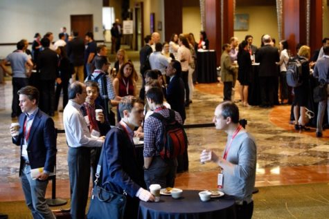
Fifth Year at Vascular Discovery: How an Early Career Navigates Through a Virtual Event

It is not news anymore that after World Health Organization (WHO) classified novel coronavirus disease (COVID-19) as a pandemic, the real-life impact of the “new normal” started to show itself. We started to see the impact by pausing the research and of course, cancelation of all scientific events. As scientists shifted their focus toward the data analysis, manuscript preparation, grants or any other means of remote work, the American Heart Association also shifted its focus to keep events happening, virtually. This new mode of attending a conference has many perks including petting your dog when the field’s connoisseur is giving a talk, drinking coffee from your favorite mug, or keeping the PJs on. However, there is a trade-off. Navigating through a professional event such as #VascularDisovery20 is challenging by itself, let alone going after it when you are not physically present. The following tips will help you to turn this year into a great opportunity.

1: Networking with Peers and Mentors: Online Presence

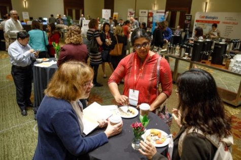
Now that there is no opportunity to join a table for lunch, mingle during the dinner event or grab a coffee with your colleagues from other countries, increase your online presence by interacting with attendees on Twitter. Use the chat mode during virtual sessions to ask questions or chat with others. If you have a burning question about a talk, either tweet at the presenter or email them. This is a great practice for increasing your “professional” online activity.

2: Utilize the Home Stay

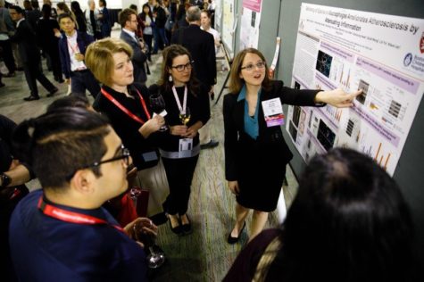
Now that you have a desk in front of you instead of sitting on conference room chairs (which are really uncomfortable from time to time), take notes with ease, have your screen open with relevant papers to the talk, take high-quality images from the slides you find important and download the available contents in advance from the #HeartHub. In addition, you can now have a comprehensive look at your favorite posters and get connected with the presenters.

3: Plan Ahead

Although you may think that you can easily jump to the laptop and login to the talk that is “live”, the reality may be different. There are still concurrent sessions that you need to choose which one you attend. Also, there is always a possibility of a technical problem, so make sure that everything is set and you know exactly which talk you are going after. Also, pay attention to the time-zone listed in the schedule.

4: Hangouts at Conference Evenings: Say Hello to Face Time or Zoom

It is an unwritten tradition that many attendees get together after a long day of scientific endeavors to sit down, chit chat and grab a drink. Use the ATVB Journal virtual happy hour with EIC Dr. Alan Daugherty as an example. If you would like to hang out with the “conference buddies”, reach out to them, set up a private virtual meeting and catch up. This is a great practice to break those shyness barriers, especially if you are at the early career stage.

It is obvious that nothing compares to be physically present among your peers, colleagues and mentors. However, during these uncertain times, we can still manage to make the most out of the opportunities we are offered. As scientists, we are always learning to overcome new challenges and come up with new solutions, therefore, navigating through a virtual event not only is a fun challenge but also is a great learning experience with many opportunities.

“The views, opinions and positions expressed within this blog are those of the author(s) alone and do not represent those of the American Heart Association. The accuracy, completeness and validity of any statements made within this article are not guaranteed. We accept no liability for any errors, omissions or representations. The copyright of this content belongs to the author and any liability with regards to infringement of intellectual property rights remains with them. The Early Career Voice blog is not intended to provide medical advice or treatment. Only your healthcare provider can provide that. The American Heart Association recommends that you consult your healthcare provider regarding your personal health matters. If you think you are having a heart attack, stroke or another emergency, please call 911 immediately.”

hidden

A Common Problem Among Scientists: Not Being the Best Presenters – Lesson One

With many conferences, symposiums and seminars always lined up throughout the year, a common shortcoming in scientists’ lives is the fear of public speaking and, consequently, a poor presentation.

As a public speaking coach and presentation skills teacher who happens to be a scientist too, I cannot emphasize how much of difference it can make when you present your data (most of the time, complicated data!) clearly and effectively. Presentations skills and public speaking skills are very useful in many aspects of work and life. Effective presentations and public speaking skills are important in business, sales and selling, training, teaching, lecturing, and generally feeling comfortable speaking to a group of people. Let’s not forget that having the confidence and capability to give good presentations and to stand up in front of an audience are extremely helpful competencies for self-development and social situations.

In a series of blog posts, my goal will be to discuss some details of how you can improve your scientific presentations and the key points that can help your presentation to stand out. Throughout the upcoming blog posts, the focus will be on the delivery aspects of the talk and visuals (slides).

Whenever I am starting a new presentation skills class, I always bring up what William Yeats, one of the foremost figures of 20th-century literature, said years ago:

“I always think great speakers convince us not by force of reasoning but because they are visibly enjoying the beliefs they want us to accept.” – William Yeats

This brings us up to the first rule, which happens to be the most common presentation problem:

Not Being Boring – The Opening

It is a common bias to feel more interested in presentations when the speaker is passionate and excited to share the results. So how can you ensure not to be boring? Here are some tips:


1) Icebreaking Polls

Live polls are a great way to “break the ice” and capture the audience’s attention, especially in bigger crowds. As part of your opening remarks, you can use a fun poll to enliven the atmosphere and also to set the tone for your event (a good live example is what usually Dr. Kiran Musunuru does in his talks).

Here are a few examples that I personally like:

  • How energized are you feeling right now?
  • As a child, what did you want to be when you grew up?
  • If age is only a state of mind, what is YOUR state of mind right now?

Remember, funny answer options are also part of the polls.


2) Make a joke at your own expense

Before making a joke, remember to always be sensitive to your crowd. People have different values, beliefs, and experiences.

As it is beautifully mentioned by Public Speaking Powers: “You don’t want to make a joke at the expense of anyone in the audience, a joke at the expense of the company, or a joke at the expense who’s introduced you, but the joke at the expense of yourself tends to work really well because you’re pointing at yourself so people can just laugh along with that.”

For example, you could say: “Look, I have a bad feeling about this. I was talking to [whoever introduced you], and they said they were going to tell a joke before I spoke but instead they just introduced me.” So you’re implying you’re the joke.


3) Ask “raise your hand” questions

My personal favorite is this type of icebreaker. It shows confidence, it boosts up your stage presence and it makes your audience to physically move. When thinking about the question you want to ask, consider the following:

Do not create a negative environment with your question. Never ask questions that may put the audience in the spot. Negative examples include, “How many of you are suffering from dyslipidemia?” or, “Have you ever been into a Cath lab as a patient?”

Instead, ask questions that are more relevant and questions that most people are going to raise their hand to: “How many of you have read the CONSORT trial results?” or, “How many of you read the new hypertension guidelines?”

Keep in mind, the whole idea of the “raise your hand” questions is to get audience’s engagement and group involvement, so the people on the outskirts who aren’t really getting into your talk feel like they should get into your talk.


4) Start a story without finishing it right away

Open up your talk by simple phrases like:  “I want to tell you a story that I think it is very important for my speech today.” Or you go on and tell your story, but you leave the conclusion out and you say: “I’ll get back to that towards the end.”

This allows you to draw people into your talk with stories, but you’re not finishing your story right away so it keeps them engaged.


5) Start by breaking some news

A good way to keep the audience engaged is to talk about a recent news/paper/article that is relevant to your presentation. I was recently in an AHA’s Strategically Focused Research Network meeting about sex differences in aortapathies and the speaker opened up her talk by discussing an article from Times Magazine, which came out on the same day, discussing sex differences.


It can be tricky to know how to start a meeting. In fact, the introduction is often the hardest part to get right. But with a great start, you can relax yourself and your audience, making them more alert and receptive. In the next upcoming blog post, I will discuss the problems throughout the delivery and body of the talk. Stay tuned!


hidden

Inflammation: A Hallmark of Abdominal Aortic Aneurysm Pathophysiology

In my previous blog post, I discussed the animal models used for studying abdominal aortic aneurysms (AAAs), the progressive dilation of the aorta which if left untreated, will lead to fatal aortic rupture. Currently, there are no pharmacological treatments available for this devastating condition and the current clinical approach is to monitor the aortic dimension and eventually, the patient will undergo open or endovascular surgical repair when the aorta attains sufficient expansion. Given the complexity of the disease and the fact that human AAA tissue are acquired at the advanced stages of the disease, researchers have relied on animal models to better understand the disease process and to facilitate development of effective non-invasive therapies.

In general, AAAs are associated with aged population, male gender and lifestyle risk factors such as hypercholesterolemia and smoking. Lessons learned from such studies show that AAA pathological hallmarks include an increased local inflammation in the aortic tissue which can be further augmented by proteolytic degradation of extracellular matrix and depletion of vascular smooth muscle cells.

Inflammation is a common characteristic of AAA, which is manifested by accumulation of inflammatory cells and a wide range of related molecular signaling changes. Macrophages are the most common cell type present in AAA tissue which are localized in media and to a higher extent, in adventitia layer.


Our laboratory’s previous studies were among the first to show that whole body deficiency of TLR4, a key player in proinflammatory signaling, can ablate the angiotensin II (angII)-induced AAA in low-density lipoprotein receptor deficient (LDLR-/-) mice. Other research endeavors using different mice background or other AAA animal models also further confirmed the critical role of TLR4, or its ligands in AAAs. Although these studies clearly show the role of TLR4 in AAAs, further research is needed to understand which cell types are responsible for TLR4 protective effects and via which mechanisms. As a researcher in this field, my current focus is on targeting aortic cell-specific TLR4 (adventitia), understanding how these cell-specific receptors contribute to the pathology, and the role of potential ligands that activate this cascade of events.

These understandings have the potential to contribute to the development of pharmaceutical approaches that can specifically target the cell receptors and prevent/slow the progression of the pathology.


Resources for Further Reading:

  1. Renin-Angiotensin System and Cardiovascular Functions: Chia-Hua Wu, Shayan Mohammadmoradi, Jeff Z. Chen, Hisashi Sawada, Alan Daugherty and Hong S. Lu, ATVB, 2018


hidden

Abdominal Aortic Aneurysm: How to Study It in Mice

Aortic aneurysms are pathological dilations with high risk of mortality due to rupture. Abdominal aortic aneurysms (AAA) are the most common form of this condition with the dilation being mainly present in the infrarenal region. Histological analysis of human AAA tissue have shed lights on pathological hallmarks of AAAs, however, such tissues are acquired at the advanced stages of the disease and do not provide clear information about the AAA initiation. Therefore, the research has relied extensively on AAA animal models to better define the underlying mechanism of AAA. In general, there are three main mouse models to study AAA: 1) perfusion of elastase into the infrarenal aorta, 2) periaortic application of calcium chloride, or 3) subcutaneous infusion of Angiotensin (Ang) II. The purpose of this blog post is to provide a short description about the AngII-induced AAA.

AngII is the major bioactive peptide of the renin-angiotensin system. The AngII-induced AAA model is probably the most commonly used animal model nowadays, which was developed at the University of Kentucky. In this model, a small osmotic mini pump is subcutaneously implanted in the animal, which will infuse AngII (mainly at the dose of 1,000 ng/kg/min) for 28 days. AngII-induced AAA is augmented by hypercholesterolemia; therefore, ApoE-/- and LDLr-/- mice are the two most common animal models used for AngII-induced AAA studies. AngII also induces AAA in normolipidemic mice, however, at much lower incidence. The AngII-induced AAA formation is independent of the modestly increased systolic blood pressure after AngII infusion and the AAA induced by chronic AngII infusion is mainly located at suprarenal aortic region. This model exhibits progressive luminal expansion, increased leukocytic infiltration, elastin fiber disruption, loss of extracellular matrix, and consequently, aortic wall remodeling – all of which are hallmarks of human AAA.

Although AngII-infused mouse models are very popular due to similarities to human AAA and reproducibility, difficulties in breeding mice to a hypercholesterolemic background has hampered its wide use. However, a recent study from our laboratory provided a rapid approach for increasing plasma cholesterol and Ang II–induced AAA incidence in C57BL/6 mice by applying a gain-of-function mutation of mouse PCSK9 protein using an adeno-associated viral method.

Aortic aneurysms are lethal asymptomatic conditions and thanks to development of animal models, considerable increase in research on aneurysm pathogenesis have shed lights on the undelaying mechanisms of the disease. Despite the progress, there are still many perplexities regarding the AAA initiation and development, and this uncertainty highlights the important of understanding the mouse models to further research this devastating condition.

Resources for Further Reading:

  1. Renin-Angiotensin System and Cardiovascular Functions
    Chia-Hua Wu, Shayan Mohammadmoradi, Jeff Z. Chen, Hisashi Sawada, Alan Daugherty and Hong S. Lu, ATVB, 2018
  2. Hypercholesterolemia Induced by a PCSK9 Gain-of-Function Mutation Augments Angiotensin II–Induced Abdominal Aortic Aneurysms in C57BL/6 Mice—Brief Report
    Hong Lu, Deborah A. Howatt, Anju Balakrishnan, Mark J. Graham, Adam E. Mullick, and Alan Daugherty, ATVB, 2016


hidden

Gut Microbiota Modulation: From Bench to Bedside

In a series of previous blog posts, I delved into the role of the gut microbiome and its contribution to cardiovascular health. As it is almost time to wrap up this year’s blogging series, I thought to provide some final points about this topic.

Large lines of evidence that shows gut microbiota is a major player in host metabolism homeostasis, has led to increased interests in leveraging findings for therapeutic aims in cardiometabolic complications. Here, I propose a framework for modulation of gut microbiota with therapeutic purposes (figure):

Schematic presentation of microbiota study frame-work. This simple representation suggests three major steps for conducting a microbiome study with the aim of investigating a disease phenotype and possible therapeutic outcome.

1. The first step would be characterizing the microbiome of disease phenotypes illustrating the alterations of specific bacterial taxa and metabolites.

2. Secondly, Koch’s postulation should be fulfilled. In short, the specific taxa should be found in abundance (or indicate specific ratio/levels) in the organism with disease phenotype but not in the healthy phenotype. Secondly, the responsible taxa (or the specific ratio/level of abundance) should be isolatable (or reproducible) and finally, transfer the disease-related taxa (or creating specific ratio/level responsible for disease) to the healthy host microbiome should introduce the disease.

3. After identifying the responsible bacteria or produced metabolites that fulfilled the Koch’s postulation, the third step would be designing an intervention based on the cardiometabolic complication, its progression level and personalization of intervention for each patient. Approaches to therapeutically modulate gut microbiota would be using probiotics, prebiotics, dietary constituents and drugging the microbiome for more specific targeting. Jamming microbiota communication, microbiome programming with modified smart bacteria and the introduction of RNA-guided nuclease CRISPR using bacteriophage carrier are among the new approaches that are starting to form for modulation of the gut microbial endocrine organ. Moreover, fecal microbiota transplantation (FMT) is also among the new approaches in treating metabolic anomalies and recently initiated clinical trial “Fecal microbiota transplant for obesity and metabolism” (ClinicalTrials.gov NCT02530385) is expected to show interesting results in the near future. Still, as mentioned throughout the series of blog posts, our understanding from complex interactions and functions of gut microbiome is in infancy and further animal and human studies are required to shed light on precise microbial targets and prevent the unforeseen consequences of long-term microbial disruption.

Conclusion and Closing Thoughts

Indeed, the community of bacteria residing in the human body was ignored for many years. But, recent evidence started to shape the idea that human’s microbial symbionts play multiple functional roles in maintaining normal metabolic functions. Successful improvement of metabolic syndrome and obesity that was discussed throughout these blog series indicate that future treatments may be, at least partially, based on microbiota interventions. More precise interventions should be developed to address the desired modulatory effect, yet, it raises new challenges since a major portion of gut bacteria is still uncultured. Also, regulatory aspects of current interventions including FMT, probiotics, prebiotics and bacterial metabolite inhibitors should be addressed in more detail since neither formulations development nor quality control guidelines are available. Moreover, it is time to move forward from small cross-sectional studies to more large-scale epidemiological investigations to understand better whether the microbial alterations cause disease development or the complications itself results in such alterations. In order to make the results of small and large microbial studies more clinically implantable, the field needs to generate universal standards for sample collection, data analysis, and sequencing to allow reproducibility and unbiased comparisons between different studies.

Despite all the hurdles in microbiome studies and translation of findings, there has been a bloom in researchers and companies looking for diagnostic and therapeutic strains and approaches to modulate them and track such modulations. The latter is more emphasized by the recent announcement from White House Office of Science and Technology Policy for the launch of United States National Microbiome Initiative (NMI) that aims to foster microbiome studies in different ecosystems. NMI aims to expand the microbiome workforce, develop platform technologies and support research to advance our understanding of microbiome and restoring its healthy function in different complications.

In the end, it is vivid that this novel area of research may impact medicine in the very near future and by addressing the current challenges, incorporated microbiome-based diagnostic and therapeutic protocols into patient care starts to emerge.

Shayan Mohammad Moradi Headshot

Shayan is a caffeine-dependent Ph.D. Candidate at the Saha Cardiovascular Research Center, University of Kentucky. His research area is focused on vascular biology and lipid metabolism. He tweets @MoradiShayan, blogs at shayanmoradi.com and he is the Winner of World’s Best Husband Award (Category: nagging).

hidden

A Short Look on Microbiome and Lipid Metabolism

Previously, I discussed how important gut microbiota is in disease condition and how it may contribute to TLR4 signaling pathway. In today’s post, I will discuss how gut microbiome can affect lipid metabolism.

Several studies supported a role for gut microbiota in the modulation of lipid metabolism1-3. In one of the early studies on this topic, Jeffrey Gordon group were able to show that conventialization of adult GF C57BL/6 mice with a normal microbiota resulted in a 60% increase in body fat content despite reduced food intake2. Further, they were able to demonstrate that Fasting-induced adipocyte factor (Fiaf) – also known as Angiopoietin-like 4 (ANGPTL4), a member of angiopoietin-like family of proteins, is suppressed in the intestine of normal mice after conventialization2. Their data from fiaf -/- mice also revealed that Fiaf is a LPL inhibitor and its suppression is needed for gut microbiota-induced deposition of triglyceride in adipose tissue. Continuing to further support the merging view of gut microbiota role in lipid metabolism, Backhed group compared the serum metabolome and the lipidomes of serum, adipose tissue and liver of CONV-R and GF mice3. CONV-R serum metabolome showed increased levels of energy metabolites while levels of fatty acids and cholesterol were reduced. Their results further showed that triglyceride levels were lower in the serum but higher in the adipose tissue and liver of CONV-R mice, consisted with increased lipid clearance3.

Although the aforementioned studies support the role of gut microbes and modulation of lipid levels, their study design did not allow identification of specific microbes responsible for the observed phenotype changes after conventialization. However, a recent novel study by Fu and her colleagues showed some of the very first human evidence that variations in gut microbiota are associated with blood lipid levels and what would the possible microbes involved1. By using a sub-cohort of LifeLines population based cohort, they investigated the impact of gut microbiome on BMI and blood lipid levels in 893 human subjects and determined the fecal microbial composition by assessing variations of bacterial 16S rRNA gene. The results of this study showed that microbial diversity was negatively correlated with body weight and triglyceride levels while positively correlated with HDL levels. Although most of the associated taxonomies were shared across BMI and lipid levels, they reported several specific taxa associated with lipids. The family Clostridiaceae/Lachnospiracease was specially associated with LDL, the family Pasteurellaceae (Proteobacteria), genus Coprococcus (Firmicutes) and genus Collinsella species Stercoris showed strong association to TG levels. Finally, they also demonstrated that microbiota contributes to lipid variations independent of age, gender and genetics. Notably, the findings of Fu et al. study are in line with previously described TwinUK population4 confirming lower abundance of families Christensenellaceae, Rikenellaceae, class Mollicutes, genus Dehalobacterium and kingdom Archaea that were associated to a high BMI. It is interesting to mention that several of the bacteria identified by Fu et al. study are also involved in BAs metabolism, thereby, it can be suggested that gut microbiota-associated changes in BA composition may play a partial role in the association of identified fecal taxa proportions and lipid levels. Although Fu et al. study supports the potential role of gut microbiota programming/modulation to improve dyslipidemia, the proof of causality for the presence of gut microbiome-lipids axis should be validated by functional studies.

With growing evidence for important role of gut microbiota in inflammation and lipid metabolism, an in-depth investigation of potential role of this commensal super organism on CVD is starting to emerge. A good example would be the role of trimethylamine N-oxide (TMAO), a gut microbiota-dependent metabolite that has been implicated in CVD, specifically in atherosclerosis development5. Recent lines of evidence show that “drugging the microbiome” to inhibit microbial TMAO production may serve as a potential therapeutic approach for prevention/treatment of CVD5,6. However, whether TMAO producing microbiota transplant into GF mice and whether TMAO administration to atherogenic mouse models will affect atherosclerosis development should be studied in far more details. Moreover, mono-specific gut microbiota manipulation by Akkermensia muciniphila recently showed antiatherogenic properties in atherosclerotic mouse model7 in agreement with results of a study in ApoE -/- mice that showed a sustained alteration of whole gut microbiota with antibiotics, improved lipoprotein profile and reduced atherosclerosis plaque development8.

References:

  1. Fu J, Bonder MJ, Cenit MC, Tigchelaar E, Maatman A, Dekens JAM, Brandsma E, Marczynska J, Imhann F, Weersma RK, Franke L, Poon TW, Xavier RJ, Gevers D, Hofker MH, Wijmenga C, Zhernakova A. The gut microbiome contributes to a substantial proportion of the variation in blood lipids. Circulation research. 2015
  2. Backhed F, Ding H, Wang T, Hooper LV, Koh GY, Nagy A, Semenkovich CF, Gordon JI. The gut microbiota as an environmental factor that regulates fat storage. Proc Natl Acad Sci U S A. 2004;101:15718-15723
  3. Velagapudi VR, Hezaveh R, Reigstad CS, Gopalacharyulu P, Yetukuri L, Islam S, Felin J, Perkins R, Boren J, Oresic M, Backhed F. The gut microbiota modulates host energy and lipid metabolism in mice. J Lipid Res. 2010;51:1101-1112
  4. Goodrich JK, Waters JL, Poole AC, Sutter JL, Koren O, Blekhman R, Beaumont M, Van Treuren W, Knight R, Bell JT, Spector TD, Clark AG, Ley RE. Human genetics shape the gut microbiome. Cell. 2014;159:789-799
  5. Wang Z, Klipfell E, Bennett BJ, Koeth R, Levison BS, Dugar B, Feldstein AE, Britt EB, Fu X, Chung YM, Wu Y, Schauer P, Smith JD, Allayee H, Tang WH, DiDonato JA, Lusis AJ, Hazen SL. Gut flora metabolism of phosphatidylcholine promotes cardiovascular disease. Nature. 2011;472:57-63
  6. Wang Z, Roberts AB, Buffa JA, Levison BS, Zhu W, Org E, Gu X, Huang Y, Zamanian-Daryoush M, Culley MK, DiDonato AJ, Fu X, Hazen JE, Krajcik D, DiDonato JA, Lusis AJ, Hazen SL. Non-lethal inhibition of gut microbial trimethylamine production for the treatment of atherosclerosis. Cell. 2015;163:1585-1595
  7. Li J, Lin S, Vanhoutte PM, Woo CW, Xu A. Akkermansia muciniphila protects against atherosclerosis by preventing metabolic endotoxemia-induced inflammation in apoe-/- mice. Circulation. 2016;133:2434-2446
  8. Rune I, Rolin B, Larsen C, Nielsen DS, Kanter JE, Bornfeldt KE, Lykkesfeldt J, Buschard K, Kirk RK, Christoffersen B, Fels JJ, Josefsen K, Kihl P, Hansen AK. Modulating the gut microbiota improves glucose tolerance, lipoprotein profile and atherosclerotic plaque development in apoe-deficient mice. PLoS ONE. 2016;11:e0146439

Shayan Mohammad Moradi Headshot

Shayan is a caffeine-dependent Ph.D. Candidate at the Saha Cardiovascular Research Center, University of Kentucky. His research area is focused on vascular biology and lipid metabolism. He tweets @MoradiShayan, blogs at shayanmoradi.com and he is the Winner of World’s Best Husband Award (Category: nagging).

hidden

Lipopolysaccharide, TLR4 and Obesity: How They Relate

In my previous blog posts, I started to discuss the importance of toll-like receptor 4 (TLR4) and how it contributes to aortic aneurysms and how microbiota-derived lipopolysaccharide (LPS) can activate the whole signaling pathway. In today’s post, I thought to discuss the TLR4 pathway, in far more details and how it contributes to obesity and metabolic disturbances.

Production of LPS and secretion from intestinal epithelial cells results in LPS binding to cytokine receptors on hepatocytes/adipocytes and as a consequence, activation of a network of signaling pathways1. Upon binding of TRL4 to its co-receptor, myeloid differentiation factor 2 (MD2), a molecular complex is formed at surface level that becomes the binding site of LPS. LPS forms a complex with lipoprotein binding protein (LBP) that binds to cell surface CD142. Upon binding of LPS and its co-receptor CD14, the subsequent transfer of LPS to the TRL4-MD2 complex starts a cascade of events leading to the activation of transcription factors that enhances the expression of many proinflammatory cytokines. TLR4-MD2 complex signals through two major pathways: myeloid differentiation factor 88 (MyD88) and TIR domain containing adaptor-inducing IFNβ (TRIF; also known as TICAM1)2. Upon ligand recognition in the MyD88 dependent pathway, it is recruited to the cytoplasmic domain of TLR. Then, protein families of TNF-α receptor associated factor 6 (TRAF6), IL-1 receptor associated kinase 1 (IRAK1) and IRAK2 are recruited by MyD883. TRAF6 activates the transforming growth factor β-activated kinase 1 (TAK1) which promotes phosphorylation of kappa beta kinase (IKK) inhibitors α, β and γ. Phosphorylated IKK complex leads to the degradation of inhibitory kappa B (IκB) and as a consequence, translocation of NFκB to the nucleus resulting in the induction of proinflammatory cytokines4. Severe reactions to the LPS are attributed to the MyD88 activation pathway resulting in production of IL-12, IL-6 and TNF-α 2. Activation of TRIF (or MyD88 independent) pathway occurs after endocytosis of TLR4-MD2 complex and is characterized by the activation of mitogen activated protein kinases (MAPKs) such as p38, ERK1/2 and c-Jun N-terminal Kinases (JNK). In the independent pathway, the induction of IFNβ and IFN inducible proteins such as monocyte chemoattractant protein 1 (MCP-1 also known as CCL2), IFNγ-induced protein (IP10 also known as CXCL10) and RANTES (also known and CCL5) are triggered5. Cani group’s study in CD14 deficient mice showed that HFD or administration of LPS showed no effect on any parameters of metabolic syndrome symptoms, further suggesting a role for TLR4 in mediating metabolic endotoxemia, adiposity and insulinemia6. Another study confirmed the aforementioned suggestion by showing a less effect on adiposity of TLR4 deficient mice challenged with HFD7. The authors also reported a higher LPS content in the cecal samples in HFD mice compared to LFD, with a close link to TLR4 induction and NFκB activation. The latter induced the expression of iNOS and COX2 while HFD challenged TLR4 deficient mice did not show activation of NFκB and changes in the mRNA levels of proinflammatory cytokines. It is also worthy to mention that the metabolic endotoxemia induced by LPS is associated with insulin resistance by activation of JNK8. This activation has the potential to promote phosphorylation of insulin receptor substrate 1 (IRS-1) at serine sites which may inhibit the normal signal transduction through insulin receptor/IRS-1 axis resulting in insulin resistance9. In addition, activation of signaling cascade induced by LPS-TLR4 increases the expression of inducible nitric oxide synthase10. The latter reacts with cysteine residues to form adducts of S-nitrosothiols which inhibits insulin signal transduction via phosphorylation of IRS-1 in serine leading to insulin resistance in hepatic, muscle and adipose tissue10.

How LPS-derived metabolic endotoxemia results in obesity onset

How LPS-derived metabolic endotoxemia results in obesity onset. A high fat diet can result in a shift in gut microbiota composition which can contribute to increased gut permeability and metabolic endotoxemia. The gut microbiota-derived LPS activates TLRs which produce proinflammatory cytokines that can contribute to onset of obesity. Abbreviations: LPS, lipopolysaccharide; TLR, toll like receptor.

In a recent study on both Myd88-/- and Trif-/- mice by Fredrik Backhed group, authors investigated the effect of lard diet (rich in saturated lipids) on gut microbiota composition compared with fish oil fed (enriched in polyunsaturated fatty acids) mice11. Their result demonstrated that mice lacking MyD88 and TRIF are protected against lard-induced white adipose tissue (WAT) inflammation and metabolic perturbations and the saturated dietary lipids interact with gut microbiota to induce inflammation in WAT. Authors reported that lard fed mice showed an increase in the serum levels of LPS compared to fish oil fed group, indicating that microbial factors may be present in the periphery that may affect WAT inflammation. Moreover, they showed that fish oil diet had increased the levels of taxa from the genera Lactobacillus and Akkermansia. Also, studies on Akkermansia muciniphila have been shown a reduction in fat mass gain and WAT macrophage infiltration alongside improvement of gut barrier function when administered to mice with HFD-induced obesity12. Microbiota transplantation from fish oil fed mice into antibiotic treated mice also showed an increase in the levels of Akkermansia with partial protection against adiposity and inflammation after 3 weeks of lard diet11.

Additionally, Vijay-Kumar and his colleagues have shown that TLR5-deficinet C57Bl/6J mice exhibit hyperphagia and develop characteristics of metabolic syndrome such as increased adiposity, insulin resistance and hyperlipidemia13. They reported that loss of TLR5 and the observed metabolic changes correlated with microbiota compositional changes and induction of inflammatory signaling. TLR5 is a component of innate immune system that is expressed in the gut mucosa and flagellated bacteria can interact with TLR5 to induce activation of pro-inflammatory gene programs for host protection14.

Taken all together, current data suggest that intestinal inflammation could be the early consequence of HFD and may induce obesity via increased levels of LPS, suggesting a causative role for gut inflammation in the onset of obesity. In addition, TLR4 is the primary receptor mediating the proinflammatory effects of LPS, therefore regulating levels of LPS and/or ligand binding capacity of TLR4 may be a target to stop progression of obesity and metabolic syndrome.

Shayan Mohammad Moradi Headshot

Shayan is a caffeine-dependent Ph.D. Candidate at the Saha Cardiovascular Research Center, University of Kentucky. His research area is focused on vascular biology and lipid metabolism. He tweets @MoradiShayan, blogs at shayanmoradi.com and he is the Winner of World’s Best Husband Award (Category: nagging).

References:

  1. Park DY, Ahn YT, Park SH, Huh CS, Yoo SR, Yu R, Sung MK, McGregor RA, Choi MS. Supplementation of lactobacillus curvatus hy7601 and lactobacillus plantarum ky1032 in diet-induced obese mice is associated with gut microbial changes and reduction in obesity. PLoS One. 2013;8:e59470
  2. Needham BD, Trent MS. Fortifying the barrier: The impact of lipid a remodelling on bacterial pathogenesis. Nat Rev Micro. 2013;11:467-481
  3. Akashi-Takamura S, Miyake K. Tlr accessory molecules. Current Opinion in Immunology. 2008;20:420-425
  4. Kawai T, Akira S. Tlr signaling. Cell Death Differ. 2006;13:816-825
  5. Albiger B, Dahlberg S, Henriques-Normark B, Normark S. Role of the innate immune system in host defence against bacterial infections: Focus on the toll-like receptors. Journal of Internal Medicine. 2007;261:511-528
  6. Cani PD, Amar J, Iglesias MA, Poggi M, Knauf C, Bastelica D, Neyrinck AM, Fava F, Tuohy KM, Chabo C, Waget A, Delmée E, Cousin B, Sulpice T, Chamontin B, Ferrières J, Tanti J-F, Gibson GR, Casteilla L, Delzenne NM, Alessi MC, Burcelin R. Metabolic endotoxemia initiates obesity and insulin resistance. Diabetes. 2007;56:1761-177
  7. Kim K-A, Gu W, Lee I-A, Joh E-H, Kim D-H. High fat diet-induced gut microbiota exacerbates inflammation and obesity in mice via the tlr4 signaling pathway. PLoS ONE. 2012;7:e47713
  8. Khan Muhammad T, Nieuwdorp M, Bäckhed F. Microbial modulation of insulin sensitivity. Cell Metabolism. 2014;20:753-760
  9. Shoelson SE, Lee J, Goldfine AB. Inflammation and insulin resistance. Journal of Clinical Investigation. 2006;116:1793-1801
  10. Sugita H, Kaneki M, Tokunaga E, Sugita M, Koike C, Yasuhara S, Tompkins RG, Martyn JAJ. Inducible nitric oxide synthase plays a role in lps-induced hyperglycemia and insulin resistance. American Journal of Physiology – Endocrinology and Metabolism. 2002;282:E386-E394
  11. Caesar R, Tremaroli V, Kovatcheva-Datchary P, Cani Patrice D, Bäckhed F. Crosstalk between gut microbiota and dietary lipids aggravates wat inflammation through tlr signaling. Cell Metabolism. 2015;22:658-668
  12. Everard A, Belzer C, Geurts L, Ouwerkerk JP, Druart C, Bindels LB, Guiot Y, Derrien M, Muccioli GG, Delzenne NM, de Vos WM, Cani PD. Cross-talk between akkermansia muciniphila and intestinal epithelium controls diet-induced obesity. Proceedings of the National Academy of Sciences. 2013;110:9066-9071
  13. Vijay-Kumar M, Aitken JD, Carvalho FA, Cullender TC, Mwangi S, Srinivasan S, Sitaraman SV, Knight R, Ley RE, Gewirtz AT. Metabolic syndrome and altered gut microbiota in mice lacking toll-like receptor 5. Science. 2010;328:228-231
  14. Vijay-Kumar M, Sanders CJ, Taylor RT, Kumar A, Aitken JD, Sitaraman SV, Neish AS, Uematsu S, Akira S, Williams IR. Deletion of tlr5 results in spontaneous colitis in mice. J Clin Invest. 2007;117
hidden

On The Front Line Of Vascular Discovery 18: Highlights From An Early Career Point Of View

The 3-day #VascularDiscovery18 was filled with cutting edge research in different areas of vascular biology. Great exposures to senior scientists made it an outstanding opportunity for networking and starting new collaborations. In this short video, I tried to share a real-time experience of the event with an early career perspective in mind, asking senior and junior scientists about their ideas about the event and science in general.

Shayan Mohammad Moradi Headshot

Shayan is a caffeine-dependent Ph.D. Candidate at the Saha Cardiovascular Research Center, University of Kentucky. His research area is focused on vascular biology and lipid metabolism. He tweets @MoradiShayan, blogs at shayanmoradi.com and he is the Winner of World’s Best Husband Award (Category: nagging).

hidden

Vascular Discovery 2018: Prologue

This year, the previously known ATVB conference will debut its new name as the Vascular Discovery: From Genes to Medicine 2018 Scientific Sessions. The collaboration of three well-known AHA Scientific Councils, (the Council on Arteriosclerosis, Thrombosis and Vascular Biology, the Council on Peripheral Vascular Disease and the Council on Genomic and Precision Medicine) will provide a unique opportunity for attendees to make the most out of their scientific experience, network with connoisseurs in the field and establish new collaborations.

As a scientist in the early stages of my career, my conference agenda will be focused on the followings:

Early Career Training Sessions – Hallmark of the Event

Attendees talk during the Early Career Sessions at ATVB17

Based on my experience from the last few years, I will look forward to attending the Early Career Training sessions. I was always captivated by the fruitfulness of these short sessions and how it helped me to shape my moving-forward career. This year, the first and second day of the conference will start with Early Career Training sessions. On Thursday, the Early Career Committee will share insights about succeeding at every stage of your career featuring talks on starting your own lab, work-life balance and transitions to the industry. The session on Friday morning is organized in cooperation with the ATVB Early Career Committee and will be focused on skills needed for difficult situations.

In my opinion, the points that will be discussed during talks in these sessions and their following Q & As will provide ample insights about how to modify your move toward future steps of your career.

Network, Network, Network!

The smaller setting of Vascular Discovery ‘18, compared to AHA Scientific Sessions or similar events, allows you to see more and to be seen more. You will have more exposure to your peers and experts in your field of interest during different segments of the event. Try to stay away, as much as possible, from peers and people from your own institution and find new connections.

Before heading to the event make sure that you ask yourself “why am I going?” Are you looking for a possible position? Is it a recommendation that you may want? Are you interested in starting collaborations? Come up with a goal and make sure you accomplish it instead of aimlessly wandering around.

Attendees use the networking opportunities during the breakfast and registration at ATVB 2017

Now that you have a networking goal, make sure to have an effective introduction when meeting someone new. Make eye contact, smile, and state your name and institution clearly then, listen (believe it or not, it is easy to miss these points when you are nervous).

Also, make sure you are not forgetting business cards and lean on “I just gave away my last one!” Moreover, have in mind that you will not remember the important details of every conversation, so be prepared to take notes. The whole purpose of networking is to connect with people in the near future and taking notes will make it easier.
 
Personally, I believe that networking is one of the priorities in attending any scientific sessions and being proactive and prepared for it will help you to make sure that you get the most out of it.

Poster Sessions – Land of Opportunities

If you are attending Vascular Discovery ‘18, you are probably aware that your research falls within the overall themes of the conference. Therefore, you find much more topics that you will be interested in, compared to more comprehensive meetings.

This point will specifically come to your realization during the poster sessions. Posters are one of the crucial currencies for communications and connections. Given the fact that how powerful posters are in making connections and receiving feedback, whether you are the presenter or the presentee, you should make sure to plan your attendance to the “land of opportunities.” Based on personal experience, visiting the posters from well-known research groups in your field of interest can help fostering strong working relationships. It would be helpful if you familiarize yourself with the names and even pictures of people who you may be interested to talk to, so you can approach them during the poster sessions.

In the case of you being the presenter, it is recommended that you prepare different versions of talking points, a short elevator pitch for less curious and a longer version for one’s with deeper interests. Finally, be open, enthusiastic and passionate during your presentations and do not be shy to ask people earlier in the day to stop by your poster.

Final Words

Vascular Discovery ‘18 will be filled with sessions discussing cutting-edge research from world-renowned scientists. Therefore, it would be worth it if you spend time on the final program and pick out the sessions/talks which you would be interested in. In case you will not be able to attend this meeting or some of the sessions, make sure to follow my special coverage of the event on my twitter (@MoradiShayan).

At the end of the day, also note that a full day of scientific quests may get overwhelming, so plan to have fun after the conference and enjoy the beauties of San Francisco.

Shayan Mohammad Moradi Headshot

Shayan is a caffeine-dependent Ph.D. Candidate at the Saha Cardiovascular Research Center, University of Kentucky. His research area is focused on vascular biology and lipid metabolism. He tweets @MoradiShayan, blogs at shayanmoradi.com and he is the Winner of World’s Best Husband Award (Category: nagging).

hidden

Einstein Died Of It In 1955; Still, We Have No Cure: Abdominal Aortic Aneurysms And Current Knowledge

It was the April of 63 years ago on a nice spring day, when the world received shocking news: Albert Einstein, one of the eminent minds of the world was dead.

The reason: a rupture in his abdominal aortic aneurysm (AAA).

AAAs are progressive luminal dilations of abdominal aorta which are asymptomatic affecting more than 3 million Americans per year with an 80% mortality rate following rupture. After all these years, we are still struggling to fully understand the mechanism behind AAAs and the main improvements have been made in surgical techniques that address the pathology in the advanced stages. Moreover, there are currently no validated pharmaceutical approaches that can address different stages of AAAs.

einstein newspaper headline

Figure 1: Einstein died of a rupture in his abdominal aortic aneurysm at 76

One of the reasons behind these shortcomings is mainly the complicated characteristics of the disease involving many different cell types at various layers of the aorta. Although analysis of human surgical samples have been able to expand our understandings about AAAs, it should be noted that these samples are acquired at the advanced stages of AAA which fail to contribute to our understanding about initiation phase of the disease. However, in the last few years, there have been many efforts to shed more light on the pathophysiology of AAAs by using animal models of AAAs.

Lesson learned from such studies show that AAA pathological hallmarks include an increased local inflammation in the aortic tissue which can be further augmented by proteolytic degradation of extracellular matrix and depletion of vascular smooth muscle cells. There are several pro-inflammatory pathways which have been reported to participate in AAA initiation, however, as I described in my previous blog post, the toll-like receptor 4 (TLR4) signaling pathway seems to be a key player.

Our laboratory’s previous studies were among the first to show that whole body deficiency of TLR4 can ablate the angiotensin II (angII)-induced AAA in low-density lipoprotein receptor deficient (LDLR-/-) mice. Other research endeavors using different mice background or other AAA animal models also further confirmed the critical role of TLR4 in AAAs.

Although these studies clearly show the role of TLR4 in AAAs, further research is needed to understand which cell types are responsible for TLR4 protective effects and via which mechanisms. As a researcher in this field, my current focus is on targeting aortic cell-specific TLR4 and how these cell-specific receptors contribute to the pathology. These understandings have the potential to contribute to the development of pharmaceutical approaches that can specifically target the cell receptors and prevent/slow the progression of the pathology.

Albert Einstein once said “The only source of knowledge is experience” and I hope after years of experience in studying AAAs, we finally get to find pharmaceutical strategies than can help millions of patients.

Shayan Mohammad Moradi Headshot

Shayan is a caffeine-dependent Ph.D. Candidate at the Saha Cardiovascular Research Center, University of Kentucky. His research area is focused on vascular biology and lipid metabolism. He tweets @MoradiShayan, blogs at shayanmoradi.com and he is the Winner of World’s Best Husband Award (Category: nagging).