hidden

Inflammation: A Hallmark of Abdominal Aortic Aneurysm Pathophysiology

In my previous blog post, I discussed the animal models used for studying abdominal aortic aneurysms (AAAs), the progressive dilation of the aorta which if left untreated, will lead to fatal aortic rupture. Currently, there are no pharmacological treatments available for this devastating condition and the current clinical approach is to monitor the aortic dimension and eventually, the patient will undergo open or endovascular surgical repair when the aorta attains sufficient expansion. Given the complexity of the disease and the fact that human AAA tissue are acquired at the advanced stages of the disease, researchers have relied on animal models to better understand the disease process and to facilitate development of effective non-invasive therapies.

In general, AAAs are associated with aged population, male gender and lifestyle risk factors such as hypercholesterolemia and smoking. Lessons learned from such studies show that AAA pathological hallmarks include an increased local inflammation in the aortic tissue which can be further augmented by proteolytic degradation of extracellular matrix and depletion of vascular smooth muscle cells.

Inflammation is a common characteristic of AAA, which is manifested by accumulation of inflammatory cells and a wide range of related molecular signaling changes. Macrophages are the most common cell type present in AAA tissue which are localized in media and to a higher extent, in adventitia layer.


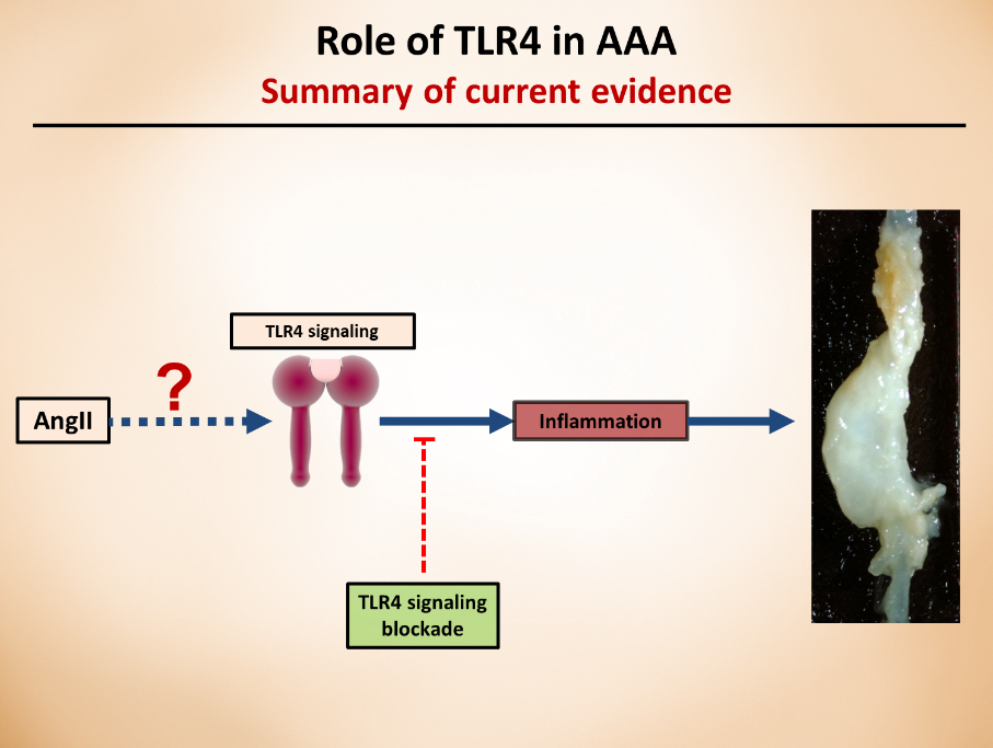
Our laboratory’s previous studies were among the first to show that whole body deficiency of TLR4, a key player in proinflammatory signaling, can ablate the angiotensin II (angII)-induced AAA in low-density lipoprotein receptor deficient (LDLR-/-) mice. Other research endeavors using different mice background or other AAA animal models also further confirmed the critical role of TLR4, or its ligands in AAAs. Although these studies clearly show the role of TLR4 in AAAs, further research is needed to understand which cell types are responsible for TLR4 protective effects and via which mechanisms. As a researcher in this field, my current focus is on targeting aortic cell-specific TLR4 (adventitia), understanding how these cell-specific receptors contribute to the pathology, and the role of potential ligands that activate this cascade of events.

These understandings have the potential to contribute to the development of pharmaceutical approaches that can specifically target the cell receptors and prevent/slow the progression of the pathology.


Resources for Further Reading:

  1. Renin-Angiotensin System and Cardiovascular Functions: Chia-Hua Wu, Shayan Mohammadmoradi, Jeff Z. Chen, Hisashi Sawada, Alan Daugherty and Hong S. Lu, ATVB, 2018


hidden

Abdominal Aortic Aneurysm: How to Study It in Mice

Aortic aneurysms are pathological dilations with high risk of mortality due to rupture. Abdominal aortic aneurysms (AAA) are the most common form of this condition with the dilation being mainly present in the infrarenal region. Histological analysis of human AAA tissue have shed lights on pathological hallmarks of AAAs, however, such tissues are acquired at the advanced stages of the disease and do not provide clear information about the AAA initiation. Therefore, the research has relied extensively on AAA animal models to better define the underlying mechanism of AAA. In general, there are three main mouse models to study AAA: 1) perfusion of elastase into the infrarenal aorta, 2) periaortic application of calcium chloride, or 3) subcutaneous infusion of Angiotensin (Ang) II. The purpose of this blog post is to provide a short description about the AngII-induced AAA.

AngII is the major bioactive peptide of the renin-angiotensin system. The AngII-induced AAA model is probably the most commonly used animal model nowadays, which was developed at the University of Kentucky. In this model, a small osmotic mini pump is subcutaneously implanted in the animal, which will infuse AngII (mainly at the dose of 1,000 ng/kg/min) for 28 days. AngII-induced AAA is augmented by hypercholesterolemia; therefore, ApoE-/- and LDLr-/- mice are the two most common animal models used for AngII-induced AAA studies. AngII also induces AAA in normolipidemic mice, however, at much lower incidence. The AngII-induced AAA formation is independent of the modestly increased systolic blood pressure after AngII infusion and the AAA induced by chronic AngII infusion is mainly located at suprarenal aortic region. This model exhibits progressive luminal expansion, increased leukocytic infiltration, elastin fiber disruption, loss of extracellular matrix, and consequently, aortic wall remodeling – all of which are hallmarks of human AAA.

Although AngII-infused mouse models are very popular due to similarities to human AAA and reproducibility, difficulties in breeding mice to a hypercholesterolemic background has hampered its wide use. However, a recent study from our laboratory provided a rapid approach for increasing plasma cholesterol and Ang II–induced AAA incidence in C57BL/6 mice by applying a gain-of-function mutation of mouse PCSK9 protein using an adeno-associated viral method.

Aortic aneurysms are lethal asymptomatic conditions and thanks to development of animal models, considerable increase in research on aneurysm pathogenesis have shed lights on the undelaying mechanisms of the disease. Despite the progress, there are still many perplexities regarding the AAA initiation and development, and this uncertainty highlights the important of understanding the mouse models to further research this devastating condition.

Resources for Further Reading:

  1. Renin-Angiotensin System and Cardiovascular Functions
    Chia-Hua Wu, Shayan Mohammadmoradi, Jeff Z. Chen, Hisashi Sawada, Alan Daugherty and Hong S. Lu, ATVB, 2018
  2. Hypercholesterolemia Induced by a PCSK9 Gain-of-Function Mutation Augments Angiotensin II–Induced Abdominal Aortic Aneurysms in C57BL/6 Mice—Brief Report
    Hong Lu, Deborah A. Howatt, Anju Balakrishnan, Mark J. Graham, Adam E. Mullick, and Alan Daugherty, ATVB, 2016