hidden

Gut Microbiota Modulation: From Bench to Bedside

In a series of previous blog posts, I delved into the role of the gut microbiome and its contribution to cardiovascular health. As it is almost time to wrap up this year’s blogging series, I thought to provide some final points about this topic.

Large lines of evidence that shows gut microbiota is a major player in host metabolism homeostasis, has led to increased interests in leveraging findings for therapeutic aims in cardiometabolic complications. Here, I propose a framework for modulation of gut microbiota with therapeutic purposes (figure):

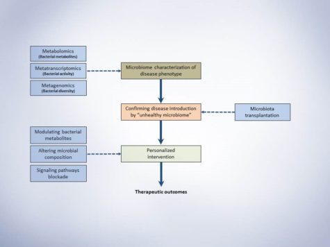
Schematic presentation of microbiota study frame-work. This simple representation suggests three major steps for conducting a microbiome study with the aim of investigating a disease phenotype and possible therapeutic outcome.

1. The first step would be characterizing the microbiome of disease phenotypes illustrating the alterations of specific bacterial taxa and metabolites.

2. Secondly, Koch’s postulation should be fulfilled. In short, the specific taxa should be found in abundance (or indicate specific ratio/levels) in the organism with disease phenotype but not in the healthy phenotype. Secondly, the responsible taxa (or the specific ratio/level of abundance) should be isolatable (or reproducible) and finally, transfer the disease-related taxa (or creating specific ratio/level responsible for disease) to the healthy host microbiome should introduce the disease.

3. After identifying the responsible bacteria or produced metabolites that fulfilled the Koch’s postulation, the third step would be designing an intervention based on the cardiometabolic complication, its progression level and personalization of intervention for each patient. Approaches to therapeutically modulate gut microbiota would be using probiotics, prebiotics, dietary constituents and drugging the microbiome for more specific targeting. Jamming microbiota communication, microbiome programming with modified smart bacteria and the introduction of RNA-guided nuclease CRISPR using bacteriophage carrier are among the new approaches that are starting to form for modulation of the gut microbial endocrine organ. Moreover, fecal microbiota transplantation (FMT) is also among the new approaches in treating metabolic anomalies and recently initiated clinical trial “Fecal microbiota transplant for obesity and metabolism” (ClinicalTrials.gov NCT02530385) is expected to show interesting results in the near future. Still, as mentioned throughout the series of blog posts, our understanding from complex interactions and functions of gut microbiome is in infancy and further animal and human studies are required to shed light on precise microbial targets and prevent the unforeseen consequences of long-term microbial disruption.

Conclusion and Closing Thoughts

Indeed, the community of bacteria residing in the human body was ignored for many years. But, recent evidence started to shape the idea that human’s microbial symbionts play multiple functional roles in maintaining normal metabolic functions. Successful improvement of metabolic syndrome and obesity that was discussed throughout these blog series indicate that future treatments may be, at least partially, based on microbiota interventions. More precise interventions should be developed to address the desired modulatory effect, yet, it raises new challenges since a major portion of gut bacteria is still uncultured. Also, regulatory aspects of current interventions including FMT, probiotics, prebiotics and bacterial metabolite inhibitors should be addressed in more detail since neither formulations development nor quality control guidelines are available. Moreover, it is time to move forward from small cross-sectional studies to more large-scale epidemiological investigations to understand better whether the microbial alterations cause disease development or the complications itself results in such alterations. In order to make the results of small and large microbial studies more clinically implantable, the field needs to generate universal standards for sample collection, data analysis, and sequencing to allow reproducibility and unbiased comparisons between different studies.

Despite all the hurdles in microbiome studies and translation of findings, there has been a bloom in researchers and companies looking for diagnostic and therapeutic strains and approaches to modulate them and track such modulations. The latter is more emphasized by the recent announcement from White House Office of Science and Technology Policy for the launch of United States National Microbiome Initiative (NMI) that aims to foster microbiome studies in different ecosystems. NMI aims to expand the microbiome workforce, develop platform technologies and support research to advance our understanding of microbiome and restoring its healthy function in different complications.

In the end, it is vivid that this novel area of research may impact medicine in the very near future and by addressing the current challenges, incorporated microbiome-based diagnostic and therapeutic protocols into patient care starts to emerge.

Shayan Mohammad Moradi Headshot

Shayan is a caffeine-dependent Ph.D. Candidate at the Saha Cardiovascular Research Center, University of Kentucky. His research area is focused on vascular biology and lipid metabolism. He tweets @MoradiShayan, blogs at shayanmoradi.com and he is the Winner of World’s Best Husband Award (Category: nagging).

hidden

Lipopolysaccharide, TLR4 and Obesity: How They Relate

In my previous blog posts, I started to discuss the importance of toll-like receptor 4 (TLR4) and how it contributes to aortic aneurysms and how microbiota-derived lipopolysaccharide (LPS) can activate the whole signaling pathway. In today’s post, I thought to discuss the TLR4 pathway, in far more details and how it contributes to obesity and metabolic disturbances.

Production of LPS and secretion from intestinal epithelial cells results in LPS binding to cytokine receptors on hepatocytes/adipocytes and as a consequence, activation of a network of signaling pathways1. Upon binding of TRL4 to its co-receptor, myeloid differentiation factor 2 (MD2), a molecular complex is formed at surface level that becomes the binding site of LPS. LPS forms a complex with lipoprotein binding protein (LBP) that binds to cell surface CD142. Upon binding of LPS and its co-receptor CD14, the subsequent transfer of LPS to the TRL4-MD2 complex starts a cascade of events leading to the activation of transcription factors that enhances the expression of many proinflammatory cytokines. TLR4-MD2 complex signals through two major pathways: myeloid differentiation factor 88 (MyD88) and TIR domain containing adaptor-inducing IFNβ (TRIF; also known as TICAM1)2. Upon ligand recognition in the MyD88 dependent pathway, it is recruited to the cytoplasmic domain of TLR. Then, protein families of TNF-α receptor associated factor 6 (TRAF6), IL-1 receptor associated kinase 1 (IRAK1) and IRAK2 are recruited by MyD883. TRAF6 activates the transforming growth factor β-activated kinase 1 (TAK1) which promotes phosphorylation of kappa beta kinase (IKK) inhibitors α, β and γ. Phosphorylated IKK complex leads to the degradation of inhibitory kappa B (IκB) and as a consequence, translocation of NFκB to the nucleus resulting in the induction of proinflammatory cytokines4. Severe reactions to the LPS are attributed to the MyD88 activation pathway resulting in production of IL-12, IL-6 and TNF-α 2. Activation of TRIF (or MyD88 independent) pathway occurs after endocytosis of TLR4-MD2 complex and is characterized by the activation of mitogen activated protein kinases (MAPKs) such as p38, ERK1/2 and c-Jun N-terminal Kinases (JNK). In the independent pathway, the induction of IFNβ and IFN inducible proteins such as monocyte chemoattractant protein 1 (MCP-1 also known as CCL2), IFNγ-induced protein (IP10 also known as CXCL10) and RANTES (also known and CCL5) are triggered5. Cani group’s study in CD14 deficient mice showed that HFD or administration of LPS showed no effect on any parameters of metabolic syndrome symptoms, further suggesting a role for TLR4 in mediating metabolic endotoxemia, adiposity and insulinemia6. Another study confirmed the aforementioned suggestion by showing a less effect on adiposity of TLR4 deficient mice challenged with HFD7. The authors also reported a higher LPS content in the cecal samples in HFD mice compared to LFD, with a close link to TLR4 induction and NFκB activation. The latter induced the expression of iNOS and COX2 while HFD challenged TLR4 deficient mice did not show activation of NFκB and changes in the mRNA levels of proinflammatory cytokines. It is also worthy to mention that the metabolic endotoxemia induced by LPS is associated with insulin resistance by activation of JNK8. This activation has the potential to promote phosphorylation of insulin receptor substrate 1 (IRS-1) at serine sites which may inhibit the normal signal transduction through insulin receptor/IRS-1 axis resulting in insulin resistance9. In addition, activation of signaling cascade induced by LPS-TLR4 increases the expression of inducible nitric oxide synthase10. The latter reacts with cysteine residues to form adducts of S-nitrosothiols which inhibits insulin signal transduction via phosphorylation of IRS-1 in serine leading to insulin resistance in hepatic, muscle and adipose tissue10.

How LPS-derived metabolic endotoxemia results in obesity onset

How LPS-derived metabolic endotoxemia results in obesity onset. A high fat diet can result in a shift in gut microbiota composition which can contribute to increased gut permeability and metabolic endotoxemia. The gut microbiota-derived LPS activates TLRs which produce proinflammatory cytokines that can contribute to onset of obesity. Abbreviations: LPS, lipopolysaccharide; TLR, toll like receptor.

In a recent study on both Myd88-/- and Trif-/- mice by Fredrik Backhed group, authors investigated the effect of lard diet (rich in saturated lipids) on gut microbiota composition compared with fish oil fed (enriched in polyunsaturated fatty acids) mice11. Their result demonstrated that mice lacking MyD88 and TRIF are protected against lard-induced white adipose tissue (WAT) inflammation and metabolic perturbations and the saturated dietary lipids interact with gut microbiota to induce inflammation in WAT. Authors reported that lard fed mice showed an increase in the serum levels of LPS compared to fish oil fed group, indicating that microbial factors may be present in the periphery that may affect WAT inflammation. Moreover, they showed that fish oil diet had increased the levels of taxa from the genera Lactobacillus and Akkermansia. Also, studies on Akkermansia muciniphila have been shown a reduction in fat mass gain and WAT macrophage infiltration alongside improvement of gut barrier function when administered to mice with HFD-induced obesity12. Microbiota transplantation from fish oil fed mice into antibiotic treated mice also showed an increase in the levels of Akkermansia with partial protection against adiposity and inflammation after 3 weeks of lard diet11.

Additionally, Vijay-Kumar and his colleagues have shown that TLR5-deficinet C57Bl/6J mice exhibit hyperphagia and develop characteristics of metabolic syndrome such as increased adiposity, insulin resistance and hyperlipidemia13. They reported that loss of TLR5 and the observed metabolic changes correlated with microbiota compositional changes and induction of inflammatory signaling. TLR5 is a component of innate immune system that is expressed in the gut mucosa and flagellated bacteria can interact with TLR5 to induce activation of pro-inflammatory gene programs for host protection14.

Taken all together, current data suggest that intestinal inflammation could be the early consequence of HFD and may induce obesity via increased levels of LPS, suggesting a causative role for gut inflammation in the onset of obesity. In addition, TLR4 is the primary receptor mediating the proinflammatory effects of LPS, therefore regulating levels of LPS and/or ligand binding capacity of TLR4 may be a target to stop progression of obesity and metabolic syndrome.

Shayan Mohammad Moradi Headshot

Shayan is a caffeine-dependent Ph.D. Candidate at the Saha Cardiovascular Research Center, University of Kentucky. His research area is focused on vascular biology and lipid metabolism. He tweets @MoradiShayan, blogs at shayanmoradi.com and he is the Winner of World’s Best Husband Award (Category: nagging).

References:

  1. Park DY, Ahn YT, Park SH, Huh CS, Yoo SR, Yu R, Sung MK, McGregor RA, Choi MS. Supplementation of lactobacillus curvatus hy7601 and lactobacillus plantarum ky1032 in diet-induced obese mice is associated with gut microbial changes and reduction in obesity. PLoS One. 2013;8:e59470
  2. Needham BD, Trent MS. Fortifying the barrier: The impact of lipid a remodelling on bacterial pathogenesis. Nat Rev Micro. 2013;11:467-481
  3. Akashi-Takamura S, Miyake K. Tlr accessory molecules. Current Opinion in Immunology. 2008;20:420-425
  4. Kawai T, Akira S. Tlr signaling. Cell Death Differ. 2006;13:816-825
  5. Albiger B, Dahlberg S, Henriques-Normark B, Normark S. Role of the innate immune system in host defence against bacterial infections: Focus on the toll-like receptors. Journal of Internal Medicine. 2007;261:511-528
  6. Cani PD, Amar J, Iglesias MA, Poggi M, Knauf C, Bastelica D, Neyrinck AM, Fava F, Tuohy KM, Chabo C, Waget A, Delmée E, Cousin B, Sulpice T, Chamontin B, Ferrières J, Tanti J-F, Gibson GR, Casteilla L, Delzenne NM, Alessi MC, Burcelin R. Metabolic endotoxemia initiates obesity and insulin resistance. Diabetes. 2007;56:1761-177
  7. Kim K-A, Gu W, Lee I-A, Joh E-H, Kim D-H. High fat diet-induced gut microbiota exacerbates inflammation and obesity in mice via the tlr4 signaling pathway. PLoS ONE. 2012;7:e47713
  8. Khan Muhammad T, Nieuwdorp M, Bäckhed F. Microbial modulation of insulin sensitivity. Cell Metabolism. 2014;20:753-760
  9. Shoelson SE, Lee J, Goldfine AB. Inflammation and insulin resistance. Journal of Clinical Investigation. 2006;116:1793-1801
  10. Sugita H, Kaneki M, Tokunaga E, Sugita M, Koike C, Yasuhara S, Tompkins RG, Martyn JAJ. Inducible nitric oxide synthase plays a role in lps-induced hyperglycemia and insulin resistance. American Journal of Physiology – Endocrinology and Metabolism. 2002;282:E386-E394
  11. Caesar R, Tremaroli V, Kovatcheva-Datchary P, Cani Patrice D, Bäckhed F. Crosstalk between gut microbiota and dietary lipids aggravates wat inflammation through tlr signaling. Cell Metabolism. 2015;22:658-668
  12. Everard A, Belzer C, Geurts L, Ouwerkerk JP, Druart C, Bindels LB, Guiot Y, Derrien M, Muccioli GG, Delzenne NM, de Vos WM, Cani PD. Cross-talk between akkermansia muciniphila and intestinal epithelium controls diet-induced obesity. Proceedings of the National Academy of Sciences. 2013;110:9066-9071
  13. Vijay-Kumar M, Aitken JD, Carvalho FA, Cullender TC, Mwangi S, Srinivasan S, Sitaraman SV, Knight R, Ley RE, Gewirtz AT. Metabolic syndrome and altered gut microbiota in mice lacking toll-like receptor 5. Science. 2010;328:228-231
  14. Vijay-Kumar M, Sanders CJ, Taylor RT, Kumar A, Aitken JD, Sitaraman SV, Neish AS, Uematsu S, Akira S, Williams IR. Deletion of tlr5 results in spontaneous colitis in mice. J Clin Invest. 2007;117
hidden

Microbiota Alterations In Obesity And Sister Complications

In my previous blog post, I briefly discussed the importance of microbiome research and how to take the first steps in conducting a microbiome study. In today’s post, I will continue to discuss the importance of this area of research with a focus on obesity.

The worldwide epidemic of metabolic syndrome and obesity in different age groups both in the United States and around the world is considered as a major public health concern. Moreover, the human gut microbiome has been linked to metabolic disease and adiposity and it is not only a marker of disease, but also contributes to pathology.

Intestinal microbiota plays a critical role in the host metabolism and immune system that extents its related physiological functions to other organs including brains, liver and adipose tissue. Metagenomic-wide association studies indicate significant changes between gut microbiota metagenome of metabolically healthy versus unhealthy individuals. Such microbiota changes are thought to be a possible cause of obesity and therefore, intestinal microbiota represents a potential therapeutic target to manage obesity.

Overview of gut microbiota role in host metabolism

Overview of gut microbiota role in host metabolism. The shift in gut bacteria can affect host metabolism via several pathways in different tissues.

Study results have illustrated alterations in the dominant gut phyla of obese subjects/animals, reporting significant reduction in Bacteroidetes and significant increase in Firmicuts and Actinobacteria. The consequence of this shift in gut microbiota is the increased potential of harvesting energy from food and a low-level inflammation. Obesity leads to a low level inflammation, specifically in adipose tissue, that results in the production of several inflammatory cytokines, which may lead to insulin resistance as well. TNF-α, IL-1β and CCL2/MCP1 are among the important inflammatory cytokines that are induced in obese state accompanied by increased macrophages, T cells and mast cells. Presence of the aforementioned cells not only correlates with the gene expressions that control inflammation, but also indicates the possible role of innate immunity in obesity and insulin resistance. Moreover, Pattern recognition receptors such as Toll-like receptors (TRLs) are activated by bacterial endotoxins such as lipopolysaccharide (LPS), which results in innate immune response and inflammation. Also, gut microbiota produce wide range of molecules, such as flagellins and peptidoglycans, which activate inflammatory pathways leading to obesity and insulin resistance. Recent data from mice genetically deficient in TLR5 reported significant changes in their microbiota and development of metabolic syndrome characteristics. Results from germ free mice also illustrated possible effects of gut microbiota on host metabolism. High fat – high sugar diet fed mice did not show same metabolic disturbance in comparison with not germ free littermates. Microbiota transplantation from obese mice also resulted in greater adiposity in comparison with lean donor recipients. It is also suggested that short chain fatty acids (SCFAs) may contribute to regulation of gut dysbiosis. These compounds such as butyrate, acetate and propionate are produced by intestinal microbiota as a result of diet-derived fibers fermentation. SCFAs are thought to be the energy source for intestinal epithelium and liver, whereas they can also play a modulatory role in immune response via reducing gut permeability.

Shayan Mohammad Moradi Headshot

Shayan is a caffeine-dependent Ph.D. Candidate at the Saha Cardiovascular Research Center, University of Kentucky. His research area is focused on vascular biology and lipid metabolism. He tweets @MoradiShayan, blogs at shayanmoradi.com and he is the Winner of World’s Best Husband Award (Category: nagging).