hidden

“Run, Forrest, Run!” – Effects of Cardiovascular Exercising on Mental Health

running

Source:pixabay

If life gave me a box of chocolates, I am pretty sure I would eat them all. As a basic science researcher, I am all too familiar with burn-outs and stress, and more recently to the effects of stress-induced anxiety. So, I started running. I ran when my worries got too overwhelming, I ran when I had a bad day in the lab. Next thing I knew, I was running for pleasure. And this is something I did not see coming, especially because I hated running before!

Long before medications were available or even prescribed for mental or emotional disorders, exercising remained the only prescription for tacking problems of mental health by doctors.1 The AHA recommends 150 minutes of moderate-high intensity aerobic exercising a week for adults. Researchers have found this to improve balance of neurotransmitters and show effects as early as the first thirty minutes.

Here are some evidence-based effects of exercising on mental health –

  1. Stress and anxiety – Chronic stress can shrivel parts of the brain. Exercises have found to reverse this effect and even induce growth of neurons and improve synaptic plasticity in the brain.1,2
  2. Depression – Regular aerobic exercising can improve blood circulation to the brain and it is known to positively influence hypothalamus-pituitary-adrenal axis of the brain. All of this bounce back the balance of neurotransmitters, elevate mood, attenuate stress and fight back fear.3
  3. Addiction – While dealing with addiction, exercising has been shown to give a sense of control. Individuals with a tendency to be obsessive, need to fill a void quickly and exercising has shown to be effective in this regard.
  4. Hormonal fluctuations in women – Hormones estrogen and progesterone play an important role for neurotransmitter in the brain, by providing receptors for them to bind. In some women, this complex pathway can behave in a way that increases aggressive behavior which is found to be dramatically reduced by exercise. Exercising is certainly known to increase levels of tryptophan, the precursor to the happy chemical serotonin. This helps in dealing with the constant fluctuations of hormones during a cycle.1
  5. Attention deficits – Aerobic exercising has been successful with disorders of distractions, improving focus, concentration and memory of a task.

As with any given task, starting it – is always a challenge. If you are someone like me who is a novice at running, or the thought of running itself induces fear and anxiety – then start with small steps. Take a walk, slowly increase that to brisk walking and when you feel comfortable start jogging. It doesn’t necessarily even need to be running – jumping rope, biking, throwing ball – any activity that spikes your heart rate are good.

So, will you run away from your problems?


References:

  1. John Ratey, Spark: The Revolutionary New Science of Exercise and the Brain, ISBN:0316028355
  2. Carek PJ, Laibstain SE, Carek SM, Exercise for the treatment of depression and anxiety. Int J Psychiatry Med. 2011;41(1):15-28.
  3. Toups M, Carmody T, Greer T, Rethorst C, Grannemann B, and Trivedi MH. Exercise is an effective treatment for positive valence symptoms in major depression. J Affect Disord 2017; 209: pp. 188-194


hidden

5 Things To Consider Before Choosing Your Dissertation Topic

As I am inching towards, what I hope would be my final year of PhD research, I have been thinking and analyzing a lot of my actions in retrospect. I thought of putting together a list of things I learned and things I wish I had considered in my first year.


1) Finding the “right” mentor.

We spend a lot of time in deciding the right lab or the best PhD supervisor. A lot has been said and done about finding the right fit. One thing I have learned is that apart from the usual parameters we set in finding the best supervisor for us personally, sometimes we forget to consider if the supervisor is right for the project. Sometimes the project may expand in an area beyond your and your mentor’s expertise. In such cases, it is important to consider whether your mentor will make the right resources available to you. Putting together a good research advisory committee, scientists who would have expertise in that specific topic, will come in handy. Research can be quite daunting and grad students deal with intense pressure and stress on a daily basis. Your time should be spent researching and not trying to find the right instrument in the cheapest core facility and definitely NOT YouTubing the workings of a new technique. Make sure to find someone to train you, attend workshops, shadow a technician and make sure your supervisor makes these available to you when needed. A mark of a good mentor is when they don’t hesitate to seek consultation or advise from an external or senior scientist who is an expert in the field.


2) Is this a good career investment?

Turns out most students forget about the crucial thing about spending years in grad school – landing the job! Most of us don’t think about job search or the next move until our final year, which I think is too late. While choosing a topic, you may want to consider things like job market, skill requirement, funding agencies and so on. For example, researching therapeutic drug targets for a disease that has no cure is far better than investing in a project discovering drug targets for a disease with multiple FDA approved drugs. Weigh the pros and cons carefully. Will your project help you acquire technical skills that are translatable to the industry? If you live in a city or country that is in dire need of science policy advisors or climate crisis advocates or good science communicators, will your PhD program give you enough skills to apply to these jobs?


3) Is there scope for collaborations?

Collaborations are a unique way to expand into different research topics in your field, whether it’s a collaboration within your group or research with a different research group altogether. This lets you become more versatile, get a flavor of how other researchers approach their science and if nothing else, learn a new scientific topic up-close. A productive collaboration is one which will take your expertise and enhance another project, without taking too much time away from your project. Inter-lab collaborations are a great way to demonstrate your negotiating, team management and interpersonal skills. Oh, and did I mention it’s good for networking? So finding aims in your proposal early on, that are good for teaming up with other groups is a good idea, especially while conferencing.


4) Will it help you AND your science grow?

I will start with the science part first. Obviously, we all want to learn and become an expert in the respective field when we started off, so what do I mean by growth here? If your research topic is only going to be a repeat of your previous techniques and scientific concepts, chances are, halfway through the project, you’re going to lose interest. It is great to start off the project with something familiar, but if it isn’t exploring in areas that are uncomfortable and challenging to you, is it really worth a PhD? Test new ideas, push your boundaries and give yourself a deadline to fully delve into answering these questions. But be wary not to spend too much time and get distracted. It is good to spend the first two years (in a five-year program) to be adventurous, but if it gets too challenging it really should not be pursued at the expense of your time.

I stress on personal growth next. PhD project is a LOT of time commitment. Especially to one very specific thing, that more often than not, will consume most, if not all of your time. This means one must consider having room for co-curricular activities that will in turn be an asset for your own research project. For example, I love to read about popular science, wildlife, climate science, conservation, policies and history. My program had a structured graduate minor alongside my PhD major and I decided to study science communication for this minor. Now, I get to write, read or watch other popular forms of science, engage with community, organize local events and dissect science policies as part of my curriculum. I have also gotten opportunities where I talked about my own research to strangers and thus, honed in my craft of communicating science. All of this will ultimately reflect in your resume and you know that apart from spending long hours fine-tuning your experiments, you will leave with heaps of useful skills for future jobs. So, I would recommend finding things that compliment your science early on, this will go a long way!


5) Will you need a backup plan?

If you are diving into something extremely challenging, let’s say it will not only require you to learn new, field-specific techniques, but it will mean questioning the dogma – make sure you have another small project to safely rely back on. If your program has at least one first-author paper requirement for dissertation, it is imperative you sit with your supervisor and make sure you will get a paper out in time. No dogma is worth challenging at the cost of your degree!


These are some pointers that I thought of, from personal experience. I hope that you will find it useful and informative.

hidden

Can A Defect In Your Heart Leave You Blind?

You must have heard about a congenital heart complication, where babies are born with a hole in their hearts or bluish-purple discoloration of tissues. Nearly 10-12 in every 1000 babies in the US are born with a congenital birth defect, most of them needing immediate medical attention. Another complication of this defect is retinopathy which can progress into severe blindness.

Tetralogy of fallot (TOF), is a heart disease where lack of oxygen led obstruction to blood flow, causes neonates to varying intensities of discoloration. In case of retinopathy prematurity, prematurely born babies are found to have unusual blood vessel development in the back of the eye. This usually happens due to high oxygen levels that babies are exposed to in the incubator, that aberrantly gives cues for insufficient in some areas and overgrowth of the blood vessels in other areas of the retina. This form of retinopathy can progress to irreversible blindness, as therapy is quite limited for patients currently.

A few cases have recently been reported where patients diagnosed with TOF showed symptoms of retinopathy and some ocular abnormalities. A pair of sisters with a history of repaired TOF showed signs of retinal abnormalities as early as two years of age.1 In another case study, even adults aged between 27 and 47 years, with a history of similar heart defects showed abnormalities in retinal blood vessels.2

Given very few case reports published so far, it is difficult to conclude a possible correlation. The only possible reason given for development of these symptoms is ischemia or lack of oxygen to the tissues, a plausible area that needs to be chalked out further. Clinicians and researchers need to investigate the incidence rate and such patients should be monitored with follow-up ocular exams.


References:

  1. Zanolli et al., Unusual retinal abnormalities in sisters with tetralogy of Fallot. Journal of AAPOS, vol. 18, no. 6, pp. 601–604, 2014.
  2. Tsui et al., Retinal vascular patterns in adults with cyanotic congenital heart disease. Seminars in Ophthalmology, vol. 24, no. 6, pp. 262–265, 2009.
hidden

Circadian Clock and Retina

Shift workers, especially those that work all night and sleep during the day, have long since been associated with many cardiovascular diseases like hypertension, diabetes, obesity and even sleep disorders.1 Many of the associated risks arise from a disrupted circadian rhythm and our well-oiled biological clocks. But, our disturbed circadian cycles also affect our vision and make us susceptible to ocular diseases. In my previous blogs, I detailed how retinal signs are early indicators of hypertension, stroke and cardiovascular disorders. Here, I decided to highlight some recent findings that connected vascular complications to our lifestyle choices, vis à vis circadian rhythm.

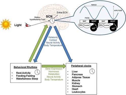
One of the most photosensitive cells in the retina are the rods and the cones, receptors of light, because of which the eye is also under regulation by the circadian clock – day and night. The master regulator of this clock is in the brain called suprachiasmatic nuclei (SCN), that operate after receiving light through the retina. At the molecular level, not surprisingly, researchers have observed different clock regulating proteins across different cell types throughout the retina. Although research is currently underway and it is still not clearly understood, how the retina is affected by disruptions in the light/dark cycle.

Source: Covassin et al., Hypertension. 2016;68:1081–1090

Source: Covassin et al., Hypertension. 2016;68:1081–1090

The figure illustrates the control of SCN by light and how this at the molecular level affects clock protein genes like CLOCK, BMAL and in turn affects peripheral and behavioral patterns.2

One study tried to look at whether one of the clock proteins that was associated with diabetes, had similar effects in the retina. They saw in their animal model, changes in the retina as well as bone marrow, comparable to those seen in diabetic animals.3 Not only this, advanced form of diabetic retinopathy, that is usually caused by abnormally growing blood vessels in the retina, show a reduced expression of clock protein genes in experimental models.4 Another study reported significant changes in the cones of mice with dysfunctional clock gene.5

Many research studies and review articles have documented the effects of disrupted circadian cycles in research settings and shown a correlation. However, direct evidence or causation factors is still under-studied in human patients – calling attention to a gap that beckons researchers to fill this void.


References:

  1. Covassin et al., Keeping up with the clock. Hypertension. 2016;68:1081–1090
  2. Liu et al., Heterogeneous expression of the core circadian clock proteins among neuronal cell types in mouse retina. PLoS One. 2012;7(11):e50602
  3. Bhatwadekar et al., Per2Mutation Recapitulates the Vascular Phenotype of Diabetes in the Retina and Bone Marrow. Diabetes. 2013 Jan; 62(1): 273–282.
  4. Busik et al., Diabetic retinopathy is associated with bone marrow neuropathy and a depressed peripheral clock. Exp Med. 2009 Dec 21; 206(13):2897-906.
  5. Ait-Hmyed et al., Mice lacking Period 1 and Period 2 circadian clock genes exhibit blue cone photoreceptor defects. Eur J Neurosci. 2013 Apr; 37(7):1048-60.


hidden

Under Pressure: What Does Retina Say About Hypertension?

An eye oftentimes feels like the most underappreciated systems in the field of vascular biology. An eye is a highly vascular organ then it gets credit for and here’s why – ranging from high blood pressure or diabetes to early signs of stroke, an eye exam can, in fact, tell a physician a lot about one’s health. In a series of blog posts, I decided to highlight these key connections between the eye and the human body. This article will focus on the current knowledge linking eye and hypertension.


Hypertension or high blood pressure is predominantly caused due to increased resistance to the walls of the blood vessels. What this leads to is increased chances of developing diseases of the cerebral, cardiovascular or even peripheral arteries. Risk factors can range from dietary habits to genetics and ethnicity, and less than half of those with hypertension are unaware of their condition. Interestingly, the eye offers a very useful set-up to get a closer look at blood vessels – without even having to inject or cut open anything. This non-invasiveness of the eye has been widely used by clinicians and researchers to diagnose diseases of the blood vessels – hypertension being one of them. This article highlights some interesting findings that researchers derived simply by examining the retinal blood vessels.

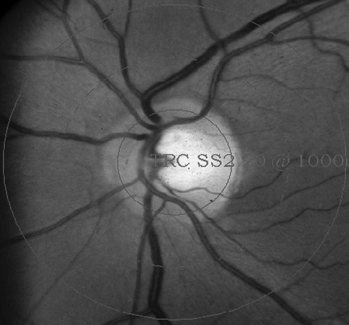
A fundoscopic photograph of the back of the eye (like seen in the image below), allows to capture the retinal blood vessels. These blood vessels share many physiological and anatomical similarities with vessels in other systems, like the brain and the heart. Naturally, any changes in the structure or integrity of these vessels have been documented and researchers have found many links and associations with the pathology of hypertension5. I previously discussed how the retinal vessels gave a sneak peek into the brain and heart, where dimensions like the diameter or tortuosity were able to indicate early signs of stroke or cardiovascular diseases.

Source: Cheung et al., Hypertension. 2012;60:1094–1103


As early as the 1960s, scientists learned that narrowing of retinal arteries were important signs of hypertension. The population-based Rotterdam study published in 2005 looked at individuals in over 55 years of age and were “pre-hypertensive.” Their findings suggested that the narrowing of both retinal arterioles and venules were associated with increased risk of hypertension and preceded development of high blood pressure2. Similarly, the Blue Mountains Eye study in Sydney found that these abnormalities in the retinal vessels predicted a 5-year incidence of severe hypertension in a patient population of older cohort3.

Source: M. Kamran Ikram et al., Hypertension. 2005;47:189–194 

This image of an eye fundus shows a semi-automated system used to measure the diameters of arterioles and venules in the retina.


Making use of this unique retinal fundoscopic tool, another group explored measurement of blood flow to the retina, in response to light-flicker in patients with high blood pressure3. They found that hypertensive patients had impaired blood flow in the retina, possibly caused due to prolonged constricted vessels. This approach is among the first to test blood flow to the retinal, instead of measuring the vessel itself – adding another asset to retinal fundus images.

Retinal images have also been used in genetic linkage studies. Large population data sets are analyzed for tracing genes and variations of the genes associated with diseases among different individuals. It is clear that changes in the diameter of retinal vessels can precede hypertension, but are there genetic predeterminants to an individual’s retinal diameters? In 2006, the Beaver Dam Study found that apart from genetic linkages found between retinal diameters and hypertension and other associated diseases, there are genetic factors that predetermine the retinal diameters – independent of hypertension4.

This simply means that there are other factors present in our systems that are genetically related to the structure and size of one’s retinal vessels. Interestingly, another research group looked retinal vessels of 6-year-old students with hypertensive parents6. They found that only the girls (not boys) had narrowing retinal vessels and were predisposed to developing hypertension later in life. This also suggests a genetic link between retinal vessels and blood pressure.

Researchers around the world have used retinal parameters as indicators of hypertension. Evidently, retinal imaging provides for a powerful tool in identifying markers of cardiovascular complications. However, this still remains a tool widely used only among researchers, and validation of retinal imaging for clinical use still remains to be seen. With emerging advanced technology, clinicians should consider a non-invasive method like this one as a diagnostic tool.


References:

  1. M. Kamran Ikram et al., Retinal Vessel Diameters and Risk of Hypertension. Hypertension. 2005;47:189–194
  2. Smith et al., Retinal Arteriolar Narrowing Is Associated With 5-Year Incident Severe Hypertension. Hypertension. 2004;44:442–447
  3. Ritt et al., Impaired Increase of Retinal Capillary Blood Flow to Flicker Light Exposure in Arterial Hypertension. Hypertension. 2012;60:871–876.
  4. Xing et al., Genome-Wide Linkage Study of Retinal Vessel Diameters in the Beaver Dam Eye Study. Hypertension. 2006;47:797–802
  5. Cheung et al., Retinal Microvasculature as a Model to Study the Manifestations of Hypertension. Hypertension. 2012;60:1094–1103.
  6. Gopinath et al., Parental History of Hypertension Is Associated With Narrower Retinal Arteriolar Caliber in Young Girls. Hypertension. 2011;58:425–430.
hidden

Eye and the Brain

An eye oftentimes feels like the most underappreciated systems in the field of vascular biology. An eye is a highly vascular organ than it gets credit for, and here’s why – ranging from high blood pressure or diabetes to early signs of stroke, an eye exam can, in fact, tell a physician a lot about one’s health. In a series of blog posts, I decided to highlight these key connections between the eye and the human body. This article will focus on the current knowledge linking eye and the brain.


One of the common cerebral diseases caused by blood vessels is stroke. Stroke, typically caused by a blood clot, can either be a chronic or acute ischemic complication. This eventually leads to loss of function in the brain and hemorrhaging (bleeding) in the related part of the brain, retina, or spinal cord. Many risk factors shared by cardiovascular diseases converge with risk factors for stroke. For example – hypertension, diabetes, and alcohol consumption can all increase incidence of stroke related problems.

In my previous blog, I explained how retinal microvessels can be a window to the vessels in the body. Dimensions like caliber (diameter) of retinal vessels can be measured noninvasively, and how they strongly correlated with cardiovascular risks. This article will focus on how, this is also resonant in case of vessels of the brain (cerebrovascular). Physiologically and anatomically, vessels in the retina and the brain share many similarities. Most commonly known would be the blood-brain barrier and the retinal-blood barrier, maintained by tight junctions that are essentially bouncers found outside a club. Even with advanced imaging technologies, capturing these neurovessels can be very challenging. Serendipitously, retinal photographs can thus be used to provide information like signs of stroke or predictors of stroke or dementia, as detailed in this article. Retinal fundus photograph of a patient (as seen in picture) shows embolism or blockage caused by cholesterol-like deposits, and are strongly associated with increased risk of stroke-related death as detailed in a study.

(source: https://doi.org/10.1161/STROKEAHA.107.496091)

One study, specifically the measurements of retinal vessels and compared its association with acute ischemic stroke in a population study. Even the subtlest changes in the retinal vessels of stroke patients were reported, which stood out on comparison with healthy patients. Interestingly, changes in dimensions of retinal vessels were also found in patients with dysfunction in cognitive behavior, suggesting similar tests can be used to diagnosing diseases like dementia and Alzheimer’s (more on this in future posts). Another common indicator between stroke and the retina was found in the retinal nerve fiber layer. Although this is slightly different than comparing the blood vessels, a research group found strong correlation between defects in the nerve fiber layer and acute stroke.

In conclusion, many research groups are now considering using tools to assess retinal vessels for diagnosis of cerebrovascular diseases. More studies in this area, can then suggest a very powerful and noninvasive diagnostic method, which could help both the patient population and the clinicians.


hidden

Eye: A Window To The Heart

An eye oftentimes feels like the most underappreciated systems in the field of vascular biology. An eye is a highly vascular organ then it gets credit for and here’s why – ranging from high blood pressure or diabetes to early signs of stroke, an eye exam can tell a physician a lot about one’s health.

In a series of blog posts, I decided to highlight these key connections between the eye and the human body. This article will focus on the current knowledge linking eye and the heart.

There are quite a few similarities between the vasculature (or simply put, blood vessels) of the eye and heart. Not only are there functional and structural similarities, but the eye and heart also share many of the common risk factors. For example, risk factors like high cholesterol, high glucose, hypertension that contribute to atherosclerosis, can also lead to eye diseases like macular degeneration and retinal vein occlusions. Photo taken of the back of the eye, that clinicians refer to as a fundus image, lets ophthalmologists look at blood vessels directly (as shown in the picture) – an eye is possibly the only organ that gives one an all-access backstage pass to its performances. Researchers and clinicians, rightfully call the eye as the window to one’s heart (this could probably apply beyond just biomedical sciences!).

Changes in the small micro-vessels of the eye can be directly correlated to underlying cardiovascular disorders. In the late 1970’s, clinical researchers learned that atherosclerotic lesions in the retinal vessels were indications of coronary artery disease and this was found by simple observation of fundoscopic images of patients. It is also possible to measure vessel dynamics like tortuosity (twists and turns) and caliber (diameter) with retinal exams coupled with flicker-light. With advanced imaging techniques, researchers are also able to calculate small changes in the microcirculation by simply imaging the retinal vessels. An interesting study performed in twin children, measuring the retinal arteriole, was able to predict signs of myocardial infarction as well.

The non-invasiveness of imaging the retinal vessels can certainly be an appeal to clinicians who otherwise rely on angiography to diagnose coronary complications. The retinal vessels can be quite information rich, but one only needs to look closely.


hidden

Live Streaming Into Scientific Sessions 2018

AHA Scientific Sessions 2018 was a unique experience for me – unable to attend the meeting, I live-streamed the sessions (first time ever for a conference!). Two of my most favorite sessions this year were the panel discussion for advanced heart failure (HF) patients, “The Metabolic Face of Heart Failure,” and the mini-symposium on “Cutting Edge in Cardiovascular Science.”

One of the main highlights in the session Metabolic Face of HF, moderated by Dr Lynne Stevenson, was the talk by cardiovascular stalwart Dr. E. Braunwald, Brigham and Women’s Hospital. Dr. Braunwald spoke of the significance and latest practices in the use of Sodium-Glucose Cotransporter-2 (SGLT2) inhibitors, a class of FDA-approved drugs for type-2 diabetes. He indicated how SGLT2 inhibitors should be explored beyond diabetes treatment and these class of drugs can benefit HF patients as well. “I had to learn about blood clotting 30 years ago, which was difficult,” he modestly admitted as he clarified the renal effects of SGLT2 inhibition. His views also seemed to resonate with Dr. Subodh Verma, St. Michael’s Hospital, Toronto and Dr. John McMurray, Glasglow University, as they covered SGLT2 inhibitors in HF, as well.

Other speakers at this session, Dr. Neha Pagidipati, Duke University and Dr. Lewandowski, Ohio State University, touched upon aspects of stroke and metabolism regulating HF, respectively. While Dr. Pagidipati compared the risk of cardiovascular diseases and stroke with the risks of diabetes, Dr. Lewandowski explained how metabolic regulator PPAR-a (transcriber of genes in fat metabolism) could be a player explored in targeted therapy.

The session ‘Cutting Edge in Cardiovascular Science’ had presenters covering diverse strategies in dealing with cardiovascular therapy, ranging from computational screening to identifying small molecule compounds, to decoding neurovascular networks and the gut microbiome. Dr. Stanley Hazen from Cleveland Clinic presented his work on understanding the microbes in the gut and their role in driving cardiovascular diseases. Dr. Hazen explained how food like red meat, which are rich in components like phosphatidyl serine, activates the gut microbiome. He described the significance of trimethylamine N-oxide (TMAO) pathway in liver and its association with HF, stroke and cardiovascular diseases. He also strategized the use of enzyme in TMAO pathway as targets of small molecule inhibitors.

Dr. Joseph Loscalzo, Brigham and Women’s Hospital, explained how repurposing drugs and finding drug targets computationally could help precision medicine vastly. He also offered his expertise and tools as open access to AHA members. Finally, Dr. Costantino Iadecola, Cornell, elaborated on the heart-brain connectome. He brought attention to the fact that dementia, known to cause hardening of arteries, led to Alzheimer’s, but we all forgot about the vascular complications of this. He bridged this connection between neurovascular dysfunction and cognitive impairment and went on to explain his research on the intake of high salt in diet caused dementia in mice models. To learn of such versatile range of topics in a session was illuminating, to say the least!

Researchers must spend time thinking about applications of their current projects beyond their own niche – this is the only way we can widen our horizons with existing tools.