Under Pressure: What Does Retina Say About Hypertension?
An eye oftentimes feels like the most underappreciated systems in the field of vascular biology. An eye is a highly vascular organ then it gets credit for and here’s why – ranging from high blood pressure or diabetes to early signs of stroke, an eye exam can, in fact, tell a physician a lot about one’s health. In a series of blog posts, I decided to highlight these key connections between the eye and the human body. This article will focus on the current knowledge linking eye and hypertension.
Hypertension or high blood pressure is predominantly caused due to increased resistance to the walls of the blood vessels. What this leads to is increased chances of developing diseases of the cerebral, cardiovascular or even peripheral arteries. Risk factors can range from dietary habits to genetics and ethnicity, and less than half of those with hypertension are unaware of their condition. Interestingly, the eye offers a very useful set-up to get a closer look at blood vessels – without even having to inject or cut open anything. This non-invasiveness of the eye has been widely used by clinicians and researchers to diagnose diseases of the blood vessels – hypertension being one of them. This article highlights some interesting findings that researchers derived simply by examining the retinal blood vessels.
A fundoscopic photograph of the back of the eye (like seen in the image below), allows to capture the retinal blood vessels. These blood vessels share many physiological and anatomical similarities with vessels in other systems, like the brain and the heart. Naturally, any changes in the structure or integrity of these vessels have been documented and researchers have found many links and associations with the pathology of hypertension5. I previously discussed how the retinal vessels gave a sneak peek into the brain and heart, where dimensions like the diameter or tortuosity were able to indicate early signs of stroke or cardiovascular diseases.
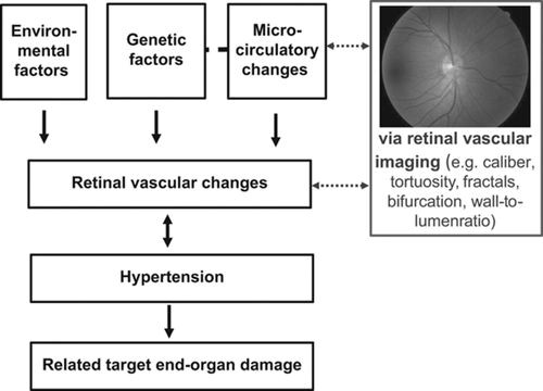

Source: Cheung et al., Hypertension. 2012;60:1094–1103
As early as the 1960s, scientists learned that narrowing of retinal arteries were important signs of hypertension. The population-based Rotterdam study published in 2005 looked at individuals in over 55 years of age and were “pre-hypertensive.” Their findings suggested that the narrowing of both retinal arterioles and venules were associated with increased risk of hypertension and preceded development of high blood pressure2. Similarly, the Blue Mountains Eye study in Sydney found that these abnormalities in the retinal vessels predicted a 5-year incidence of severe hypertension in a patient population of older cohort3.
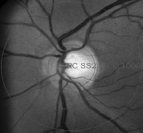
Source: M. Kamran Ikram et al., Hypertension. 2005;47:189–194
This image of an eye fundus shows a semi-automated system used to measure the diameters of arterioles and venules in the retina.
Making use of this unique retinal fundoscopic tool, another group explored measurement of blood flow to the retina, in response to light-flicker in patients with high blood pressure3. They found that hypertensive patients had impaired blood flow in the retina, possibly caused due to prolonged constricted vessels. This approach is among the first to test blood flow to the retinal, instead of measuring the vessel itself – adding another asset to retinal fundus images.
Retinal images have also been used in genetic linkage studies. Large population data sets are analyzed for tracing genes and variations of the genes associated with diseases among different individuals. It is clear that changes in the diameter of retinal vessels can precede hypertension, but are there genetic predeterminants to an individual’s retinal diameters? In 2006, the Beaver Dam Study found that apart from genetic linkages found between retinal diameters and hypertension and other associated diseases, there are genetic factors that predetermine the retinal diameters – independent of hypertension4.
This simply means that there are other factors present in our systems that are genetically related to the structure and size of one’s retinal vessels. Interestingly, another research group looked retinal vessels of 6-year-old students with hypertensive parents6. They found that only the girls (not boys) had narrowing retinal vessels and were predisposed to developing hypertension later in life. This also suggests a genetic link between retinal vessels and blood pressure.
Researchers around the world have used retinal parameters as indicators of hypertension. Evidently, retinal imaging provides for a powerful tool in identifying markers of cardiovascular complications. However, this still remains a tool widely used only among researchers, and validation of retinal imaging for clinical use still remains to be seen. With emerging advanced technology, clinicians should consider a non-invasive method like this one as a diagnostic tool.
References:
- M. Kamran Ikram et al., Retinal Vessel Diameters and Risk of Hypertension. Hypertension. 2005;47:189–194
- Smith et al., Retinal Arteriolar Narrowing Is Associated With 5-Year Incident Severe Hypertension. Hypertension. 2004;44:442–447
- Ritt et al., Impaired Increase of Retinal Capillary Blood Flow to Flicker Light Exposure in Arterial Hypertension. Hypertension. 2012;60:871–876.
- Xing et al., Genome-Wide Linkage Study of Retinal Vessel Diameters in the Beaver Dam Eye Study. Hypertension. 2006;47:797–802
- Cheung et al., Retinal Microvasculature as a Model to Study the Manifestations of Hypertension. Hypertension. 2012;60:1094–1103.
- Gopinath et al., Parental History of Hypertension Is Associated With Narrower Retinal Arteriolar Caliber in Young Girls. Hypertension. 2011;58:425–430.