hidden

Fellow Focus: Peer Mentorship Program

During my first year of general cardiology fellowship, our program underwent an exciting transition – our incoming fellowship class increased from 6 fellows the previous year to 10 fellows in my class, nearly doubling the size of the fellowship. This growth was necessitated by the welcome addition of the West LA VA as a rotation site for house staff, which meant spreading our fellowship across 4 different clinical sites. While this gives our fellows the opportunity to train in a wide variety of clinical settings with different patient populations and pathologies, it also creates a very large fellowship program, with 30 fellows altogether. Although there are advantages to having a large program, a program this size poses the risk that fellows ultimately may not get to build the camaraderie that many acknowledge is a pivotal aspect of their medical training. Indeed, much of the education received during clinical training occurs through our peers.

At the beginning of my chief fellowship year, my co-chiefs and I chose to implement a new program within our fellowship to promote peer mentorship among our fellows. Inspired by an outstanding JACC article describing a peer mentorship program that was instituted in Columbia University’s Pediatric Cardiology fellowship,1 we created a similar program in which “Houses” were formed consisting of one fellow from each class. Each House would be led by its senior 3rd year fellow, who would also choose a faculty mentor for the group (Figure 1).

Figure 1: Schematic representation of the House system. Each pillar represents one “House” consistent of 3 fellows and 1 faculty mentor. Adapted from Reference 1.

Each House was advised to meet at least once each quarter, with each meeting organized by the senior fellow. Meetings were suggested to be informal, but discussion items, such as research opportunities, balancing family and fellowship, and exploring career paths, were recommended. Further, senior fellows were encouraged to take ownership of mentoring their respective 1st year fellows, particularly during the first half of the year.

After surveying fellows before and after the implementation of the House program, the results of the program were rather impressive. For instance, prior to the program, only 44% (4/9) first-year fellows said a senior fellow helped them during an early-year overnight call. Post-implementation, this number improved to 100% (10/10 first-year fellows from the next class). Further, first-year fellows were asked to assess their satisfaction with “Peer Mentorship” within the fellowship, and these ratings improved significantly after the House program implementation (Figure 2).

Figure 2: First-year fellow satisfaction for peer mentorship within the fellowship program.

Many more aspects were assessed in these surveys, as well as in surveys given to senior fellows and faculty members. These included measures of feedback frequency given by senior fellows and faculty on conference presentations and measures of academic productivity (abstract submissions to national meetings, peer-reviewed publications). While many other factors besides the House program may have contributed to the improved scores we observed on these surveys (perhaps even a placebo effect), the feedback we received on this program from fellows and faculty were overwhelmingly positive. As the program is far from perfect, we have found that it was easy to implement, sustainable, and effective at improving fellow satisfaction with our training program. As it continues into its second year, we hope that it will continue to improve, and more importantly, that it will further improve the fellowship experience within our program.

This past weekend, I shared our experience with this peer mentorship program with others as a moderated poster abstract at ACC.19 in New Orleans, Louisiana.2 For other large training programs looking for methods to improve peer mentorship, we believe our House system was effective and would be happy to share more details from our experience with those who are interested.


References:

  1. Flyer JN and Joong AN. Improving Peer Mentorship: A Novel Fellow “House” Program. J Am Coll Cardiol. 68:2907-10. 2016
  2. Hsu JJ, Flyer JN, Joong A, Small A, Vampola S, Yang EH, Watson KE. House of Cards: Implementation of a Formal Peer Mentorship System in an Adult Cardiology Fellowship Program. American College of Cardiology, Cardiovascular Training Section, New Orleans, LA. March 2019


hidden

Tech in Cardiology

Tech in Cardiology

On a recent flight from San Francisco, I found myself sitting in a dreaded middle seat.  To my left was a programmer typing way in Python, and to my right was an oncologist flipping through a slide set on chemotherapy trials.  While this may sound like the beginning of a bad joke, I remember this moment because it got me thinking about the influence of tech on medicine.  The purpose of my trip, by the way, was to interview for a fellowship position in cardiology, a specialty with arguably some of the most impressive tech.


Wearables

Not to discount advances in medical devices (e.g. leadless pacemakers, bioprosthetic valves), the emergence of consumer-facing wearable devices is as trendy as ever.  Google recently collaborated with AHA to build its fitness app (Google Fit), which uses algorithms to quantify physical activity in terms of “heart points.”1  The Apple Health app now incorporates EKG capabilities, allowing patients to record episodes of arrhythmias—something I have certainly witnessed in cardiology clinic.2


Big data

Big data is an increasingly prominent component of clinical research, and a number of joint ventures with medical and tech leaders have emerged.  One Brave Idea3 is a research collaboration between AHA and Verily (Alphabet’s life sciences division) which uses genomics to study coronary artery disease.  Meanwhile, Verily’s Project Baseline4 is a massive longitudinal observational study—a modern version of the Framingham Heart Study.


Artificial intelligence

AI could eventually play a prominent role in medical diagnosis and decision-making.  The Stanford Machine Learning Group5 has developed a neural network that outperforms cardiologists in diagnosing arrhythmias on EKG—a significant improvement on existing algorithms which are often unreliable.  AI also carries vast potential in radiologic interpretation.  Already, Veril is using machine learning to interpret retinal images not only to detect diabetic retinopathy and macular edema but also to extrapolate information about cardiovascular risk.6


EMR

Electronic medical records represent an obvious space for tech innovation.  Fast Healthcare Interoperability Resources (FHIR) are making it easier to share health information across our disjointed EMR systems.  Providers are now able to push health data directly to patients’ iPhones using Apple Health Records.7  One can only speculate whether we will see a legacy software giant compete directly in the EMR space.


Cardiology and the rest of medicine has long excelled at basic science and translational research, but digital tech is increasingly creeping in.  We are in a tech zeitgeist, and this is good for both patients and providers.


References:

  1. https://www.heart.org/en/news/2018/08/21/google-just-launched-heart-points-here-are-5-things-you-need-to-know
  2. https://www.apple.com/healthcare/site/docs/Apple_Watch_Arrhythmia_Detection.pdf
  3. https://www.onebraveidea.org/
  4. https://verily.com/projects/precision-medicine/baseline-study/
  5. https://stanfordmlgroup.github.io/projects/ecg/
  6. https://blog.verily.com/2018/02/eyes-window-into-heart-health.htm
  7. https://www.apple.com/healthcare/health-records/
hidden

Behind Our Backs: A Flurry of Complementary Health Approaches 

As a cardiologist who trained in a quaternary care hospital, I am used to treating the sickest patients, such as those with large heart attacks, shock and cardiac arrest.  When I go to my weekly clinic, I have to suddenly shift my focus. Much healthier people walk through the door and we spend the majority of the time discussing preventive strategies to reduce their risk of future heart events through prescription treatments and lifestyle changes.

In my clinic, I am exclusively focused on treating or preventing heart disease using a defined armamentarium of evidence-based approaches that I’ve learned over my years of training.  As healthcare providers, we set a cut-off of patient conditions and respective treatments that are “doctor-worthy.” Those are health complaints that are serious enough for us to treat, and their treatments have met high thresholds of evidence to make recommendation guidelines. The reality however is that health is not merely the absence of disease, and patient priorities regarding their health are not always aligned with our recommended item list. They realize that their priorities might be “non-doctor worthy,” so they turn behind our backs to online and community resources for guidance.

If you’re a healthcare provider, the next time you review a patient’s medication list, I encourage you to look at the number of naturals, vitamins, and supplements on it. When I did this exercise myself, I found that around 90% of my patients take at least one non-prescribed item, and often several of them. I then researched the statistics and found that my patients are not far off from the general U.S. population. More than two-thirds of Americans take a vitamin, mineral or supplement. Nearly half of older Americans take vitamins and minerals. Almost 18% of adults take a natural product, including the 7.8% of Americans who take fish oil.  This does not include complementary therapies such as acupuncture, massage, and mind-body practices which are used by more than 30% of adults.

This flurry of complementary health approaches is happening behind our backs. As a result, people are left unguided and sometimes misguided by a flourishing market. For example, there are thousands of ingredients, each being packaged and marketed in hundreds of products. For a single health condition, people can choose from a list of nearly a thousand products. The result is a fruit salad containing the effective and the ineffective, the safe and the unsafe, the appropriate and the inappropriate…

https://nccih.nih.gov/health/meditation/overview.htm

https://nccih.nih.gov/health/meditation/overview.htm

Turning our backs is not the answer. Complementary health approaches could be powerful resource to help with patient’s wellness. Integrating those approaches into mainstream medicine is key. This is why many top academic centers now have integrative medicine departments, and the NIH dedicates an agency for scientific research on the subject.

Guidance is critical for three reasons. First, it ensures that people only use complementary approaches when appropriate. This means that they do not replace treatments by their doctors with less potent or effective approaches and they do not not delay seeking medical care when necessary. Second, it is important to distinguish ingredients and practices with the highest level of evidence for effectiveness for a condition (the minority), from those with evidence for lack of effectiveness or those with insufficient evidence (the majority). Third, guidance regarding safety of naturals, vitamins, and supplements as well as their interactions with prescription medications would help avoid detrimental consequences.

Climbing the ladder starts with a first step. I recently started asking my patients about their vitamins, naturals, and supplements, including why they take them, how they learned about them, and what are the results they’re achieving. You should do the same. You’ll be surprised!

hidden

Precision Medicine: Is It The Future For Heart Diseases?

Cardiovascular disease is famously known as a disease that “rose from relative oblivion to the uno numero killer worldwide.” Globally, there were an estimated 422.7 million cases of cardiovascular diseases (CVD) and 17.92 million deaths in 2015. Over the past 50 years, significant efforts have been made to suppress or even eradicate cardiovascular diseases. The figure1 to the left is adopted from Havlik and Feinleib illustrates the various strategies applied to reduce the deaths associated with coronary heart disease, which are also relevant for CVD more broadly.

While discussions are aimed towards who or what deserves the credit for this decline, the prevention and cure still remain obscure, highlighting that we need a shift in management of these patients. Currently, the approach to CVD treatment is evidence-based medicine. This supposes “one size fits all,” that individuals with common symptoms share the same disease and will respond to similar management strategies, and ignores that patients are unique at large. Moreover, current healthcare is expensive and inefficient at most part.


Precision Medicine

Precision medicine represents a new approach where patient care is targeted towards prevention and cure considering individual differences of patients. The goal is to identify what’s best for a particular patient than what benefits the average population. As figure to the left shows, it is aimed to achieve through the accumulation of personalised data (clinical, biological, environmental & genetic) and computed predictive models that will inform logical therapy for each patient2.

The success of precision medicine relies on extensive clinical testing, electronic health records, genetic profiling, big data sets, and novel analytical and implementation methods to create a person-specific information that can then be used to identify an optimal intervention with minimal risk.

The benefits of precision medicine included better medical management, safer dosing options, reduced adverse events, reduce inappropriate procedures and medical interventions, and improved patient management.


Precision Medicine in Cardiology and Challenges

Cardiology has been slower than other disciplines in pursuing precision medicine. This is now changing as several attempts are beginning to take shape. Efforts are in place to define distinctive patient groups, identify molecular targets, develop risk models and evaluate the effects of drugs through genome scale metabolic models.  But there are several barriers in precision medicine that also limits the widespread application and advancement of it in modern medicine. First of all, the multidisciplinary approach requires synchronisation between several departments, calls for advances in technology, regulatory oversights, big data storage, and ethical concerns with the use of genetic information storage.

There is also a large concern that precision medicine is just like stem cell revolution, where the promise to become what it is may not be achievable but will have incremental gains on a case by case basis. But by comprehensive understanding, united efforts, clinical application, evidence-based practices and technological advancements, precision medicine could change the entire landscape of cardiovascular health care system in the near future.


References:

  1. Jones DS and Greene JA. The decline and rise of coronary heart disease: understanding public health catastrophism. American journal of public health. 2013;103:1207-1218.
  2. Duffy DJ. Problems, challenges and promises: perspectives on precision medicine. Briefings in Bioinformatics. 2015;17:494-504.


hidden

The Invaluable Importance of Mentorship Throughout Your Career as a Female Cardiologist

Featuring an Interview with Dr. Stacey Rosen recipient of the American Heart Association (AHA) 2018 “Women in Cardiology Mentoring Award”


Lack of Females in the Cardiology Field

Over 36% percent of Internal Medicine residents are females. Despite this fact, females account for less than 20% of the Adult Cardiologist workforce in the United States and account of less than 10% of Interventional Cardiologists in the United States1. Recruitment and retention of many talented female cardiologists remain a constant challenge and is due to a variety of reasons. Some of these reasons include the thought that cardiology is a grueling field that does not allow for work life balance and is often inhospitable for females desiring to start a family. There is also difficulty in retaining females in the field due to increased gender discrimination in the field1. In fact the Professional Life Survey conducted by the American College of Cardiology had reported that many female cardiologists in the field report a high level of career satisfaction which has not changed over the last 20 years1. However, there are many challenges that have remained the same for female cardiologists over the last 2 decades, such as gender discrimination, the need to arrange for paid or unpaid childcare, being single and not having any children1. In addition, there has been aging of the workforce and there are increasingly more female cardiologists practicing in an academic and/or hospital employed setting rather than in private practiceand therefore having less autonomy over their work schedule and environment.


Need for Effective Mentorship For Female Cardiologists and the American Heart Association Women in Cardiology Mentoring Award


There is an ever increasing need to not only recruit more females in the field of Cardiology, but to also retain many talented female cardiologists in the field. Finding a good mentor and fostering good mentorship is invaluable for many females throughout their career in Cardiology. The Women in Cardiology Committee of the American Heart Association (AHA) values the importance of good mentorship and as such bestows the Women in Cardiology Mentor Award that is sponsored by the AHA Council on Clinical Cardiology to Cardiologists who have been recognised as having an outstanding record of effectively mentoring and supporting female cardiologists. Dr. Stacey E. Rosen,  Endowed Chair and Vice President for the Katz Institute for Women’s Health at Northwell Health, Partners Council Professor of Women’s Health at Hofstra North Shore-LIJ School of Medicine at Hofstra University and Professor of Cardiology at the Donald and Barbara Zucker School of Medicine at Hofstra/Northwell is the recipient of the 2018 AHA Women in Cardiology Mentoring Award. She has mentored and supported numerous female cardiologists, and in the following interview, I had the honor of discussing the following questions with Dr. Rosen.

Courtney could we add a hyperlink to the AHA WIC mentor award nomination site?


What attracted you to the field of Cardiology?

Dr. Rosen: “I am the first member of my family to become a doctor. I am the daughter of educators and while in high school , I volunteered at Memorial Sloan Kettering in the pediatric child-life center. I thought that the medical field would allow me to pursue various professional options including clinical care, education, advocacy and research. I felt one could never be bored in medicine! As a student in the 6-year medical program at Boston University, we dissected a bovine heart and I was so amazed by the “simplicity” of the structure. Nothing else seemed intriguing after that – my only important decision was between pediatric cardiology and adult cardiology.”

Who were the inspirational persons that influenced this decision?

Dr. Rosen: “The Division of Cardiology at Boston University School of Medicine in the 1980’s – and ever since – was extraordinary. The faculty at Boston City Hospital (BCH) demonstrated commitment and passion for those in the underserved neighborhoods near BCH and the faculty at University Hospital were national respected clinicians , investigators and educators.”

Who were your mentors in Cardiology and how did they contribute to the advancement of your career?

Dr. Rosen: I completed Internal Medicine residency and a chief resident year at Montefiore Medical Center in the Bronx, and was privileged to work with an impressive Division of Cardiology. Hildrud S. Mueller, MD and James Scheuer , MD taught me the importance of rigor and attention to detail in both clinical work and research. I presented my first oral presentation with John Fisher, MD – the recently retired division chief – and learned to perform a complete and thorough cardiac exam with Mark Menegus, MD.

As a fellow at Cornell – New York Hospital, Mary Roman, MD, Peter Okin, MD, Paul Kligfiled, MD, and Richard Devereux, MD fostered my fascination with imaging and non-invasive cardiology. Doppler physics was replacing hemodynamic assessment in the cardiac catheterization lab and I was hooked!

Finally, my first faculty position was at Mount Sinai Medical Center. Working in the echo lab with Martin Goldman, MD was an extraordinary opportunity. He inspired me to view echocardiography as a critical tool to enhance optimal patient care and helped me to develop the skills to become a lab director in my next position.

You have mentored many colleagues in the Cardiology field and have been valued by many as a great mentor, which led to your selection for the 2018 AHA Women in Cardiology Mentorship Award. What are the factors that mentees should consider when selecting a mentor?

Dr. Rosen:Mentees should consider several critical factors:

  • Compatibility – Choose someone with whom you are compatible but not someone who is a “mini me“ of yourself. You do want a mentor who will challenge you, be comfortable providing feedback, and teach you to internalize and utilize feedback to advance your goals.
  • Trust – You want a mentor whom you can trust – you will likely be sharing important and perhaps confidential conversations. It is also critical to know that you must earn a sense of trust from your mentor.
  • Expertise – Your mentor does not have to have the most senior titles or positions, but should have the requisite expertise to help you advance your career and help navigate challenges.
  • Willingness – A great mentor is one who is devoted to helping you develop a vision and is delighted to share knowledge and wisdom. It should be someone who is a good listener and has sufficient time to commit to the relationship.


Would you recommend having more than one mentor?

Dr. Rosen:  “Absolutely! But do remember that the mentee needs to commit sufficient time to each relationship in order to optimize the value of the partnership


How can mentees truly harness the power of strong mentorship?

Dr. Rosen:First – choose your mentor wisely and respect the relationship. Meet with individuals who you think may be good mentors and ask questions that will help you make a decision and get advice from friends and colleagues. Networking is often the key to identifying good choices.

Second – do the work and always respect the relationship! Discuss the goals and expectations of the partnership, as well as the process for communication, meetings and feedback. Be respectful of your mentor’s time and build trust immediately.


What are the differences between mentorship and sponsorship?

Dr. Rosen: Simply put – mentors advise you, while sponsors advance your career. A mentor is someone who can offer support, guidance and feedback, and allow you to develop your personal vision. A sponsor is earned – not chosen. A sponsor connects us to opportunities and advocates for our career advancement. It has been said that women are over-mentored and under-sponsored.


How has the experience of being a great mentor for so many colleagues contributed to your own career and personal growth?

Dr. Rosen: My late father was a middle school principal and my mother is a retired school teacher who taught in an underserved community in Brooklyn, NY. I learned from them the deep satisfaction one gets from teaching and advancing someone’s skills and abilities. I am also so grateful to MY mentors and get great satisfaction from “paying it forward.”  I am certain that I have learned as much from my mentors as they have learned from me!

What have been the 3 most rewarding experiences you have had throughout your career?

Dr. Rosen: As the director of the fellowship program at Northwell, I have had the privilege to work with dozens of trainees. I have helped impact their professional success and hopefully, the joy they find from practicing cardiology.

As chief of cardiology at Long Island Jewish Medical Center, I was able to rebuild a division that had weathered a hospital merger. Together with my colleagues, we completely revamped the division.

As a lifelong advocate for women’s heart health, I get enormous pride seeing the impact we have had on women’s health through improved clinical care, advances in gender-specific investigation and through advocacy and changes in policy. I know that our work is not done and that we still need to continue to advance women’s heart health agenda.

How has the field of Cardiology evolved over the duration of your career with regards to gender diversity and inclusivity of women in Cardiology?

Dr. Rosen: Unfortunately, our field has not advanced sufficiently when it comes to gender diversity and inclusion in Cardiology. Currently, fewer than 25% of cardiology fellows are female and fewer than 15% of board-certified cardiologists are women. Now that women are 50% of medical school graduates, the importance of developing a strategic approach to this lack of diversity is critical, or we will see a true talent drain in the near future. The good news is that both the American Heart Association and the American College of Cardiology have focused on lessening this disparity by better understanding the barriers facing female cardiologists and by making changes that will encourage young women to choose Cardiology.

What advice would you give to females considering a career in Cardiology?

Dr. Rosen: I can honestly think of no better choice! As clinicians, we can develop long-term longitudinal relationships and have an enormous impact on health and longevity. I believe that cardiology combines the best features of primary care and subspecialty medicine. As investigators, we can have a lasting impact on individuals and communities. Cardiology is also a field that is perfect for those who enjoy advocacy and advancing health policy improvements. I urge women NOT to eliminate Cardiology as a possibility because of concern about the challenges. Find the area of the field that you love, without fear or compromise, and then make decisions that will allow you to fulfill your vision of work – life integration.

References:

  1. Lewis SJ, Mehta LS, Douglas PS, et al. Changes in the Professional Lives of
    Cardiologists Over 2 Decades on behalf of the American College of Cardiology Women in Cardiology Leadership Council. J Am Coll Cardiol 2017


hidden

Participating in Science Outreach is a Win-Win For Everyone

Last month I wrote about the January is for Advocacy AHA initiative and discussed the importance of physicians and scientists getting involved in science advocacy. Since I mentioned I wanted this New Year’s resolution to stick, I am continuing this theme for February because there are so many different ways to get involved with our communities and advocate for change.

One way I personally enjoy engaging with my community is by participating in science outreach activities. Now, the best part about this type of extra-curricular community engagement is that it comes in a variety of different forms, whether it be judging a local science fair, visiting an elementary classroom to talk about your science and do an experiment (my favorite is isolating DNA from strawberries with them), or even Skyping with a classroom of students through the fantastic Skype A Scientist program (you don’t even have to leave work!).

Before I highlight a fantastic cardiology outreach program that just recently happened, I want to take a moment to discuss why science outreach programs are so critical for our both our local and scientific communities. We are all busy, so finding time to fit something like this into our schedules feels like a scary game of Tetris. However, science and particularly the cardiology field, has diversity/inclusivity issues that need attention. Even though this is an issue that some may feel like has “been addressed,” women still make up around only a third of scientific researchers across the world. While this varies across disciplines, only around 13% of cardiologists are women – even though roughly 50% of medical students are women. All of these numbers are even lower for people of color.

While I understand that these issues are insanely complex and speak to the need of a re-vamp of how our scientific and medical institutions are structured, we need to continue to flame the excitement for science in students of every background, gender and race. The easiest way to do this is by getting involved and not just in your neighborhood, but also underserved communities. I highlighted a variety of ways to get involved in science outreach in last month’s post. The STEM ecosystem is a particularly good resource for getting in contact with underserved communities you may have not been aware of before.

I mentioned before that I completely understand that getting involved or organizing a science outreach event feels overwhelming. However, working with other colleagues within your network who are also passionate about this issue is the key to really making an impact. Just like with everything else in science, you don’t have to do this alone!

This is exactly the approach Dr. Kathryn Berlacher and Diana Rodgers took to organize their recent She Looks Like A Cardiologist event last month in Pittsburgh. Both women took their passion for increasing the diversity in cardiology into creating a fantastic event where 28 female high school students interested in becoming cardiologists got to meet with local women in cardiology. The day was filled with a mix of lectures, group discussions and some simulation, as well as one-on-one lunch with paired mentors. The best part of this event is that it’s not over – the organizers didn’t want this to be a “one and done” day, so every girl got paired with one of the mentors who will help answer college application questions, advise on jobs and summer experiences, and just be a resource for them in the future. You can find a great breakdown of the day on Dr. Berlacher’s twitter page (@KBerlacher). Seeing this event on Twitter is actually what inspired me to write this blog post and I emailed Dr. Berlacher right away to talk about it. I asked her if she thinks outreach activities are valued within our field and I loved her response:

Definitely – many of my colleagues do it and love it. Almost all of my fellows do it. The fellows who came on Saturday raved about the event afterwards, saying they thought it was going to be a great event for the high school girls, but at the end of the day they felt inspired and invigorated too.  Things like this (and all our other volunteering), keeps us grounded and really provides perspective. I honestly think it’s a great way to bond – AND to combat burnout in the field, which is another hot topic. If you feel valued and feel that what you’re doing is making a difference, then you’re much less likely to get burnt out at your job.”

I hope this event inspires you to get involved in science outreach within your community as much as it inspired me. Here’s to seeing many more events like this in the future.


hidden Coronary Slow Flow Phenomenon: Myth or Fact?

Coronary Slow Flow Phenomenon: Myth or Fact?

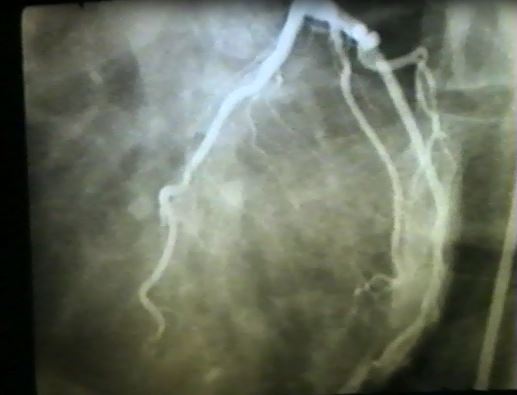
One of the most challenging clinical scenarios to a cardiologist is the patient presenting with symptoms suggestive of obstructive coronary artery disease (i.e. angina), in whom coronary angiography reveals patent coronary vessels. Due to the seemingly ‘normal’ arteries, current clinical practice tends to underestimate the impact of these presentations, but there are subsequent difficulties in their management. To many cardiologists, angina in the absence of CAD is a myth rather than a fact — “what you don’t know, you don’t miss.” One such presentation often being missed or ignored is the “coronary slow flow phenomenon.” A classic example of slow flow angiogram is shown here.


What is coronary slow flow?

Nearly 50 years ago, Tambe and colleagues1 initially described this angiographic entity in patients with angina symptoms where they noted the injected contrast during coronary angiography moved very slowly through the coronary arteries, and aptly named “coronary slow flow phenomenon.” The prevalence is estimated at approximately 1-7% of elective angiograms2,3. The condition was largely neglected until Professor John Beltrame identified the distinct clinical features associated with this intriguing entity and thus concluded the coronary slow flow phenomenon was a new coronary disorder rather than angiographic curiosity. Evidence suggests that the coronary slow flow phenomenon leads to clinical manifestations of ischemia, arrhythmias, acute coronary syndromes and even sudden cardiac death.


How is coronary slow flow diagnosed?

Coronary slow flow phenomenon is usually identified subjectively by visual judgment.

  • Thrombolysis in myocardial infarction (TIMI) flow grade reflects the speed and completeness of the passage of the injected contrast through the coronary tree. In the setting of coronary slow flow, diagnosis can be made on the basis of TIMI 2 flow grade (ie: requiring ≥ 3 beats to opacify the vessel)4.
  • Corrected TIMI frame count (CTFC) facilitates the standardization of TIMI flow grades and flow assessment. It represents the number of cine-frames required for contrast to first reach standard distal coronary landmarks. TIMI frame count > 27 frames have been frequently used to diagnose slow flow5.


What is the underlying cause of this presentation?

The coronary circulation consists of epicardial vessels and microvasculature. In the absence of epicardial stenosis, microvascular dysfunction may explain the pathophysiology of coronary slow flow phenomenon. Supporting this hypothesis, biopsy studies have revealed structural microvascular coronary abnormalities in slow flow patients. Reduced endothelium dependent flow-mediated dilatation (FMD) of the brachial artery has been detected in patients with coronary slow flow phenomenon, suggesting that endothelial dysfunction is implicated in the aetiology. However, there are still multiple questions and controversies regarding the underlying pathophysiology and whether this pathology is limited to coronary arteries or is a manifestation of systemic vascular or endothelial disease remains to be answered.


What is the medical management for coronary slow flow phenomenon?

Although coronary slow flow phenomenon patients have good overall prognosis, ongoing anginal episodes results in considerable impairment of their quality of life. Professor Beltrame has been long fighting the battle of identifying appropriate management for these patients, in particular, therapies that limiting the anginal episodes. His group has shown dipyridamole and mibefradil has some benefit in this setting, yet larger studies are required to confirm these findings. Currently available anti-anginal agents are of limited clinical value. To date, no large trial testing pharmacological approaches has been conducted, and the evidence available is derived from small studies, some with inhomogeneous inclusion criteria.


So, is it a myth or fact?

Over the past 50 years, the coronary slow flow phenomenon has evolved from a curious ‘myth’ to an identified coronary disease entity. Despite this progression of thinking, significant efforts are still required to unpack this intriguing condition, particularly in relation to effective therapies to improve symptoms and quality of life.


References:

  1. Tambe AA, Demany MA, Zimmerman HA and Mascarenhas E. Angina pectoris and slow flow velocity of dye in coronary arteries–a new angiographic finding. Am Heart J. 1972;84:66-71.
  2. Beltrame JF, Limaye SB and Horowitz JD. The coronary slow flow phenomenon–a new coronary microvascular disorder. Cardiology. 2002;97:197-202.
  3. Hawkins BM, Stavrakis S, Rousan TA, Abu-Fadel M and Schechter E. Coronary Slow Flow– Prevalence and Clinical Correlations &ndash. Circulation Journal. 2012;76:936-942.
  4. Chesebro JH, Knatterud G, Roberts R, Borer J, Cohen LS, Dalen J, Dodge HT, Francis CK, Hillis D, Ludbrook P and et al. Thrombolysis in Myocardial Infarction (TIMI) Trial, Phase I: A comparison between intravenous tissue plasminogen activator and intravenous streptokinase. Clinical findings through hospital discharge. Circulation. 1987;76:142-54.
  5. Gibson CM, Cannon CP, Daley WL, Dodge JT, Jr., Alexander B, Jr., Marble SJ, McCabe CH, Raymond L, Fortin T, Poole WK and Braunwald E. TIMI frame count: a quantitative method of assessing coronary artery flow. Circulation. 1996;93:879-88.


hidden

The Powerful Role of Social Media in the Field of Cardiology

The growth and use of social media have grown exponentially over the last decade with an eight fold increase since 20051. Social media is generally defined as an Internet-based platform that allows individuals and/or communities to gather virtually to communicate ideas, collaborate, share information, share pictures and videos, either as a direct message or general post in real time1. There are several social media platforms that healthcare professionals may use, such as Twitter, LinkedIn, Doximity, and Facebook. Twitter has been a very popular platform in the field of Cardiology with many Cardiologists, Cardiovascular research scientists, Cardiology providers, professional journals and Cardiology-based professional societies using this platform to expand their reach to their colleagues, professional society members, and the public in an effort to educate, advocate and raise awareness. There are several powerful roles that social media serves in the Cardiology field. These include networking, sharing meaningful opinions, fostering educational discussions centered around a cardiology topic or paper of interest, promoting or raising awareness of the latest research or guideline publication, promoting a professional meeting or event, promotion of healthy initiatives, collaboration among colleagues and support of colleagues.


Beneficial Uses of Social Media

a) Networking

Social media platforms allow many professionals in the cardiology field to connect with other colleagues and follow prominent cardiologists and research scientists. This connection transcends geographic borders, and therefore allows users to extend their networking reach internationally. This ability to network provides a sense of community and serves as one’s professional village where colleagues are able to share their professional ideas and share opinions on various topics.

b) Sharing important opinions and educational discussions on topics or publications relevant to Cardiology

Twitter also allows users to discuss topics and publications relevant to cardiology. Many times these are threads of a conversation joined by several colleagues. However, several professional organizations such as the American Society of Echocardiography (ASE) and the American College of Cardiology (ACC) have virtual tutorials, called “tweetorials,” which allows users to present and discuss a topic of interest in real time. These discussions can be very educational and serve as great learning tool. These discussions may also include reference to relevant publications and allows users to stay up to date with the scientific literature, as well.

c) Promotion of professional meetings and events

Many professional societies, such as the American Heart Association (AHA), American College of Cardiology (ACC), American Society of Echocardiography (ASE), American Society of Nuclear Cardiology (ASNC), Society of Nuclear Cardiology (SCCT), and the Society of of Cardiac Magnetic Resonance Imaging (SCMR), use social media platforms to promote their annual scientific meetings and events at these meetings. In fact, many of these meetings have social media (SoMe) ambassadors to help in promoting their meeting and to share important educational slides and messages from the meeting with other social media users, which is an excellent educational tool in getting important points out to the the rest of the cardiology community and the public.  Additionally, these professional meetings allow for cardiology colleagues who have connected virtually on Social Media to meet in person, as well. A hashtag (#) is a metadata tag that is used on social media platforms that allows posted content associated with a specific theme or content to be easily found2. Useful and popular hashtags used in the field of cardiology on Twitter are #CardioTwitter and #Cardiology. In fact many of the annual scientific meetings for several professional organizations will use hashtags for their meeting to allow social media users to readily identify posted social media content related to the meeting. This usually generates a significant degree of social media traffic and commentary related to the meeting and this further promotes the meeting and the professional organization globally. In fact at the 2018 annual American Heart Association’s Scientific Sessions, (#AHA18) there were over 300 million impressions generated globally2 on Twitter using the #AHA18 hashtag.

d) Discussion and promotion of latest research papers and guidelines

Many professional medical journals post important publications such as research papers and guidelines on social media to assist in promotion of important educational documents. In addition social media users also post their latest research papers and invited talks to help in promoting their scientific work and in the sharing of important educational information. With regards to posted research papers, it has been suggested that citations of research papers on Twitter can increase the citation rate of the paper and can also increase the impact factor of the the publication journal2. A prior analysis3 has shown that social media activity related to a publication paper increases the citation rate of the paper and therefore helps to promote published academic work. In fact the latest 2018 AHA/ACC Cholesterol management guidelines4, as well as the latest Physical Activity guidelines5, were released at the recently concluded American Heart Association meeting (#AHA18) and there was a significant amount of social media activity and discussion related to these two manuscripts. This therefore assisted to raise awareness of these guidelines within the cardiovascular community.

e) Starting healthy initiatives and sharing health promoting information with peers and the public

Promotion of healthy initiatives, such as heart healthy eating and increasing physical activity, have also been done on social media. Many cardiologists have used social media to share health educational material with the public and their colleagues.

f) Collaboration with and Providing Support for colleagues

Social media, especially Twitter, can help to create your professional community with colleagues who have similar professional interests. It provides a platform for collaboration with peers for various initiatives and opens the door for opportunities to collaborate with colleagues on research projects. This social media village creates a network that can be supportive with regards to helping to promote your professional interests and your academic publications through retweets and commentary.


Responsible use of social media

Responsible use of social media is very important, Always ensure that there is adherence to patient privacy regulation and ensure that social media posts are free of any patient identifying information. It is also vital that you maintain a high level of professionalism and avoid posting any social media information or pictures that can be professionally and ethically compromising for both yourself and others. It is very important not to tarnish your professional brand.6


Conclusion

The benefits of social media platforms such as Twitter are numerous and proves to be an increasingly relevant  learning tool that assists in keeping one abreast of the medical literature. Twitter is also very useful for one’s career  growth and provides a great opportunity for networking with peers globally. Social media helps in building your professional brand.


References:

  1. Ventola CL. Social Media and Health Care Professionals: Benefits, Risks, and Best Practices. P T. 2014 Jul; 39(7): 491-499, 520.
  2. American Heart Association Scientific Sessions 2018 meeting metrics provided by the AHA
  3. Eysenbach G. (2011) Can Tweets predict citations? Metrics of social impact based on Twitter and correlation with traditional metrics of scientific impact. J Med Internet Res 13:e123.
  4. Grundy SM, Stone NJ, Bailey AL, Beam LT, Birtcher KK, et al. 2018AHA/ACC/AACVPR/AAPA/ABC/ACPM/ADA/AGS/APhA/ASPC/NLA/PCNA Guideline on the Management of Blood Cholesterol. JACC Nov 2018, 25709; DOI: 10.1016/j.jacc.2018.11.003
  5. The Physical Activity Guidelines for Americans: THe HHS Roadmap for an Active Healthy Nation. Second Edition. ADM Brett P. Giroir, MD
  6. Bullock-Palmer RP. You Are Now a Board-Certified Cardiologist and Cardiac Imager…Now What? The Importance of Lifelong Learning and Career Growth
    May 2018 https://www.acc.org/membership/sections-and-councils/imaging-section/section-updates/2018/05/17/09/44/you-are-now-a-boardcertified-cardiologist-and-cardiac-imager


hidden

Deep Learning in Cardiology

Thirteen years ago in my first anatomy class of Med School, the instructor asked us to make sure our learning is “deep.”

“You need to memorize the names of every single tiny nerve and muscle, because they all will be on your exam. One day you could be surgeons and if you cut out the wrong structure, you can kill someone!” he asserted as we all stood there in fear.

Later in Med School, we were told that half of what we’re learning will be wrong by the time we practice. But one thing we were not told is that the way we learn and the entire premise of what makes a good doctor would also change. For example, “deep learning” itself now means something different to me and to most healthcare professionals. If you are reading this article so far, then you are likely to have seen the term floating around in medical journals.

Deep learning is a type of machine learning in which the computer uses multiple layers of processing to extract features from otherwise vague data input, such as an ECG or a slice of an MRI.  Each layer uses outputs from the previous layer. Through deep learning, the computer simulates the neural network of the brain and is able to learn and make sense of abstraction.

Working at the Broad Institute of Harvard and MIT allowed me to recently be part of a team that uses deep learning to solve important problems in cardiology.  Over several weeks, cardiologists, scientists, and machine learning experts worked in teams to train computers on deep learning models so that they understand data such as medications, ECGs, genomic architecture, and cardiac MRIs of tens to hundreds of thousands of people.

The insights I gained were incredible.  Just like medical students perform better on their exam if they learn “deeper,” the longer you train a computer model, the more it learns and the better it performs in predicting – but it does so at much faster rates than any doctor could ever match. For example, in only two days, we trained a computer to read the ejection fraction from a cardiac MRI as good as a doctor would. Using the MRI, the computer could also predict with reasonable precision the presence of hypertension and coronary artery disease, without knowing anything else about the patient. The power of computer vision is beyond imagination. While it could take you a full day to read 100 ECGs, a well-trained deep learning model could read them in only few seconds. It could also identify patterns in the data that the human eye could not discern, which might or might not be biologically or clinically relevant.

As data availability and computing power continue to grow, we will be seeing more and more applications of deep learning in cardiology.  While we do so, we should stay mindful that human supervision and our role as doctors in charge of our patient’s health is more important than ever. This requires us to understand how computers work and how those models are built through working with multidisciplinary teams. If we do this right, we can probably do less deep learning ourselves by delegating to computers, and gain a whole lot of extra time that we can invest in taking care of our patients.


hidden

The Significant Areas of Interest in the Field of Cardiac Imaging in 2018

There were several exciting developments in 2018 with regards to cardiac imaging. The role of the cardiac imager is becoming increasingly relevant in today’s cardiology practice environment and bridges across several subspecialties in Cardiology, such as electrophysiology with the use of transesophageal echocardiography (TEE) in the placement of left atrial appendage closure devices. These devices include Watchman and interventional cardiology with structural and valvular heart disease and echocardiographic guidance with transaortic valve replacement (TAVR), percutaneous mitral valve repair with MitralClip, as well as atrial septal and ventricular septal closure devices. The field of cardiac imaging has matured over the years and not only includes echocardiography and nuclear cardiology, but also includes advanced imaging with cardiac magnetic resonance imaging (cMRI) and cardiac computed tomography. In addition, there has been the rise of the interventional echocardiographer specializing in the use of echocardiography in guiding percutaneous and surgical treatment of structural heart disease. In fact, there has been recognition of cardiac imaging by several professional societies such as the American College of Cardiology with publication of the state-of-the-art paper, The Future of Cardiac Imaging Report of a Think Tank Convened by the American College of Cardiology1. There have also been several disease states that have been positively influenced by the development of new diagnostic technology in cardiac imaging, such as cardiac amyloidosis. Cardiac imaging has also positively influenced preventive cardiology with release of the latest American Heart Association (AHA)/ American College of Cardiology (ACC) 2018 Cholesterol Management Guidelines2. The following areas were, in my opinion, considered topics of great interest in 2018 in the field of cardiac imaging.


Nuclear Imaging

Cardiac Amyloidosis. For several years, cardiac amyloidosis, particularly transthyretin type (ATTR type), was thought to be a diagnosis that was very difficult to make with endomyocardial biopsy being the only method to confirm the diagnosis. However, nuclear cardiac imaging has changed the landscape of this disease with the novel application of old technology with the use of technetium 99m pyrophosphate (Tc-99m PYP) in the diagnosis of ATTR type cardiac amyloidosis3. The sensitivity and specificity of this technique in diagnosing this disease state is >95%, and oftentimes avoids the need for endomyocardial biopsy to make this diagnosis4. The development of this technique in diagnosing the disease has increased the recognition of this disease in many patients with diastolic heart failure, and even in patients with severe aortic valve stenosis undergoing TAVR. This has also led to greater research and development of new treatments for this disease, such as tafamidis, patisiran and inotersen. The development of these medications will hopefully improve the overall prognosis for patients with this disease.


Echocardiography

The Rise of the Interventional Echocardiographer in Structural Cardiac Imaging. There has been increasingly relevant areas of interest in structural heart disease, such as percutaneous mitral valve repair with MitralClip, especially with the release of the study findings from the COAPT trial5.  In addition, transaortic valve replacement (TAVR) has become increasingly available for many patients with severe aortic valve stenosis, and many institutions have began offering this therapy to many of their patients. Additionally, left atrial appendage occlusive devices such as the Watchman device are being increasingly used in patients with atrial fibrillation who are at high risk for hemorrhagic complications with anticoagulation, despite having indications for thromboembolic prophylaxis. With these new developments, there has been the rise of the interventional echocardiographer, who serves a vital role with the use of echocardiography in guiding the placement of these devices in the treatment of structural heart disease. Many fellows are now seeking additional training in this field to meet this demand, as this area has invited a growing interest in the cardiology field and has attracted many trainees.


Cardiac Computed Tomography

The revisiting of Coronary Calcium Score as a Powerful Tool in Preventive Cardiology. The release of the latest AHA/ACC Cholesterol Management Guidelines has been an area of great interest in the field. The latest guidelines have included the use of coronary calcium scoring with cardiac CT as a tool to further risk stratify patients to guide the use of pharmacologic therapy for patients with hyperlipidemia2. This has led to  the resurgence of Cardiac CT for coronary calcium scoring as a valuable tool for cardiologists in the field of preventive cardiology.

Utility of Cardiac CT in the assessment of Women with suspected Cardiovascular Disease.  There has also been the increasing recognition of Cardiac CT as a useful diagnostic tool for women suspected of having cardiovascular disease (CVD)6. Hopefully, this will result in the increased appropriate use of Cardiac CT in the management of CVD in women.


Cardiac MRI

The complementary role of Cardiac MRI with echocardiography and assessment of valvular and structural heart disease. Cardiac MRI has become an established imaging modality in the assessment of valvular heart disease. This has been embraced by the American Society of Echocardiography’s latest Valvular Guidelines, which includes cardiac MRI as playing a complementary role in the assessment of the severity and etiology of valvular heart disease7. The use of Cardiac MRI is also useful in the assessment of other disease states, such hypertrophic cardiomyopathy and risk assessment for sudden death8.

The rise of Cardiac MRI compatible devices. There has also been the development of Cardiac MRI compatible devices which now allows many patients with these devices to be able to have cardiac MRIs performed safely. Cardiac MRI is therefore a viable diagnostic tool for these patients.

The first administration of certification board exam in cardiovascular magnetic resonance imaging (CBCMR).  With the maturation of Cardiac MRI as a viable imaging modality, 2019 will see the inaugural administration of the first certification exam in cardiovascular magnetic resonance imaging (CBCMR), which will occur between May 7 – June 7, 2019, and the 2019 application window will open on January 15, 2019. ​​


Conclusion:

With the dawn of a new year in 2019, it is clear that the future of cardiac imaging is very bright. I am looking forward to many more promising developments in this field and hope that this field will continue to attract many more talented cardiologists in this area of cardiology.


References:

  1. Douglas PS, Cerqueira MD, Berman DS, Chinnaiyan K, Cohen MS, Lundbye JB, et al. The Future of Cardiac Imaging Report of a Think Tank Convened by the American College of Cardiology. J Am Coll Cardiol Img 2016;9:1211–23.
  2. Grundy SM, Stone NJ, Bailey AL, Beam LT, Birtcher KK, et al. 2018AHA/ACC/AACVPR/AAPA/ABC/ACPM/ADA/AGS/APhA/ASPC/NLA/PCNA Guideline on the Management of Blood Cholesterol. JACC Nov 2018, 25709; DOI: 10.1016/j.jacc.2018.11.003.
  3. Dorbala S, Bokhari S, Miller E, Bullock-Palmer RP, Soman P, Thompson R. ASNC Practice Points: 99mTechnetium-Pyrophosphate Imaging for Transthyretin Cardiac Amyloidosis (American Society of Nuclear Cardiology website). 2018. Available at: https://www.asnc.org/Files/Practice%20Resources/Practice%20Points/ASNC%20Practice%20Point-99mTechnetiumPyrophosphateImaging2016.pdf.
  4. Gillmore JD, Maurer MS, Falk RH, Merlini G, Damy T, Dispenzieri A, et al. Nonbiopsy Diagnosis of Cardiac Transthyretin Amyloidosis. Circulation. 2016 Jun 14;133(24):2404-12. Doi: 10.1161/CIRCULATIONAHA.116.021612. Epub 2016 Apr 22.
  5. Stone GW, Lindenfeld J, Abraham WT, Kar S, Lim DS, Mishell JM,et al. COAPT Investigators.Transcatheter Mitral-Valve Repair in Patients with Heart Failure. N Engl J Med. 2018 Dec 13;379(24):2307-2318. doi: 10.1056/NEJMoa1806640. Epub 2018 Sep 23.
  6. Truong QA, Rinehart S, Abbara S, Achenbach S, Berman DS, Bullock-Palmer R,et al. SCCT Women’s Committee.Coronary computed tomographic imaging in women: An expert consensus statement from the Society of Cardiovascular Computed Tomography. J Cardiovasc Comput Tomogr. 2018 Nov – Dec;12(6):451-466. doi: 10.1016/j.jcct.2018.10.019. Epub 2018 Oct 23.
  7. Zoghbi WA, Adams D, Bonow RO, Enriquez-Sarano M, Foster E, Grayburn PA, et al. Recommendations for Noninvasive Evaluation of Native Valvular Regurgitation A Report from the American Society of Echocardiography Developed in Collaboration with the Society for Cardiovascular Magnetic Resonance. J Am Soc Echocardiogr. 2017 Apr;30(4):303-371. doi: 10.1016/j.echo.2017.01.007. Epub 2017 Mar 14.
  8. Weng Z, Yao J, Chan RH, He J, Yang X, Zhou Y, He Y.Prognostic Value of LGE-CMR in HCM: A Meta-Analysis. JACC Cardiovasc Imaging. 2016 Dec;9(12):1392-1402. doi: 10.1016/j.jcmg.2016.02.031. Epub 2016 Jul 20. Review.