hidden

FAQs about Dialysis Patients

We’ve just started a new academic year, so now’s a good time to review some uncertainties and myths surrounding the care of dialysis patients.

Hey the patient is on chronic dialysis = total kidney failure, therefore iodinated CT contrast is A-Okay?
It depends.
Residual kidney function (the patient still has urine output) means better outcomes in terms of survival, nutrition, quality of life and better control of electrolytes and anemia. The kidneys work 24/7 to clear toxins and volume, and are better than dialysis at clearing middle molecules like phosphorus and protein-bound uremic toxins.
So if the dialysis patient still makes urine, avoid nephrotoxins including iodinated CT contrast unless there is strong medical necessity. Protect those kidneys!

Well we really need to give the CT contrast, so we’ll just coordinate dialysis afterward and that will save the day right?
Evidence is mixed but suggests that dialyzing out IV contrast is ineffective. You sucker punched the kidneys, and there’s no take-backsies.
However if there is concern that the amount of contrast led to volume overload and is compromising lung function – dialysis can remove volume and help with that.

How about MRI gadolinium contrast? Is the answer also “it depends”?
Gadolinium = never ok! Nephrogenic systemic fibrosis. Rare, but BAD. Look it up.

The nephrologist threw a fit when I asked for a PICC line for IV antibiotics. Why is she so loco?
Please recite daily: NO PICC LINES IN DIALYSIS PATIENTS. This also applies to predialysis patients with advanced chronic kidney disease who are heading toward dialysis dependence.
To do dialysis requires a dialysis access. For hemodialysis patients, the preferred access is an arm arteriovenous fistula (AVF). For peritoneal dialysis patients, peritoneal membrane failure eventually occurs and they will have to switch to hemodialysis. PICC lines cause vein trauma thus predisposing to future AVF failure. Along the same lines, radial artery approach for left heart cath should be avoided in patients with advanced chronic kidney disease.
Switch to PO antibiotics if possible. Or pick IV antibiotics that can be given during dialysis (cefazolin, vancomycin, cefepime are examples). If prolonged IV access is absolutely necessary, a small-bore internal jugular tunneled catheter (like a Hickman) can be considered.
There will be exceptions based on an individual patient’s clinical situation and life expectancy – please discuss with your favorite nephrologist!

The dialysis diet is low sodium, low phosphorus, low potassium. My patient is diabetic so she can’t even have carbs. Can she eat anything??
We’re not saying zero. Daily limits are sodium 2 gm, potassium 2 gm, phosphorus 800 mg. The dialysis diet is complex – that’s why every dialysis unit has dedicated dietician(s).
She should eat a lot of protein. Dialysis is a catabolic treatment and patients are encouraged to eat high protein diets (1.2-1.5 g/kg/day) to avoid protein calorie malnutrition.

Are all renal supplement drinks good for all kidney disease patients?
No! The supplement drinks that are for dialysis patients (Nepro, Novasource) are HIGH protein (see above, dialysis patients are recommended to eat a lot of protein). These are NOT good for predialysis patients with advanced chronic kidney disease, who need to be on LOW protein diet which may be beneficial in slowing progression of kidney failure. An example of a low-protein supplement for predialysis patients is Suplena Carb Steady.

My patient is on chronic hemodialysis and does not have a clinic follow up scheduled with his nephrologist. What is this craziness?
Most hemodialysis patients are in-center (they go for scheduled dialysis treatments at a dialysis unit, typically 3 days per week) and their nephrologist will round on them at the dialysis center. So they don’t need a separate clinic appointment.
Patients who do home dialysis will see their nephrologist in clinic once per month.

My hemodialysis patient was started on a blood thinner. Do I need to let the nephrologist know?
Please ALWAYS let the nephrologist know when a dialysis patient is started on warfarin or apixaban. (Apixaban is currently the only FDA-approved direct oral anticoagulant for dialysis patients.) Heparin is often used in the dialyzer circuit to prevent clotting. If a patient is started on anticoagulation, heparin may be held since the oral anticoagulant may be sufficient.
From a big picture standpoint, the use of anticoagulation in dialysis remains controversial (see my prior blog).

Ok we decided anticoagulation is no longer needed (or maybe the patient self-discontinued her apixaban… true story). Everything ok now?
On the flip side, if oral anticoagulation is stopped, please notify the nephrologist so that heparin can be re-introduced. If a dialyzer circuit clots and blood cannot be returned to the patient, this is equivalent to losing a half unit of blood.

Wei Ling Lau Headshot
 
Wei Ling Lau, MD FASN is Assistant Professor in Nephrology at University of California-Irvine. She is currently funded by an AHA Innovative Research Grant, and has been a speaker for CardioRenal University and the American Society of Nephrology. Follow her on Twitter @Kidneys1st.
hidden

If you give a patient calcium…

You have to tell them when to take it.

My prior blog warned about the unknowns of natural supplements in general. This follow up piece is focused on calcium-based products, which are commonly utilized for a variety of medical indications. (Also, am feeling particularly inspired about this topic as I am putting together a National Kidney Foundation webinar for dieticians that discusses calcium in chronic kidney disease – includes a lot of fun pathophysiology and pictures of high-calcium foods!)

The timing of when to take the calcium is quite critical. General rules: calcium taken with meals serves as a binder to prevent absorption of a certain food component. Calcium taken between meals is absorbed and will boost body calcium stores. Let’s go into more specific examples…

Prevention of oxalate kidney stones (dietary calcium with meals)
Calcium oxalate is the most common type of kidney stones. While drinking plenty of fluids to maintain dilute urine is the cornerstone of stone prevention (used “stone” twice there and it’s legit), calcium is a proven adjunct intervention to lower urinary oxalate. The concept here is that dietary calcium binds oxalate, and the calcium-oxalate complex is pooped out so less oxalate is absorbed by the gut, and less oxalate reaches the kidneys. In a randomized clinical trial, normal calcium intake of 1200 mg/day with restricted oxalate, protein and salt intake decreased stone recurrence compared to a low-calcium diet of 400 mg/day.

Prevention of hyperphosphatemia in dialysis patients (calcium-based binders with meals)
Chronic kidney disease (CKD) patients have a high prevalence of vascular calcification and heart failure. As kidney function declines, blood phosphorus levels increase and hyperphosphatemia has been linked with adverse cardiovascular events and death. Calcium-based phosphate binders, such as calcium carbonate and calcium acetate, are prescribed with meals to decrease phosphorus absorption (along the same lines as for calcium use with oxalate, see above). This approach becomes a slippery slope, because of the concurrent risk of positive calcium balance in CKD patients whereby excess calcium is also a risk factor for vascular calcification. Neither blood calcium nor urinary calcium levels accurately reflect total body calcium, so there is no easy way to gauge a patient’s calcium status. Clinical trials have noted less progression of coronary artery calcification in CKD patients on non-calcium phosphate binders (as compared to the calcium-based binders), thus nephrologists have become more cautious with prescribing calcium-based phosphate binders in CKD patients.

Treatment of osteoporosis (calcium supplements with meals or between meals… it depends)
Calcium and vitamin D for osteoporosis treatment is controversial but experts agree that high-risk patients would benefit from these supplements for fracture prevention. They also agree that it is important to avoid excessive dosing, e.g., adding elemental calcium 1000 mg per day in an individual who is already taking in a normal amount of dietary calcium (1200 mg/day) would increase the risk of adverse cardiovascular events or kidney stones. When taking calcium for osteoporosis therapy, the intention is to optimize calcium absorption from the gut (very different from the two scenarios presented above). The calcium formulation dictates timing with meals; for calcium citrate, there is good absorption regardless of whether the supplement is taken with or between meals. Calcium carbonate is taken with meals as gastric acidification is needed to release the calcium for gut absorption. Magnesium, zinc and iron should not be taken at the same time with calcium supplements as each can interfere with the other’s absorption.

Supplementation in hungry bone syndrome (may require both IV and oral calcium!)
Hungry bone syndrome is defined by a significant drop in blood calcium following surgical removal of hyperfunctioning parathyroid glands. The sudden decrease in parathyroid hormone levels leads to unopposed uptake of calcium by the bones. Hypocalcemia can be sustained for a few weeks, and patients often require IV calcium repletion within the first week followed by high doses of oral calcium 1-3 grams per day with vitamin D therapy. Similar to use of calcium for osteoporosis therapy, the goal here is to optimize calcium absorption from the gut. The rule of thumb is that elemental calcium should be spread out through the day up in doses of 500 mg as this is the gut’s threshold for effective calcium absorption.

Finally, it is a good idea to inform patients that it is the “elemental calcium” portion of supplements that we are referring to when discussing calcium doses. Calcium carbonate contains 40% elemental calcium, and calcium citrate contains 21% elemental calcium. An “ultra” strength calcium carbonate 1,000 mg tablet contains 400 mg of elemental calcium which is within the absorption threshold. Calcium supplements are a prime example where how much and when can impact response to therapy.

Wei Ling Lau Headshot

Wei Ling Lau, MD is Assistant Professor in Nephrology at University of California-Irvine. She is currently funded by an AHA Innovative Research Grant, and has been a speaker for CardioRenal University and the American Society of Nephrology. Follow her on Twitter @Kidneys1st

hidden

When Survival Benefit Transcends Infection Risk

The 22-year-old single mom flags me down at the dialysis unit. “The transplant team discussed getting my consent for increased risk kidney donors – do you think this is something I should go for?” Disclaimer: I’m not a transplant nephrologist. Worrisome thoughts hit my mind… She is so young. Why take on any risk of contracting HIV or hepatitis C? Patients so young will need at least 2-3 transplants through their lifetime. Will she really get a good quality kidney that will hopefully function for 15-20 years, if it was from an increased risk for disease transmission (IRD) donor? (The “increased risk” refers to behaviors that increase risk of contracting HIV, hepatitis B or hepatitis C.)

Fortunately, transplant outcomes data has been reassuring and shows that organs from IRD donors are non-inferior to non-IRD organs. In fact, a recent report from the American Journal of Transplantation showed a survival benefit in accepting IRD kidneys. In patients who declined an IRD kidney, only 31% later received a kidney transplant (5 year follow up period) and this was of a lower quality than the initial IRD organ offered.
 
Organ transplantation from a deceased donor is the ultimate paradox of a new lease on life from someone other’s tragedy. But the reality is that most patients waiting for a transplant will die before an organ becomes available. There are ~115,000 people on the UNOS (United Network for Organ Sharing) wait list and the majority are waiting for a kidney transplant (~95,000); average wait-time for a kidney from a deceased donor is 5 years. The longer a wait-listed dialysis patient is living with kidney failure and is exposed to the uremic constellation of metabolic derangements, inflammation, anemia and blood pressure fluctuations – the higher the risk of progressive vascular injury and sudden cardiac death. End stage kidney failure is a huge public health burden, totaling $34 billion per year in Medicare spending.

Thus from the perspective of both the individual patient and tax dollars, optimizing the number and quality of transplants is a big deal. Which brings us back to the topic of IRD donors who have a history of behaviors that put them at risk of having HIV, hepatitis B or hepatitis C. Many of these donors are otherwise young and relatively healthy; this means that there is the potential to transplant high quality organs from 1 donor to as many as 8 recipients. In the U.S. we are seeing an alarmingly fast-growing segment of IRD organ donors in the opioid overdose crisis. In 2016 there were ~42,000 deaths from opioid overdose, a five-fold increase from 1999.

A recent NEJM correspondence states “the drug-abuse epidemic has been associated with a sharp increase in the recovery of organs from brain-dead donors in the United States but not in Europe”. In the U.S., the proportion of organ donors who died from drug intoxication increased from 1.2% of all donors in the year 2000 to 13.7% as of 2016. In contrast, the Eurotransplant registry from 8 European countries showed no significant change (≤1% per year). The outcomes data from this donor population is the silver lining in this tragic crisis. In an Annals of Internal Medicine analysis of almost 20,000 organs transplanted from over 7,000 drug-overdose donors, 5-year patient and graft survival was similar as compared to organs from trauma- or medical-death donors.

The majority of IRD donors who have no prior history of HIV or hepatitis won’t have a transmissible virus. But right now we don’t have tests that allow us to be 100% certain about a donor’s infectious status at time of organ procurement. The concept of “increased risk” refers to the risk of recent acquisition of HIV or viral hepatitis that is missed in the Nucleic Acid Testing (NAT) window period. (Window period = the time between potential infection and the point when the test will give an accurate result, thus you get false-negative results.) Don’t get me wrong, NAT is a remarkable improvement over prior serologic testing; for example, hepatitis C NAT has a window period of 3-5 days as compared to 70 days with traditional antibody (serologic) testing. NAT itself is a controversial topic since false-positive results can occur, and experts have raised concerns that this may result in unwarranted discard of organs. However in IRD donors NAT is a powerful tool for more accurate risk assessment. For example, donors with recent IV drug use with negative serological testing have a risk of undetected hepatitis C of 300.6 per 10,000 donors (3%). Having both negative serology and negative NAT reduces this risk to 32.4 out of 10,000 donors (0.3%).

To summarize: the U.S. is facing an epidemic of drug-overdose deaths. Simultaneously, there is a long list of patients waiting for a life-saving heart, kidney, lung or liver transplant. These two issues have become intertwined. It is an unsettling topic of discussion, but it behooves us to counsel patients that organ transplants from “increased risk” donors are non-inferior and improve survival.

Wei Ling Lau Headshot

Wei Ling Lau, MD is Assistant Professor in Nephrology at University of California-Irvine. She is currently funded by an AHA Innovative Research Grant, and has been a speaker for CardioRenal University and the American Society of Nephrology. Follow her on Twitter @Kidneys1st

hidden

Vascular Calcification… It’s Complicated

The Vascular Discovery meeting in San Francisco last week was whirlwind of learning and networking. My favorite moment was at the Friday 7 am Early Career Training session where, by coincidence, I ended up at the same table with @Ritu_Ganguly1, @MoradiShayan and @JeffHsuMD. We had all signed up to provide social media coverage of the conference via the Twitter account of the Journal of the American Heart Association (JAHA), so it was great to meet them in person. Later in the day, Ritu and I worked (read: laughed and cried) through the Genome Editing Bootcamp together, a challenging case-based workshop led by the excellent Dr. Kiran Musunuru.

As discussed in my pre-conference blog, vascular research is extremely pertinent to chronic kidney disease. Children on dialysis can manifest the same arterial calcification as a 70 year old. An established mechanism in vascular calcification is the phenotype switch where vascular smooth muscle cells start behaving like bone cells, secreting matrix vesicles filled with calcium-phosphate mineral into the extracellular matrix. At the Vascular Discovery meeting Dr. Elena Aikawa discussed advances made in the understanding of matrix vesicles, which are critical precursors of microcalcifications. In a JCI paper, her group reported co-localization of the protein sortilin with caveolin-1 and tissue nonspecific alkaline phosphatase, and defined sortilin’s role in loading mineral into vesicles. Dr. Aikawa raised a follow up question: Would future therapies that block activation of sortilin prevent microcalcifications, and thus prevent vascular calcification?

Dr. Catherine Shanahan, who described the role of programmed cell death or apoptosis in dialysis-associated vascular calcification, discussed the interaction of aging and vascular cell phenotype change at her Vascular Discovery talk. Her lab has been examining the nuclear lamina, or network of filament proteins which are a part of the cell nucleus. It turns out that the aging vascular smooth muscle cell accumulates prelamin A (see Circulation Research paper), which leads to DNA damage and triggers the osteogenic phenotype switch. This raises the intriguing question: Can we reverse cell aging to block vascular calcification?

The nature of scientific research is that more questions are raised as progress is made. Scientific meetings such as Vascular Discovery have an important role in updating investigators and clinicians, fostering new collaborations and training early career professionals.

Wei Ling Lau Headshot

Wei Ling Lau, MD is Assistant Professor in Nephrology at University of California-Irvine. She is currently funded by an AHA Innovative Research Grant, and has been a speaker for CardioRenal University and the American Society of Nephrology. Follow her on Twitter @Kidneys1st

hidden

The Vascular Discovery Meeting Is This Week!

Scientific knowledge is constantly evolving, and scientific meetings change over time to stay current with the target audience. Sometimes a revamp of the conference name is timely. For example, the American Society of Nephrology (ASN) retitled its annual Renal Week to Kidney Week in 2011 to move away from using nomenclature that might confuse the layperson (renal: late Latin adjective “relating to the kidneys”). This year, the AHA’s Arteriosclerosis, Thrombosis and Vascular Biology / Peripheral Vascular Disease (ATVB/PVD) meeting has been renamed Vascular Discovery and is happening in San Francisco May 10-12, 2018.

Thanks to the AHA Early Blogger program, I will be attending the ATVB conference for the first time, and will be live-tweeting for the Journal of the American Heart Association (JAHA). (Yes I still occasionally refer to the meeting as ATVB… not quite used to its snazzy new title yet!) I am totally stoked to be going to ATVB as vascular research has been a core interest of mine for several years, ever since fellowship training in the lab of Dr. Cecilia Giachelli at the University of Washington. Chronic kidney disease accelerates vascular disease at both the intimal and medial layers of arteries; i.e., atherosclerosis and medial vascular calcification. Medial vascular calcification is particularly intriguing as this is seen in the arteries of children on dialysis, without atherosclerosis (presumably because the children lack athero risk factors such as smoking, diabetes and aging). Dr. Giachelli identified a fascinating phenotype switch that occurs in the vascular smooth muscle cells of patients with chronic kidney disease; the cells start behaving like bone and deposit calcium-phosphate crystals in the artery wall, leading to vascular calcification and ultimately heart failure.

So, you may ask, why would a vascular cell decide to build bone in the wall of the blood vessel? This could be a defense mechanism, to avoid the cell imploding and dying. It turns out that too much intracellular calcium or phosphorus (an imbalance that can happen in chronic kidney disease) can trigger apoptosis (which also promotes vascular calcification!). By turning on genes that allow the cell to export calcium-phosphate mineral, the vascular cells avoid programmed cell death. Unfortunately, big picture-wise, this cell-centric ninja move fails the body as vascular calcification is associated with heart failure and sudden cardiac death. (Of note, Dr. Catherine Shanahan – one of the first to describe the role of apoptosis in uremic vascular calcification – will be a speaker at Vascular Discovery this week.)

ATVB is one of the smaller AHA conferences and thus the “concurrent sessions dilemma” is less of an issue. (Not like the AHA Scientific Sessions or ASN Kidney Week where one has to make a choice between 5-8 talks that are going on All At The Same Time.)  The Vascular Discovery program this week includes sessions on precision medicine, diabetes-vascular complications, lipid metabolism, gut microbiome and inflammation. For vascular biology enthusiasts, this meeting is a must.

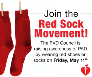
Follow #JAHAMeetingReport and #AHAEarlyCareerBlogger for live tweets from Vascular Discovery. And don’t forget to show your support for vascular disease awareness and research by wearing red socks on Friday!

Red sock movement

Wei Ling Lau Headshot

Wei Ling Lau, MD is Assistant Professor in Nephrology at University of California-Irvine, where she studies vascular calcification and brain microbleeds in chronic kidney disease. She is currently funded by an AHA Innovative Research Grant, and has been a speaker for CardioRenal University and the American Society of Nephrology.

hidden

Natural Supplements Can Be A Pain In The Kidneys

Mrs. M presented with a list of 20 natural supplements that she was taking to boost her immune system and fight off a lingering fatigue attributed to Lyme disease.  She was in nephrology clinic to evaluate a 30% decline in kidney function over the recent 3 months.

She got straight to the point.  “Are the supplements hurting my kidneys?”  The best answer I could come up with was: “Maybe”.  Which led to a discussion about why side effects can happen with herbal or other dietary supplements:

  • Metabolism of any drug or supplement involves several enzymatic pathways.  Due to genetic variability, an individual may not be able to metabolize a certain supplement, and accumulation of byproducts leads to off-target effects.  As an example, the “Asian flush” after alcohol intake is due to deficiency of acetaldehyde dehydrogenase, a key enzyme needed to break down booze in the body.  The resulting acetaldehyde accumulation causes facial redness, headache, and heart palpitations.
  • Supplements contain more than just the pure natural product.  Excipients (inactive ‘other ingredients’) are added to serve as filler, flavoring, coating, preservative, etc.1 The inactive ingredients would typically pose little to no health risk, however, again due to genetic variability some people experience side effects.  For example, potassium sorbate is sometimes used to prolong the shelf life of herbal products; sensitivity to this compound induces nausea and stomach cramps.
  • The optimal beneficial dose of many natural supplements is unknown.  Any supplement taken in excess will overwhelm metabolic pathways and result in toxicity.  Of note, many herbal products evolved as plant defenses against being eaten by insects or animals,2 thus large amounts can be viewed as natural poisons.
  • In the setting of liver or kidney disease, byproducts can accumulate and cause adverse effects.
  • Interactions with prescribed medications can be problematic.  One example is St. John’s wort which is consumed for its anti-anxiety and anti-inflammatory properties; this herb is known to inhibit warfarin and thus increases clotting risk in patients on warfarin anticoagulation.

The use of herbal supplements is growing.  In a national U.S. survey of approximately 26,000 people, more than one-third of respondents reported using herbal supplements.3 Older age and higher education were associated with higher use of herbal supplements.

Even vitamins or minerals, when over-supplemented, can cause problems.  The recommended daily vitamin C intake is 90 mg for men and 75 mg for women; these needs are easily met by a balanced diet.  Many people take vitamin C (usually 1000 mg daily) to boost the immune system.  Vitamin C supplementation in men is associated with increased kidney stones,4 likely due to increased urine levels of oxalate (a metabolite of vitamin C) and formation of calcium oxalate stones.  Vitamin D calcium combination supplements, commonly taken for prevention of bone fractures, are also associated with increased risk of kidney stones5 as high body calcium levels leads to high urine calcium.  It is interesting to note that the prevalence of kidney stones in the U.S. increased from 1 in 20 persons to 1 in 11 persons in the past 2 decades (NHANES data from 1994 and 2007-2010).6 One wonders if this trend is partly driven by use of vitamin and mineral supplements.

Research is ongoing to examine how natural supplements can be used to derive health benefits without causing harm.  For example, our group and other researchers are investigating the antioxidant effects of curcumin derivatives (from the spice turmeric) in kidney disease.7 Studies in cancer cell lines discovered that curcumin at high concentrations has a paradoxical effect of increasing oxidative stress (overwhelming the antioxidant benefits), emphasizing the importance of correct dosing to avoid harm.

So what is the best approach if you have decided to take a natural supplement?  Definitely let your doctor know so that the medication list is checked for potential interactions and blood tests can be monitored (if necessary).  Avoid taking amounts in excess of the manufacturer’s instructions.  If you have chronic kidney disease, herbal supplements should be avoided per National Kidney Foundation recommendations8 (until more data is available) given concerns for high potassium or phosphorus content, diuretic effects, or direct kidney toxicity. 

In the case of Mrs. M, she decided to quit the 20 natural supplements and her kidney function returned to normal a few weeks later.

Suggested reading:

1. Natural Healthy Concepts blog on inactive ingredients in supplements (2014).
2. Advances in Nutrition review paper about herbal extracts that evolved as plant defenses against herbivores and insects (2011).
3. Journal of Patient Experience report on prevalence of herbal supplements use in the U.S. (2017).
4. JAMA Internal Medicine report of increased kidney stones in Swedish men taking vitamin C supplements (2013).
5. JAMA meta-analysis noting increased incidence of kidney stones in persons taking vitamin D with calcium (2018).
6. European Urology article reporting increasing occurrence of kidney stones in the U.S. from analysis of National Health and Nutrition Examination Survey data (2012).
7. PubMed list of published research papers about curcumin effects in kidney disease (as of April 2018).
8. National Kidney Foundation website advising against use of herbal supplements in chronic kidney disease patients (2015).

Wei Ling Lau Headshot
Wei Ling Lau, MD is Assistant Professor in Nephrology at University of California-Irvine, where she studies vascular calcification and brain microbleeds in chronic kidney disease. She is currently funded by an AHA Innovative Research Grant, and has been a speaker for CardioRenal University and the American Society of Nephrology.

hidden

Chronic Kidney Disease: The Silent Killer

“He felt well, so he didn’t follow up with his doctor.”

Our nephrology team was gathering history about a patient who had landed in the Emergency Department with advanced kidney failure and its consequences: confusion, severe anemia, metabolic acidosis, and a high blood potassium threatening to push him into cardiac arrest.  We asked the family: Did he have any known kidney problems?

“His doctor mentioned abnormal kidney function 3 years ago.  But he’s felt really healthy, and has been too busy to go back to the clinic.”

Convincing someone they have chronic kidney disease can be tough.  “But I feel fine!” is a common response along with a look of disbelief or suspicion (like they’re not sure if I’m trying to sell them something).  This is usually followed by: “What can I do to make my kidneys better?”  This part is a real downer because they find out I actually don’t have anything to sell – there isn’t any therapy that can regenerate kidney function.  It’s all about preventative measures to preserve the functioning nephrons – we focus on improving lifestyle practices and treating comorbidities so as to avoid further kidney injury.

World Kidney Day has been recognized on the 2nd Thursday of March every year since 2006 and tends to pass by with little fanfare.  Public awareness and media coverage of kidney disease is relatively low compared to conditions such as heart disease or cancer.   However, the statistics associated with chronic kidney disease are downright scary.  Chronic kidney disease doubles the risk of mortality from cardiovascular events or infection.  Every year, more Americans die from kidney disease than from breast cancer and prostate cancer combined.  End-stage kidney failure requiring dialysis has an average survival of 10-15 years.  If an individual transitioned to dialysis at the same time that he welcomed a baby to the family, he might not live to see his child graduate from high school.

person shruggingAbout 1 in 10 people worldwide, and >20 million in the US, have chronic kidney disease.

The functioning unit in the kidney is the nephron, and humans are born with 900,000 to 1 million nephrons per kidney (or less, if born premature).  No new nephrons form after birth.  We are actually born with more kidney function than we need to maintain electrolyte and fluid balance – evolutionary proof that the kidneys are so important!

In most cases of progressive kidney disease, the body is remarkably adept at adjusting to the buildup of toxins.  The person “feels well” until the kidney function falls below 15% at which point the “crash” happens and they feel terrible.

Research is ongoing to develop saliva tests that quantify levels of toxins that have diffused from the blood, as a measure of kidney function.  These are yet to be validated and commercialized.  For now, periodic blood tests (inconvenient and painful, but necessary) are the only way we can reliably monitor chronic kidney disease to guide treatment recommendations.

Wei Ling Lau Headshot
Wei Ling Lau, MD is Assistant Professor in Nephrology at University of California-Irvine, where she studies vascular calcification and brain microbleeds in chronic kidney disease. She is currently funded by an AHA Innovative Research Grant, and has been a speaker for CardioRenal University and the American Society of Nephrology.

hidden

Patient Portals: Healthcare In The Email Era

Interactive online portals where patients can exchange messages with their physicians, in addition to viewing test results, have been around for over a decade.  The Kaiser Permanente system established its MyChart patient portal (later renamed My Health Manager) in 2003.  In 2014, the Centers for Medicare and Medicaid Services (CMS) introduced an electronic health records (EHR) Incentive Program focused on Patient Electronic Access to reward providers who could demonstrate that at least 5% of their patients were accessing their health information online.  The 2015-2017 EHR Incentive Program increased the target to >50% for eligible physicians.
 
There are many intuitive benefits to soliciting patient engagement via an online portal, as evidenced by the Kaiser Permanente experience.  Patients like being able to email their providers, and this factors into patient satisfaction scores.  Clinic visits can be more rewarding and productive, because email exchanges have kept the provider and patient on the same page.  The use of secure email has been shown to boost some clinical outcomes such as improved glycemic, cholesterol and blood pressure control.  Observational studies noted that face-to-face visits per patient per year were slightly decreased in tandem with increased use of secure emails, suggesting that virtual check-ins can promote more effective healthcare utilization.  (On a related note, e-consults between primary care docs and specialists can avert up to 40% of unnecessary in-person referrals.)
 
I have certainly had positive patient interactions via email that have facilitated timely medication refills, avoided ER visits, and alleviated patient anxiety.  But in this era where the patient-doctor relationship transcends in-person encounters, challenging shortcomings related to healthcare-via-email have emerged.
 
        Symptoms acuity.  Patients may not realize the seriousness of their symptoms and resort to email instead of seeking immediate care.  If they send out an email, then sit back and wait for a response, this can result in treatment delay.  I once got an email from a patient describing chest pain radiating down her left arm.  (ER visit was not averted in this case.)
 
        Getting the message across.  The majority of patients will not know what to make of their test results.  Email messages can similarly be misconstrued.  I once sent a follow-up email to a patient to advise that blood counts and coagulation tests (routine CBC and PT/PTT) would be checked before an outpatient kidney biopsy.  She emailed back to thank me for ruling out blood clots in her kidneys as a cause of kidney failure…
 
        Unreimbursed provider time.  Unlike lawyers who bill by the minute for phone conversations, medical providers are expected to take on patient email as just another part of the job.  Physicians already spend a significant amount of time on the computer for charting, ordering and reviewing tests, sending prescriptions, etc.  As icing on the cake, after responding to a patient’s email we sometimes have to chart a note documenting that we sent an email…
 
        Disparities in access to e-health.  Some of my older patients never warmed up to the world-wide-web, and give me a dazed/confused look at the mention of “patient portal” and “internet”.  Other barriers to e-health include lack of access to technology, illiteracy, or if the patient’s preferred language is different from the EHR platform (perhaps Google Translate can help here).
 
        I will email my doctor at 11 am on a Sunday while she’s on vacation out of state.  [patient emails] [cell phones] = [the doctor is always in].  Sometimes it’s hard to hide behind the automated “away-from-office” message because you know your on-call colleague isn’t familiar with the patient’s history.  It’s like we’re all practicing concierge medicine at some level.
 
Some of the issues mentioned above can be fixed with a simple phone call.  The more complex challenges related to increasing demands on the busy provider and sometimes disparate patient expectations will be harder to resolve.
 
 Wei Ling Lau Headshot

Wei Ling Lau, MD is Assistant Professor in Nephrology at University of California-Irvine, where she studies vascular calcification and brain microbleeds in chronic kidney disease. She is currently funded by an AHA Innovative Research Grant, and has been a speaker for CardioRenal University and the American Society of Nephrology.

hidden

Of Mice And Men

I have run into zealous naysayers from both camps. From a clinical researcher: “Human trials are the ultimate, difficult to run, very different from animal studies which may have no clinical relevance.” From a bench scientist: “Epidemiologic studies are trash in, trash out. Well designed animal studies are real science that will advance health.”
 
There’s an element of truth to both sides. Patient trials are complex and costly, liable to inter-subject variation (diabetic nephropathy is notorious), lag-time bias, selection bias, and in the case of non-blinded trials there is potential treatment effect due to patient preference (see blog by #AHAEarlyCareerBlogger @LeonieKlompstra). Thus truly successful RCTs are uncommon. At #ISC18, it was remarkable to hear the findings from the DEFUSE 3 trial which showed that in select patients with suitable brain imaging profile, thrombectomy for ischemic stroke beyond the traditional 6-hour window (a 6-16 hour timeframe was specified) conferred improved functional and mortality outcomes compared to medical therapy alone. The trial was terminated early due to efficacy superiority, with a staggering number needed to treat (NNT) of two. The NNT declaration incited spontaneous cheering from the audience – uncommon at scientific meetings where we politely applaud at the end of talks – because it is rare to see such robust positive RCT outcomes.
 
For identifying at-risk cohorts or new targets for preventative healthcare, epidemiology is essential. The inherent limitation here is that correlation is not causality. With a large database (thousands of patients) sophisticated tools for multivariate and time-varying adjustments can result in a dizzying array of associations that have to be carefully interpreted. (Simple illustrative case: positive correlation between higher number of firemen in areas with more fires. The firemen didn’t cause the fires. I hope.)
 
Animal studies have their niche in that they allow for evaluation of disease or drug pathways in a living model when a human study is not feasible. Animal studies are fraught with their own set of flaws, the most prominent being translational failure due to the model not adequately replicating human disease. Dr. Jun Chen gave the Thomas Willis Lecture at #ISC18 and pointed out the importance of streamlining integrative methods in stroke models to make them clinically relevant. The majority of animal trials will not translate into approved clinical interventions, but still serve to advance our understanding of pathophysiology and drug effects. Some major discoveries (including insulin, erythropoietin, klotho, aspirin, and numerous anti-cancer therapies) would not have been possible without judicious animal research.
 
Advancements in patient care would not be possible without both basic science and patient trials (some impressive folks out there wear both hats!). Bench and clinical research each have their strengths and limitations and deserve to be critically interpreted, while prioritizing exchange of ideas and constructive feedback to collectively move medicine forward.
 
Wei Ling Lau Headshot
Wei Ling Lau, MD is Assistant Professor in Nephrology at University of California-Irvine, where she studies vascular calcification and brain microbleeds in chronic kidney disease. She is currently funded by an AHA Innovative Research Grant, and has been a speaker for CardioRenal University and the American Society of Nephrology.

hidden

Social Media In Medical Education: #mindboggling IMHO

I have always been late in the game in terms of catching up with social media.  When I started college in Canada, it took some convincing before I joined Friendster as a way to keep in touch with friends and family in Southeast Asia.  Friendster lost popularity soon after, thus I was not jumping on the boat when Facebook surfaced a few years later.  When it became apparent that FB was here to stay, I created an account and have found it to be valuable for maintaining connections in various circles (family, friends from elementary / high school / college / medical training, and last but not least, parents of my kids’ friends because that’s how I meet people now).  But until very recently I still viewed social media as “social” and kept it separate from “work” and professional development. 
 
Social icon collageThat all changed when I was accepted into the AHA Early Career Blogger team this recent November and was given an ultimatum to start a Twitter account.  I wanted to throw a fit right there on that comfy sofa in the #AHA17 Early Career lounge.  We already spend too much time with electronics – for research, scientific writing, patient care, charting, email, Facebook lurking.  I had the impression that Twitter was an avenue for self-promotion, cutesy looping videos and sales ads.  Why would I create a social media “work” account that wouldn’t get me grants or patient referrals?
 
This is what I have learned during my first 2 months on Twitter: a lot of great information.  I was correct to have misgivings in the sense that I am spending a *little* more time with my laptop.  (Had to get over the fear-of-missing-out mentality with an obsessive need to refresh that Home icon…)  The access to medical knowledge and peer experts is really quite amazing.  For a succinct discussion on the benefits – and limitations – of social media for medical professionals, check out the recent article by #AHAEarlyCareerBlogger @chadialraies.  From a nephrologist’s standpoint, below are some of the highlights of my Twitter experience to date:
 
Doctors and scientists doing each other’s homework: Posting a query to @askrenal or @nephjc taps into the collective Twitter nephrology community.  There are dedicated educators out there who regularly provide feedback and links to helpful publications.  Personal anecdotes from fellow clinicians are also valuable.  As an example, I was curious as to how other nephrologists were monitoring for severe hypocalcemia which can happen when denosumab (a relatively new osteoporosis drug) is given to patients with advanced chronic kidney disease; @hswapnil offered his approach and @edgarvlermamd forwarded a Japanese cohort study that had a lot of useful information (but may not have popped up on my radar as it was not PubMed indexed).
 
Gender and minority representation: It is encouraging to see the diversity and achievements showing up in the posts with #WomenInNephrology, #IlookLikeADoctor or similar hashtags.  (But don’t buy into this #ILookLikeANephrologist post.)
 
Live discussion forums: @nephjc hosts journal clubs where take-home points are summarized in high-yield visual abstracts and participants can join chat forums at designated times to contribute comments and questions to a live feed.  It was especially neat when both the lead author and senior author from the PRESERVE trial were online to answer questions – equivalent to a celebrity sighting in our world.  (See my prior blog about the PRESERVE trial.)
 
Inter-disciplinary learning: Who knew there were so many smart people out there besides nephrologists?  (just kidding!)  I read about platypnea-orthodeoxia syndrome, DNA-sensor technology to diagnose rare diseases, updates to the Infectious Diseases Society of America treatment guidelines for infectious diarrhea, and fascinating @neiltyson musings such as “If you accumulate all the flora, fauna, and metal your true love gives you each day in the “Twelve Days of Christmas” song, you’ll own 12 Trees, 40 Gold Rings, 140 Humans, and 185 Birds of 6 different species.”  I know some of this info will serve me well during Internal Medicine boards recertification.
 
Of course, careful judgment is warranted since no policies are in place to guarantee that social media reflects evidence-based medicine.  This an honor system that assumes medical professionals are engaging with social media in a responsible and ethical manner.  Medical education through social media has evolved as an area of research in itself; a search on PubMed using the keywords “social media AND medicine” yields >7000 reports.  When approached correctly, there is no doubt that social media is a powerful tool that connects patients, clinicians, scientists and industry, and facilitates learning via a global collective of experiences and differences.


Wei Ling Lau Headshot
Wei Ling Lau, MD is Assistant Professor in Nephrology at University of California-Irvine, where she studies vascular calcification and brain microbleeds in chronic kidney disease. She is currently funded by an AHA Innovative Research Grant, and has been a speaker for CardioRenal University and the American Society of Nephrology.