hidden

PET or MRI, that is the question – Part 2

In our previous blogs, we discussed ASL MRI to image abnormal blood vessels in Moyamoya patients. We looked at a study that compared MRI and PET images of normal and healthy people. In this blog, we will look at the application of ASL MRI on adult Moyamoya patients.

According to the latest study by Dr. Moss Zhao (AHA Postdoctoral Fellow, 2021), ASL MRI can detect impaired blood flow and circulation in Moyamoya patients without any radiation or contrast agents.

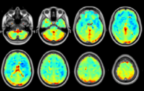
In this study, Dr. Zhao’s team enrolled nearly 30 adult Moyamoya patients without acute stroke or tissue infarcts in the brain. They scanned the brain of these patients using the advanced ASL MRI technique (multi-delay ASL) that were tested successfully on normal and healthy people before. Similar to the study designed in the previous blog, Dr. Zhao’s team collected both ASL and PET images from the Moyamoya patients at the same time before these patients underwent their bypass surgery. The results from this study were convincing that ASL can replace PET to characterize the pathophysiology of Moyamoya disease patients. The image in this blog shows the vessel occlusion and its impact on blood flow in a Moyamoya patient.

Another advantage of ASL is the elimination of contrast agents. In many MRI exams, patients need to receive a contrast agent (such as gadolinium) to enhance the image quality. However, the contrast agent can cause side effects and deposit in the brain. Although there is no evidence suggesting its impact on health, we should minimize the use of these contrast agents. In the same study, Dr. Zhao’s team also demonstrated that the advanced ASL can create images without contrast agents and that doctors favored the new ASL technique over the conventional contrast-based imaging method.

Taken together, ASL MRI will become more accessible to radiologists to diagnose Moyamoya disease without causing side effects to patients. The technique will allow safe, affordable, and fast for imaging to identify patients with a higher risk for stroke.
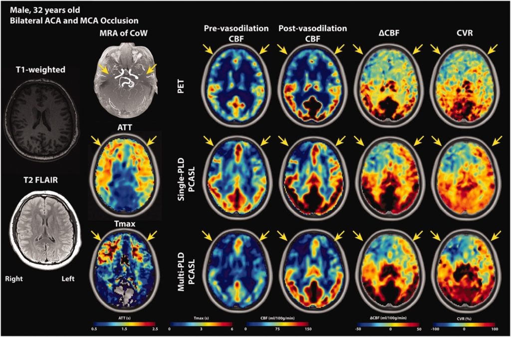
Image source: JCBFM


REFERENCE

https://doi.org/10.1177/0271678X221083471


“The views, opinions, and positions expressed within this blog are those of the author(s) alone and do not represent those of the American Heart Association. The accuracy, completeness, and validity of any statements made within this article are not guaranteed. We accept no liability for any errors, omissions, or representations. The copyright of this content belongs to the author and any liability with regards to infringement of intellectual property rights remains with them. The Early Career Voice blog is not intended to provide medical advice or treatment. Only your healthcare provider can provide that. The American Heart Association recommends that you consult your healthcare provider regarding your health matters. If you think you are having a heart attack, stroke, or another emergency, please call 911 immediately.”

hidden

PET or MRI, that is the question – Part 1

In our previous blogs, we discussed that doctors can diagnose Moyamoya disease using medical imaging systems, such as positron emission tomography (PET) and magnetic resonance imaging (MRI). But which one is better?

According to some recent studies performed by Dr. Moss Zhao (AHA Postdoctoral Fellow, 2021) at Stanford University, MRI is better thanks to its accessibility, safety, and affordability.

For decades, PET has been the gold standard technique for diagnosing Moyamoya disease based on imaging the blood flow in the abnormal blood vessels in the brain. However, PET uses radioactive tracers to create the images, making it complex and expensive. Although the amount of radiation is less than a 5-hour flight, doctors have sought for alternative and less invasive techniques to replace PET, especially for children. In recent years, arterial spin labeling (ASL), an advanced MRI technique, has emerged to replace PET imaging to measure blood flow in the brain. Because ASL MRI is more accessible at most hospitals and the procedure is less complex than PET, ASL has gained popularity in many research and clinical institutions for Moyamoya patients.

At Stanford University, Dr. Moss Zhao demonstrated that ASL could replace the conventional PET imaging technique without exposing patients to radiation and causing side effects. The image quality and measurement accuracy of ASL are compatible with PET, implying that ASL can be used to characterize the abnormal blood flow and circulation in Moyamoya patients. Among the different implementations of ASL, Dr. Zhao developed an advanced technique dubbed ‘multi-delay ASL’ that gives the best image quality with the least amount of scanning time. For less than 5 minutes, multi-delay ASL can produce images that require more than 20 minutes for PET imaging. The image in this blog shows the images collected by ASL and PET at the same time from normal and healthy people. Using the latest ASL techniques, doctors can identify patients with a high risk for stroke based on their MRI scans for just under 5 minutes without using any radioactive substances. Dr. Zhao’s team is currently testing this technique on the pediatric population to enable this non-invasive imaging technology to be accessible to patients across the lifespan.

Image source: NeuroImage

References:

https://doi.org/10.1016/j.neuroimage.2021.117955


“The views, opinions, and positions expressed within this blog are those of the author(s) alone and do not represent those of the American Heart Association. The accuracy, completeness, and validity of any statements made within this article are not guaranteed. We accept no liability for any errors, omissions, or representations. The copyright of this content belongs to the author and any liability with regards to infringement of intellectual property rights remains with them. The Early Career Voice blog is not intended to provide medical advice or treatment. Only your healthcare provider can provide that. The American Heart Association recommends that you consult your healthcare provider regarding your health matters. If you think you are having a heart attack, stroke, or another emergency, please call 911 immediately.”

hidden

A Possible Link Between That “Gut Feeling” and Heart Disease

Let’s face it- this #COVID19 pandemic has found us seesawing between embracing extra workouts and healthier homemade meals to lamenting over those extra pounds from those sourdough starters. Many of us can use a jump start to reclaim our #hearthealthy goals. #AHA20 has provided us a captivating session on the link between diet, the gut microbiome, and cardiovascular disease.

Drs. Katherine Tucker, Wilson Tang, and Caroline Genko presented the basics of how the quality of our diet affects the diversity of bacteria and level of systemic inflammation in the body, the role of the TMAO pathway in atherosclerosis, and how oral pathogens can affect both atherosclerosis and the gut microbiome:

SCIENCE: Epigenetics alter the transcription of genes through modification (DNA methylation, histone modification, and miRNAs). These processes can be affected by stress, diet, and the microbiome.

TAKEAWAY: We have some control over the expression of our genes if we adhere to healthy lifestyle changes that improve our stress, sleep, diet, and physical activity.

SCIENCE: Gut bacteria metabolize indigestible fibers into short-chain fatty acids (SCFAs) which have been associated with the prevention of chronic disease and are also important for muscle function.

TAKEAWAY: Continue eating more plants and whole grains to prevent heart disease and other chronic diseases.

SCIENCE: Animal-based diets increase bile-tolerant microorganisms that are responsible for pro-inflammatory pathways.

TAKEAWAY: Limit animal meat (especially red meat) to decrease the amount of inflammation in your body. Inflammation = heart disease!

SCIENCE: The more highly processed the diet is, the higher the risk of CV disease even after multivariable adjustment.

TAKEAWAY: Limit the middle aisles of your grocery store and go rogue on the perimeter aisles, which should be abundant in perishables (fruits and vegetables) and freshly baked whole grains.

SCIENCE: Increased TMAO levels are associated with atherosclerosis and are only one of the many pathways involved in the link between the gut and CV disease. Red meat is associated with elevated TMAO levels. There is also unique crosstalk between organs, with reduced excretion of TMAO by the kidneys with increased consumption of red meat. Lastly, caloric restriction and intermittent fasting have been associated with decreased levels of TMAO.

TAKEAWAY: As above, another reason to limit or avoid red meat!

SCIENCE: Animal studies showed those fed a Western diet and infected with P. gingivalis (oral pathogenic bacteria) had accelerated atherosclerosis. The gut microbiome composition was also affected by P. gingivalis infection.

TAKEAWAY: Be sure to take care of your oral health and see your dentist regularly for cleanings to help prevent cardiovascular disease.

After reading this, I hope you all reconsider how you think about going with your “gut” when deciding on your next meal.

Eat well, be well, and be safe. And keep posting #PetsofAHA20.


“The views, opinions and positions expressed within this blog are those of the author(s) alone and do not represent those of the American Heart Association. The accuracy, completeness and validity of any statements made within this article are not guaranteed. We accept no liability for any errors, omissions or representations. The copyright of this content belongs to the author and any liability with regards to infringement of intellectual property rights remains with them. The Early Career Voice blog is not intended to provide medical advice or treatment. Only your healthcare provider can provide that. The American Heart Association recommends that you consult your healthcare provider regarding your personal health matters. If you think you are having a heart attack, stroke or another emergency, please call 911 immediately.”