hidden

Should interventional cardiologists perform thrombectomy?

“Sutor, ne ultra crepidam” a Latin expression for shoemakers not beyond the shoe, a common saying to warn people to avoid passing a judgment beyond their expertise.

With mechanical thrombectomy changing the management of stroke and becoming the standard of care for patients with large vessel occlusion (LVO), a new challenge has emerged, adequate access for care.

A recent cross-sectional study by Aldstadt et al.1 aimed to determine the percentage of the US population with 60 min (ground or air) to a designated or non-designated endovascular capable stroke center, or percentage of non-designated endovascular centers that were 30 min from an endovascular capable center.  They reported that overall a 49.6% of the US population is within 60 min of an endovascular capable stroke center, while 37% of the US population lacked access to endovascular capable centers within 60 min. For the non-endovascular stroke centers, 84% have access within 60 min, and 45.4% are within 30 min drive from an endovascular capable stroke center.

Since time is the brain, increasing the access to care is of paramount importance and increasing the number of well-trained physicians equipped to perform and treat stroke holistically.  Since there are approximately 10 times more interventional cardiologists, radiologists, and vascular surgeons than neuro interventionalist in the USA (10.000 vs. 800-1000)2, some non-endovascular capable hospitals have explored the option of incorporating some of this workforce to contribute to patient care.

Some retrospective studies3 with low sample sizes have described that their interventional cardiologist team was able to perform a thrombectomy safely, with the guidance of a stroke neurologist. Nonetheless, they are not clear on the prior training these cardiologists have had regarding neurovasculature, the nuances of the procedure, critical care, and stroke neurology.

Endovascular Neurosurgery and Interventional Neuroradiology is a field shared by physicians with different backgrounds in training, such as neurosurgeons, neurologists, and interventional radiologists. Regardless of their background or training, they are all required to complete an additional 1-2 years of training exclusively for neurointervention. Endovascular physicians trained rigorously per ACGME4 requirements were most of the physicians involved in the clinical trials (ESCAPE and DAWN) and maintained a high caseload volume of thrombectomy. The cumulative case volume is crucial since it has been associated independently for obtaining good recanalization and outcomes.5

Even if the technical aspects have various similarities between the endovascular fields, shoemakers not beyond the shoe, cannot be translated from one field to another without proper training. To adduce that interventionalist cardiologist can inherently treat intracranial diseases would be, in my opinion, not in benefit of the care of the patient, even if they are the only option nearby where no endovascular treating center can be reached, the patient outcome of patients is directly correlated with the expertise of the treating physician.

Nonetheless, interventional cardiologists should only be allowed to perform thrombectomies if they complete a full endovascular fellowship with the requirements established by the ACGME and as the other specialties go through (interventional radiology, neurosurgery, and neurology). This formal training could contribute to those rural areas where there is no possibility to access an endovascular center. More efforts should be made to increase access to endovascular capable stroke centers, to continue training neurosurgeons, radiologists, and neurologists to meet patients’ demands requiring this life-saving treatment.  But I don’t consider converting specialists in treating myocardial infarctions to stroke being a priority in the US.

REFERENCE

  1. Aldstadt J, Waqas M, Yasumiishi M, et al. Mapping access to endovascular stroke care in the USA and implications for transport models. Journal of NeuroInterventional Surgery. 2021:neurintsurg-2020-016942.
  2. Hopkins LN, Holmes DR. Public Health Urgency Created by the Success of Mechanical Thrombectomy Studies in Stroke. Circulation. 2017;135(13):1188-1190.
  3. Hornung M, Bertog SC, Grunwald I, et al. Acute Stroke Interventions Performed by Cardiologists: Initial Experience in a Single Center. JACC Cardiovasc Interv. 2019;12(17):1703-1710.
  4. Hussain S, Fiorella D, Mocco J, et al. In defense of our patients. J Neurointerv Surg. 2017;9(6):525-526.
  5. Kim BM, Baek J-H, Heo JH, Kim DJ, Nam HS, Kim YD. Effect of Cumulative Case Volume on Procedural and Clinical Outcomes in Endovascular Thrombectomy. Stroke. 2019;50(5):1178-1183.

“The views, opinions and positions expressed within this blog are those of the author(s) alone and do not represent those of the American Heart Association. The accuracy, completeness and validity of any statements made within this article are not guaranteed. We accept no liability for any errors, omissions or representations. The copyright of this content belongs to the author and any liability with regards to infringement of intellectual property rights remains with them. The Early Career Voice blog is not intended to provide medical advice or treatment. Only your healthcare provider can provide that. The American Heart Association recommends that you consult your healthcare provider regarding your personal health matters. If you think you are having a heart attack, stroke or another emergency, please call 911 immediately.”
hidden

Bifurcations: EPISODE 3 – TAP TECHNIQUE

As the summer holidays wind down to the final few days, many of us are heading back to the routine of work, school and home.  With the end of summer, my Bifurcation Series comes to a close as well. The final episode is the TAP technique.

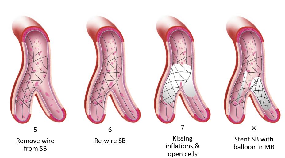
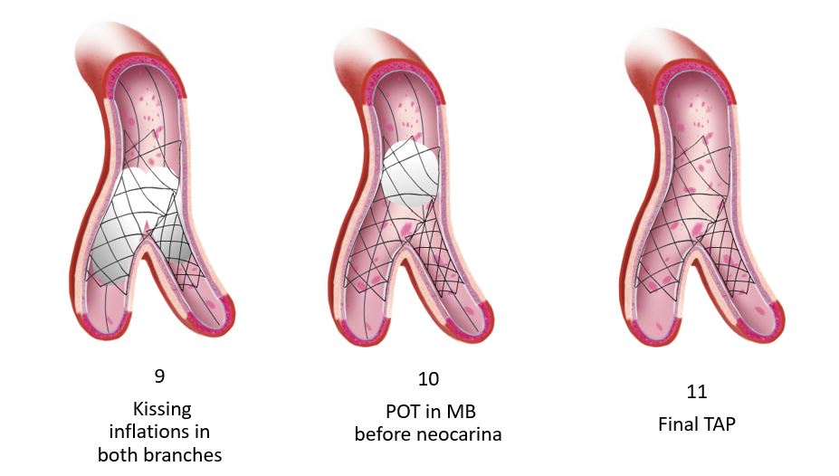
Operators find this to be the least cumbersome of all the 2-stent strategies. Many resort to it during emergencies as the access to the main branch (MB) is maintained throughout the procedure. The steps are fewer which ensures expeditious coverage of both vessels followed by the conventional optimization steps including kissing and proximal optimization with a non-compliant balloon. Similar to culotte, this strategy allows operators to start with a provisional strategy and convert to TAP should the need arise. In addition, there is minimal stent overlap. This technique is considered a modification of what was formally known as T-stenting. The primary limitation of the original T-stenting was missing the ostium of the side branch (SB). This geographic miss is what prompted many operators to perform minimal protrusion to mitigate in-stent restenosis at that missed segment. Hence the name TAP, T and small protrusion, was coined. Although this technique has been adopted worldwide, there are no large randomized trials with long term outcome data to reference. There are some published data; however, that are worth reviewing.


Study TAP strategy Patients (n) Unprotected
left main stem
Follow-up
duration
TVR Definite stent
thrombosis
Burzotta et al’ Bail-out TAP in provisional 73 37.0% 9 months 6.8% 1.40%
Al Rashdan
et al7
Systematic TAP 156 10.3  % 36 months
(range 24-48 months)
5.3% 0.06%
Burzotta et a1 Bail-out TAP in
provisional procedures
19 5.0% 12 months 5.3% none
Naganuma et al Bail-out TAP (type B dissection or
TlMI <3 or stenosis >50% in the SB)
95 18.9% 36 months 9.7% none
ARTEMIS
study10
Bail-out TAP (type B dissection or
TlMI <3 or stenosis >75% in the SB)
71 26.8% 12 months 8.5% none
 SB:side branch;TVR: target vessel revascularisation


Burzotta et al, 2007

The modification of the T-stenting was first described in 2007 by Burzotta et al.1 It was evaluated in vitro and in two independent series of patients undergoing elective drug-eluting stent (DES) implantation on a bifurcation lesion. In vitro testing demonstrated perfect coverage of the bifurcation with minimal stent’s struts overlap at the proximal segment of SB ostium with a single layer stent struts. Sirolimus, paclitaxel, or zotarolimus DES were deployed in 73 patients (67% with Medina 1,1,1 lesions and 44% of unprotected distal left main disease) using the TAP technique. The procedural success was achieved in all cases. At 9 months the clinically-driven target vessel revascularization (TVR) was 6.8%. Since this was a pilot study, the investigators recommended larger outcome trials to further evaluate this technique. No comparison arm was available in this initial trial.


Al-Rashdan et al, 2009

In 2009 Al-Rashdan et al published their series of 156 consecutive patients who underwent TAP stenting.2 This was a single center study that resulted in a 99% procedural success rate and a major adverse cardiac events (MACE) free survival rate of 88% at 36 months average follow up. The TVR rate was 5.3%. Although to date this represents the largest cohort of TAP cases, the results are limited to a single center with no randomization which precludes further conclusions.


Burzotta et al, 2009

In 2009, Burzotta’s group prospectively enrolled 266 consecutive patients requiring treatment of a bifurcation lesion.3 The MB was treated with a DES and TAP was reserved as a bailout strategy. Only 19 of the total required a bailout 2-stent strategy. Nine percent of the total had unprotected left main disease. At one year, the MACE rate was 8.2%. A non-hierarchical analysis revealed a 0.4% cardiac death, 4.1% MI, 4.5% TVR and 2 of the total had probable stent thrombosis (ST).  Given the small number of bailout 2-stent strategy arm, this study only demonstrates safety.


Naganuma et al, 2013

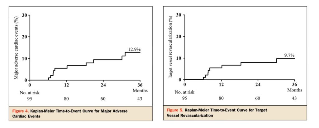
Naganuma et al retrospectively analyzed data of all patients who underwent TAP technique with DES between July 2005 and January 2012.4 A total of 95 patients were enrolled. Angiographic procedural success was achieved in all cases. A true bifurcation was found in 78.9% of those enrolled. The 3-year MACE, cardiac death or myocardial infarction, TVR and target lesion revascularization (TLR) rates were 12.9%, 3.1%, 9.7%, and 5.1%, respectively. No ST was observed in this cohort. Once again, the investigators recommend larger trials to make solid recommendations.

Naganuma et al, JACC Cardiovasc Interv. 2013;6:554-61.

Naganuma et al, JACC Cardiovasc Interv. 2013;6:554-61.


ARTEMIS Study 2014

The ARTEMIS study was published in 2014.5 It evaluated the mid-term angiographic results of TAP as the bailout strategy in symptomatic patients who were treated with one-stent strategy (DES of the MB) and kissing balloon inflation of the SB who subsequently developed impingement of the branch. TAP was performed if residual diameter stenosis of SB was ≥75%, presence of ≥type B dissection or flow impairment of the SB occurred. A total of 71 patients were enrolled with a MEDINA classification 1,1,1 lesions occurring in 60% of the total. At 9 months, restenosis was occurred in 12.5% of the total. Late lumen loss in the MB and SB was 0.22 ± 0.19 and 0.34 ± 0.37 mm, respectively.


Dzavik et al, 2014

In 2014, there was much hype revolving around bioresorbable technology. Dzavik et al performed in vitro bifurcation stenting employing different modalities on synthetic arterial models.6 The everolimus-eluting bioresorbable vascular scaffold (Abbott Vascular, Santa Clara, California) (BVS) was used. A low-pressure final kissing balloon inflation was performed to complete the procedures. The results demonstrated that a single-stent technique optimally opened the SB without deforming the BVS in the MB. T or TAP-stenting covered the SB ostium completely. Culotte and crush with 2 BVS stents was successful; however, disruption was reported after the low pressure kissing inflation in one case. Investigators concluded that it was feasible to perform bifurcation stenting with BVS in large caliber vessels. They also recommended that a provisional strategy as the default. TAP or T-stenting with a metal DES is preferable. As the overall in vivo outcome data for BVS remains cautionary at best, the use of BVS outside clinical trials is not recommended whether for focal type A lesions or complex bifurcations.

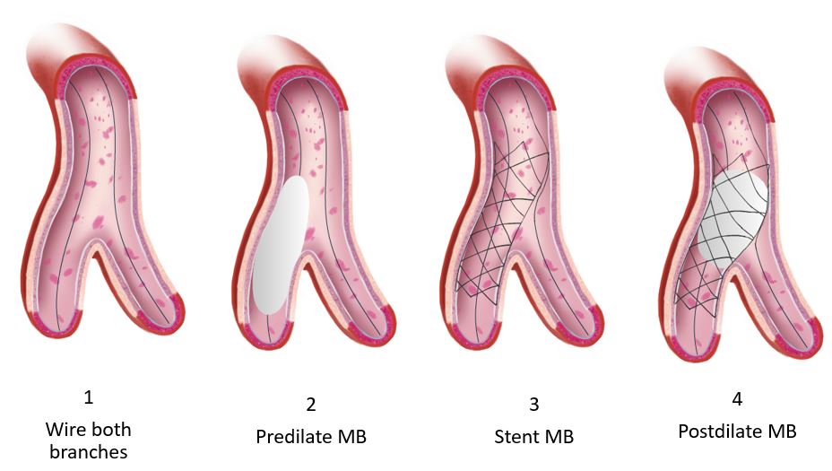
The technique itself is illustrated below. As mentioned earlier, it is one of the simpler 2-stent strategies. Like other strategies, appropriate sizing, positioning and optimization ultimately dictate the final angiographic and clinical outcomes. Intracoronary imaging facilitates these crucial steps. Yet, as with all interventions, judgment is the cornerstone of any successful procedure. When appropriate, and based on both Syntax score and clinical scores, surgical revascularization should be considered. When one opts for percutaneous revascularization, the indication for the procedure, its potential risks and complexity should be shared with the patient. For operators, judging the significance of the SB, the angle of the bifurcation, the size of both vessels and the need for mechanical circulatory support is valuable. Finally, complex bifurcation stenting is not for everyone. When appropriate, such complex procedures should be referred to expert operators for the best outcomes.

Animations/illustrations courtesy of Graphic Designer Dania Al-Shaibi

Email: [email protected]


References:

  1. Burzotta F, Gwon HC, Hahn JY, Romagnoli E, Choi JH, Trani C, Colombo A. Modified T-stenting with intentional protrusion of the side-branch stent within the main vessel stent to ensure ostial coverage and facilitate final kissing balloon: the T-stenting and small protrusion technique (TAP-stenting). Report of bench testing and first clinical Italian-Korean two-centre experience. Catheter Cardiovasc Interv.2007;70:75-82.
  2. Al Rashdan I, Amin H. Carina modification T stenting, a new bifurcation stenting technique: clinical and angiographic data from the first 156 consecutive patients. Catheter Cardiovasc Interv.2009;74:683-90.
  3. Burzotta F, Sgueglia GA, Trani C, Talarico GP, Coroleu SF, Giubilato S, Niccoli G, Giammarinaro M, Porto I, Leone AM, Mongiardo R, Mazzari MA, Schiavoni G, Crea F. Provisional TAP-stenting strategy to treat bifurcated lesions with drug-eluting stents: one-year clinical results of a prospective registry. J Invasive Cardiol.2009;21:532-7.
  4. Naganuma T, Latib A, Basavarajaiah S, Chieffo A, Figini F, Carlino M, Montorfano M, Godino C, Ferrarello S, Hasegawa T, Kawaguchi M, Nakamura S, Colombo A. The long-term clinical outcome of T-stenting and small protrusion technique for coronary bifurcation lesions. JACC Cardiovasc Interv. 2013;6:554-61.
  5. Jim MH, Wu EB, Fung RC, Ng AK, Yiu KH, Siu CW, Ho HH. Angiographic result of T-stenting with small protrusion using drug-eluting stents in the management of ischemic side branch: the ARTEMIS study. Heart Vessels.2014 Mar 14.
  6. Dzavik V, Colombo A. The absorb bioresorbable vascular scaffold in coronary bifurcations: insights from bench testing. JACC Cardiovasc Interv.2014;7:81-8.


hidden

Bifurcations: From An Interventional Cardiologist’s Perspective

Approximately 15-20% of all coronary interventions are bifurcations1. Based on the overall Syntax Score, coronary artery bypass grafting is often recommended particularly in the setting of multi-vessel disease, diabetes and impaired left ventricular function. Once a decision to proceed with percutaneous revascularization is made, it is imperative that operators select the most appropriate revascularization strategy suited for an individual patient.

Briefly, the published data still recommends a provisional strategy as the default1. Of course, a two-stent strategy is commonly employed for bail out. An up-front two stent strategy is reserved for the following:

  1. This strategy is recommended for true Bifurcation Lesions with a Medina (111,011,101). This is of importance if the lesion is long and extends > 5-10 mm beyond the ostium of a sizeable side branch (> 2.5mm).
  2. If the side branch is very large, poor myocardial reserve, and a high jeopardy score, the hemodynamic consequences may be significant warranting revascularization of the side branch.
  3. Finally, if the angle to the side branch is acute rendering access too difficult, it is advisable to proceed with a two-stent strategy.

Over the course of the next couple of blogs, I will briefly review the randomized trials and steps of the different strategies. This month, my focus will be on DK-Crush.

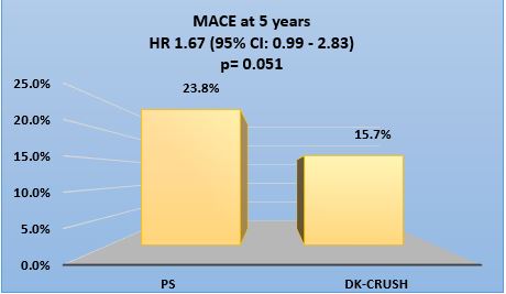
DK Crush II was a randomized trial comparing DK-Crush to provisional stenting in symptomatic patients with a Medina 1,1,1 or 0,1,1 lesion. One-hundred and eight five were enrolled in each arm. The primary endpoint was major adverse cardiac events (MACE), namely cardiac death, MI and target vessel revascularization (TVR) at 5 years. It concluded that DK-Crush was associated with a lower 5-year MACE rate compared to provisional stenting.


Chen et al. Circ Cardiovasc Interv 2017;10:e 004497

Chen et al. Circ Cardiovasc Interv 2017;10:e 004497


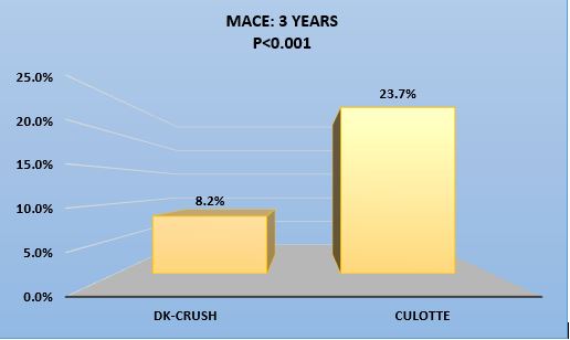
DK Crush III was a randomized trial comparing DK-Crush to Culotte stenting in symptomatic patients with a distal Left Main lesion that is Medina 1,1,1 or 0,1,1. Approximately, 210 were enrolled in each arm. The primary endpoint was a composite of MACE and TVR at 3 years. It concluded that DK-Crush was associated with a lower 3-year MACE rate compared to Culotte stenting.

Chen et al. JACC. Cardiovasc. Interv 2015;8:1335-42                                 

3-years outcome DK crush Culotte p-value
Death % 1.4 2.9 0.34
MI % 3.4 8.2 0.037
TVR % 5.8 18.8 <0.001
Definite ST % 0 3.4 0.007

Chen et al. JACC. Cardiovasc. Interv 2015;8:1335-42


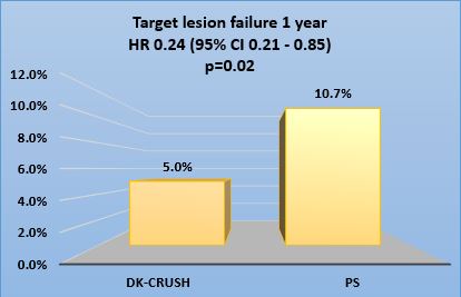
DK Crush V was a randomized trial comparing DK-Crush to a provisional strategy in symptomatic patients with a distal Left Main lesion that is Medina 1,1,1 or 0,1,1.  It enrolled approximately 240 patients in each arm. The primary endpoint was target lesion failure defined as cardiac death, target vessel myocardial infarction or target lesion revascularization at 12 months. It concluded that DK-Crush was superior at 12 months.

Chen et al. J Am Coll Cardiol. 2017;70:2605-17

Chen et al. J Am Coll Cardiol. 2017;70:2605-17


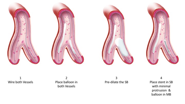
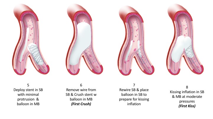
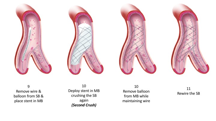
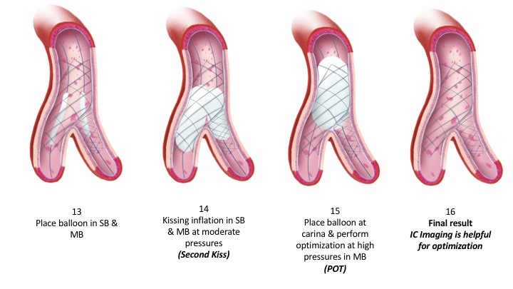
The steps of this technique can be summarized in the illustration (Image A). It is noteworthy, that critical steps in any bifurcation technique include intracoronary imaging and proximal optimization (POT). Imaging allows appropriate determination of size of the vessels in question, length of the disease and characterization of the lesion before the planned strategy. It, therefore, permits the operator to perform the necessary lesion preparation if calcified. Upon completion of the procedure, imaging allows appropriate evaluation of the stent expansion and apposition with additional post-dilatation if need be. Proximal optimization is a fundamental step irrespective of the technique adopted. It permits the operator to expand the main branch stent to facilitate the remainder of the steps, prevent the wire from entering behind the stent struts, prevent stent compression and ultimately permit appropriate stent apposition. This in itself facilitates future intervention and reduces stent thrombosis. Many have added an additional POT before the second kissing inflation to facilitate crossing into the SB. It is important that fluoroscopic imaging is sharp to allow appropriate positioning of the non-compliant balloon at the proximal stent edge and at the neo-carina. Several techniques including “Stent Boost” and “Clear Stent” specific to each vendor are readily available. Finally, given the multiple steps in bifurcation stenting, radiation safety is imperative for any and all techniques.

Image: Steps of DK Crush

Illustrations are the production of Graphic Designer Dania Al-Shaibi ([email protected])


References:

  1. Jens Flensted Lassen1* MD, PhD; Niels Ramsing Holm1, MD; Goran Stankovic2, MD, PhD; Thierry Lefèvre3, MD; Alaide Chieffo4, MD; David Hildick-Smith5, MD; Manuel Pan6, MD; Olivier Darremont7, MD; Remo Albiero8, MD; Miroslaw Ferenc9, MD; Yves Louvard3, MD. Percutaneous coronary intervention for coronary bifurcation disease: consensus from the first 10 years of the European Bifurcation Club meetings. EuroIntervention 2014;10:545-560.
  2. Hildick-Smith D, de Belder AJ, Cooter N, Curzen NP, Clayton TC, Oldroyd KG, Bennett L, Holmberg S, Cotton JM, Glennon PE, Thomas MR, Maccarthy PA, Baumbach A, Mulvihill NT, Henderson RA, Redwood SR, Starkey IR, Stables RH. Randomized trial of simple versus complex drug-eluting stenting for bifurcation lesion: the British Bifurcation Coronary Study: old, new, and evolving strategies. Circulation. 2010;121:1235-43.
  3. Behan MW, Holm NR, Curzen NP, Erglis A, Stables RH, de Belder AJ, Niemela M, Cooter N, Chew DP, Steigen TK, Oldroyd KG, Jensen JS, Lassen JF, Thuesen L, Hildick-Smith D. Simple or complex stenting for bifurcation coronary lesions: a patient-level pooled-analysis of the Nordic Bifurcation Study and the British Bifurcation Coronary Study. Circ Cardiovasc Interv. 2011;4:57-64.
  4. Colombo A, Bramucci E, Sacca S, Violini R, Lettieri C, Zanini R, Sheiban I, Paloscia L, Grube E, Schofer J, Bolognese L, Orlandi M, Niccoli G, Latib A, Airoldi F. Randomized study of the crush technique versus provisional side-branch stenting in true coronary bifurcations: the CACTUS (Coronary Bifurcations: Application of the Crushing Technique Using Sirolimus-Eluting Stents) Study. Circulation. 2009;119:71-8.
  5. Ferenc M, Gick M, Kienzle RP, Bestehorn HP, Werner KD, Comberg T, Kuebler P, Buttner HJ, Neumann FJ. Randomized trial on routine vs. provisional T-stenting in the treatment of de novo coronary bifurcation lesions. Eur Heart J. 2008;29:2859-67.
  6. Maeng M, Holm NR, Erglis A, Kumsars I, Niemela M, Kervinen K, Jensen JS, Galloe A, Steigen TK, Wiseth R, Narbute I, Gunnes P, Mannsverk J, Meyerdierks O, Rotevatn S, Nikus K, Vikman S, Ravkilde J, James S, Aaroe J, Ylitalo A, Helqvist S, Sjogren I, Thayssen P, Virtanen K, Puhakka M, Airaksinen J, Christiansen EH, Lassen JF, Thuesen L. Long-term results after simple versus complex stenting of coronary artery bifurcation lesions: Nordic Bifurcation Study 5-year follow-up results. J Am Coll Cardiol. 2013;62:30-4.
hidden

Fast and Curious: Rushing to Medical Emergencies

Guidelines in the U.S. support that any patient who presents with a possible heart attack (ST elevation myocardial infarction-STEMI) is planned to have a cardiac catheterization promptly, and ideally within 90 minutes of being diagnosed with an EKG. One of my friends who is an interventional cardiologist like me, recently found himself in a interesting situation. My friend was on call and received an urgent page from the emergency department, notifying him of a patient possibly with an acute presentation of a heart attack. He promptly responded and was rushing over to the hospital where he works, which is about 10 to 15 minutes away from where he lives. En route while rushing, he was going over the speed limit when when suddenly he saw the flashing blue lights of an approaching patrol car. He pulled over and had to wait until a trooper got out of a car and approached him. He showed the patrol man his identification and tried to explain the need for expediency for at least 5 to 10 minutes before he was able to convince the policeman of the nature of the medical emergency, and was allowed to proceed. Fortunately, the patient was promptly treated as soon as my friend arrived, and had a successful outcome without any detriment as a result of the delay. However, this incident brought some issues to my attention.

Most hospitals require interventional cardiologists to live within 15-20 minutes of driving distance – however, in crowded, urban areas, the estimates may well be off especially during crowded business hours and traffic congestion. Remaining in-house during call may circumvent the issue on occasion, but does take a toll on the personal and family life of the interventionist. In the situation of a evolving heart attack, where speed is of the essence – should/could there be some form of an alerting system to enable easy and quick passage for the care providers attending to that emergency? These thoughts crystallized more and while driving today, I pulled to the side to allow a fire truck with blaring sirens and flashing lights to pass me – potentially to respond to call where multiple other trucks were headed. It made me wonder if for a medical emergency where attendance of a physician and a team with a specific skill set maybe life-saving, if having any systems-based assistance was worthwhile if it could be provided without excess cost?

Additionally, it is also imperative for the personal safety of the care providers when their minds may be preoccupied with continuous interruptions in the form of phone calls and pagers going off to coordinate care. I was recently discussing this when it was brought to my notice that a situation like this is not without precedent, and the state of California actually provides tags which allows expedient passage for caregivers rushing to provide emergency medical assistance.

In this era of glo-signs and neon markers with even ride share services like Uber or Lyft displaying logos prominently – would it not be worthwhile for emergency medical care providers to display their mission so as to allow speedy and safe transport? Maybe the thought leaders and national societies can deliberate on this and consider future research to identify and validate the need for same.


hidden

Intermittent Fasting

Recently, there has been growing interest in intermittent fasting. In clinic, I find it is much easier to answer questions about coronary angiography and interventions than it is about fasting. This month, I invited Dr. Christian Assad to provide us with an overview of intermittent fasting in a Video Interview.

Dr. Assad is currently an Interventional Cardiologist and the Director of the CardioMetabolic Clinic at RGV Cardiology in McAllen, Texas. His previous research involves the role of the immune system with heart failure, application of google glass in tele-mentoring. Since he moved to McAllen, Texas (arguably the most obese city in United States) he started focusing on prevention and the therapeutic roles of Intermittent Fasting, Ketogenic diet, Low Carbohydrate diet and other lifestyles that have helped many of his patients revert the metabolic syndrome and put DMII in remission.

The interview addresses all the following:

  1. Is it just a fad or are there health benefits? (Are there health benefits beyond weight loss?)
  2. What are the different methods of intermittent fasting and which do you recommend from your own experience?
  3. Is intermittent fasting recommended for life or only brief periods?
  4. What are the physiological effects of intermittent fasting?
  5. In which individuals would intermittent fasting be contraindicated?
  6. Can we debunk these myths?
    • Diabetes control becomes difficult with interrupted fasting compared to regular frequent meals.
    • Pregnant women who fast can harm the fetus.
    • Elderly individuals don’t benefit from fasting. In fact, fasting may accelerate dementia.
    • Intermittent fasting curtails the effects of exercise and body building programs?