hidden

RNA, DNA, and COVID-19

As my co-blogger Jeff Hsu, MD, PhD said to me this week, the COVID-19 pandemic has created the ultimate hackathon – the world’s smartest people hyperfocused on the same problem. For this month’s blog, I am outlining few ways that genomics researchers are hoping to advance our understanding of SARS-CoV-2.

Pathogen Evolution and Transmission

Scientists around the world have pledged to openly share genetic data to aid in the understanding of pathogen spread, and one of these collections of open-source tools is Nextstrain.1 Nextstrain is a database of viral genomes, a bioinformatics pipeline for phylodynamics analysis, and an interactive visualization platform that presents a real-time view of the evolution and spread of seasonal endemic viral pathogens (e.g. influenza) and emergent viral outbreaks (e.g. SARS-CoV-2, Zika, Ebola).1 Over time, viruses naturally accumulate random mutations into their genomes, and these mutations can be used to identify infection clusters that are closely genetically related. Therefore, this can lend insight into introduction events and growth rates. The Nextstrain 2019-nCoV page shows incredible graphical displays of the inferred phylogeny, global transmission events, and genomic diversity over time. At the time of their most recent Situation Report and Executive Summary (dated 3/27/2020), the Nextstrain team had analyzed 1,495 publicly shared SARS-CoV-2 genomes and provided transmission pattern reports for North America, Europe, Central and South America, Asia, Africa, and Oceania.

For a great introduction to the importance of genomics in identifying the emergence of SARS-CoV-2, check out this Cell Leading Edge Commentary, authored by two of the scientists who were involved in the initial genomic sequencing of the virus.2

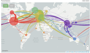
Global map of inferred 2019-nCoV transmission from Nexstrain.

Genetic Influences on Disease Outcomes

In addition to collecting data on viral genomics, researchers have come together to pool genetic data from patients to try to answer urgent questions regarding the variability in clinical outcomes across patients with COVID-19. To investigate the genetic susceptibility to disease, these researchers will be comparing the DNA of different cohorts of patients with COVID-19, for example, those with serious disease to those with more mild manifestations. The COVID-19 Host Genetics Initiative is one of the largest collaborative initiatives with now over 75 biobanks and studies from around the world listed as partners. Their aims are to facilitate sharing of COVID-19 host genetics research, identify genetic determinants of COVID-19 susceptibility and severity, and provide a platform to share the results to the scientific community. Other large national biobanks like UK Biobank and Iceland’s deCODE Genetics are also planning to add COVID-19-related data to their genomic databases.

The COVID-19 Host Genetics Initiative at http://covid19hg.org

How can you keep up with the explosion of data in this space? The Centers for Disease Control and Prevention has created an online Coronoavirus Disease Portal, which is a continuously updated database of scientific literature, CDC and NIH resources, and other materials that pertain to genomics, molecular and other precision medicine and precision public health tools in the investigation and control of coronaviruses, such as COVID-19, MERS-CoV, and SARS.

References

  1. Hadfield et al., Nextstrain: real-time tracking of pathogen evolution, Bioinformatics(2018).
  2. Zhang and Holmes, A Genomic Perspective on the Origin and Emergence of SARS-CoV-2, Cell (2020), https://doi.org/10.1016/j.cell.2020.03.035

“The views, opinions and positions expressed within this blog are those of the author(s) alone and do not represent those of the American Heart Association. The accuracy, completeness and validity of any statements made within this article are not guaranteed. We accept no liability for any errors, omissions or representations. The copyright of this content belongs to the author and any liability with regards to infringement of intellectual property rights remains with them. The Early Career Voice blog is not intended to provide medical advice or treatment. Only your healthcare provider can provide that. The American Heart Association recommends that you consult your healthcare provider regarding your personal health matters. If you think you are having a heart attack, stroke or another emergency, please call 911 immediately.”

hidden

microRNA – Small RNA, Enormous Powers

If you could peek inside one of the cells in your body, it more or less looks like a factory in which various machines are undergoing different molecular processes. The ultimate goal of this cell machinery is to synthesize a protein. Thus, to produce these complex structures, there are other entities required in the cells known as DNA and RNA.

Double stranded DNA, which holds all the blueprints for the proteins, firstly gets converted into single stranded RNA molecules by the process called transcription and these newly synthesized RNAs are further translated into proteins. In addition to this, there are some negative regulators of the gene expression at the post transcriptional levels known as microRNA (miRNA).

As the name suggests, miRNAs are small 21–25 nucleotide, single stranded, endogenously expressed RNAs which do not code into proteins. These miRNAs are transcribed from DNA in the nucleus and are processed with the help of enzymes (RNA polymerase II and Drosha) and exits the nucleus upon which its further undergoes cleavage with enzyme called dicer. Once matured, miRNA enters the RNA induced silencing complex (RISC) which guides it to target mRNA sites, where it is able to repress protein production by destabilizing the mRNA and induce translational silencing (Figure 1). miRNAs are fine tuners of the gene expression and have been reported to be critical regulators of cardiomyocyte (CM) differentiation, proliferation and other cellular processes during embryogenesis through the adult life.


Figure 1: miRNA synthesis and processing in the cell (image taken from reference 1)

So, let’s just talk about role of these powerful miRNA in cardiac regeneration. Many non-mammalian vertebrates are capable of complete regeneration of the heart after an injury throughout their life by re-entry of existing CMs into cell cycle and compensating for the loss of contracting cells. However, mammalian hearts are not blessed with such advantage and CMs undergo nuclear division without cytokines leaving most of the cells binucleated. Under normal conditions, adult mammalian CMs renew to replace the cardiomyocytes that undergo apoptosis at a rate of 0.5–1% a year, while this number is insufficient to replace 1 billion CMs lost during myocardial infarction. Thus, the most feasible strategy to overcome this loss is to coax the preexisting CMs into proliferation.

In the heart, miRNAs have been found to be closely intertwined with cardiac signaling and transcriptional pathways to regulate CM proliferation. High throughput miRNA screening has provided new insights in the role of miRNA in sending the CM back in cell cycle and thus, providing an opportunity of innovative therapies of cardiac repair.

In a recent study, Huang and colleagues reported miRNA-128 upregulation in CMs during the postnatal switch from proliferation to terminal differentiation and hence its deletion extends proliferation of postnatal CMs2. Other experiments conducted in mice indicated the increase in mRNA of 2 targets involved in CM growth and proliferation upon deletion of miRNA -133 family members mi-133a-1/miR-133a-2. miRNA-15 family presents another example for role of miRNA in CM proliferation. miR-195 (member of miR-15 family) was found to be a vital regulator of cell cycle genes and its up regulation inhibits cell cycle progression and induces mitosis arrest postnatally.

Further, studies in zebrafish model demonstrated the downregulation of miR-99 and Let-7a/c miRNA family during of regeneration of amputated ventricular apex while over expression of their target proteins resulted in CM differentiation and proliferation. On the contrary, few miRNA have been identified whose induction have led to remarkable stimulation of CM proliferation and cardiac repair. A study conducted on CMs from adult rats received a lot of appreciation when they showed miR-590 and miR-199a delivery to the cells induced their reentry into cell cycle postnatally3.

The studies mentioned above have shown exciting results which can lead to long-lasting and potent effects of miRNA in cardiac repair especially by the stimulation of post-natal CM proliferation (Figure 2). Despite the successful results obtained in vitro studies, miRNA delivery systems for transporting the molecules to the heart have yet to be optimized.

Figure 2: microRNAs in cardiac regeneration

While miRNA is an excellent therapeutic target because of its ability to target multiple genes in a single disease, further works needs to be done to improve its local targeting and reduce cost and toxicity in order to make it possible to use this excellent system to promote CM proliferation in pre-clinical or in clinical settings.


References:

  1. Beezhold KJ, Castranova V, et al. Microprocessor of microRNAs: regulation and potential for therapeutic intervention. Mol cancer. 2010;9:134
  2. Huang W, Feng Y, et al. Loss of microRNA-128 promotes cardiomyocyte proliferation and heart regeneration. Nat Commun 2018;9:700. 
  3. Katz MGFargnoli AS, et al. The role of microRNAs in cardiac development and regenerative capacity. Am J Physiol Heart Circ Physiol.2016;310(5):H528-41.