hidden

Innovations in 3-D and 4-D Technology in the Cath Lab

There have been tremendous advances in 3-dimensional (3-D) technologies in the past few years, not only in various medical and surgical fields but also in our daily lives outside of work; with more and more new features in cell phones, computer design programs, and movies!!  4-dimensional (4-D) imaging captures 3-D images over time. These technologies are particularly important in cardiology, especially in interventional cardiology. The heart is a very dynamic organ, and understanding the variation in the anatomy of vessels and geometry of cardiac structures is key to ensuring successful procedures, patient’s safety and good outcomes. More recently, newer innovations in both 3-D and 4-D technologies have been developed, so I decided to shed light on some of these innovations and how they can be potential game-changers in the cath lab.

  • 3-D Holograms

This technology was actually displayed at the Transcatheter Cardiovascular Therapeutics (TCT) 2019 meeting. It converts live transesophageal echo (TEE) imaging into real-time 3-D holographic video in the cath lab to aid structural heart procedures.  The 3-D hologram is projected on a special display screen, and the interventional cardiologist uses hand movements and a foot pedal/switch to change the image orientation without breaking the sterile field. It also allows the operator to see the tools they use in the cath lab, including catheters or devices, in real-time in a 3-D format. This technology does not even require the user to wear 3-D glasses! It was submitted for FDA regulatory review in September 2019.

  • HeartFlow Planner

This is a noninvasive, real-time virtual tool for coronary artery disease intervention. It allows interventional cardiologists to virtually map vessels on a 3-D coronary tree, with color codes indicating the fractional flow reserve-computed tomography (FFR-CT) values for each vessel as measured by a computational fluid dynamics algorithm. This seems to be a good tool for percutaneous coronary intervention (PCI) planning in vessels with significant disease; as it aims to provide us with a non-invasive way to determine whether a stenotic lesion if potentially flow limiting. However, it is important to note the CT-FFR has its own limitations, and some patients might still need invasive FFR for accurate assessment. This tool was approved by the FDA in September 2019.

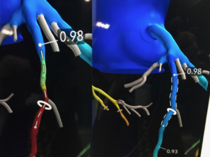
Figure 1: 3-D CT-FFR coronary tree showing both flow limiting and non-flow limiting lesions [from reference 1].

  • 3-D Printing

3-D printing has been used in the surgical fields for more than a decade. It refers to making complex 3-D objects from computer-aided designs. This technology has been increasingly utilized in structural heart procedures in the past few years, where these 3-D models can be printed from a patient’s CT, magnetic resonance imaging (MRI), or 3-D ultrasound images (Figure 1). These 3-D printed structures not only help with procedural planning and device sizing but also allow operators to practice dry runs and perform pre-procedural navigation.

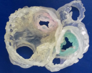
Figure 2: Image of a 3-D printed model which shows cardiac valves and major vessels with their geometric locations relative to each other (reference 3).

  • 4-D Imaging

4-D imaging adds an important component to 3-D imaging, which is the change of these 3-D images over time. 4-D flow images include the direction of blood flow, blood velocities and shear wall stress [2] (Figure 3). This is particularly important in coronary interventions, structural heart procedures and different congenital abnormalities where identification of blood flow in the 4-D view is useful, especially when the anatomy is complex. These changes in position over time help guide our procedures, not only to ensure successful outcomes but also to avoid potential complications. These 4-D images require large amounts of data, but they can be obtained from either cardiac MRI or computational fluid dynamics, which is a specialized area of mathematics and fluid mechanics in engineering [2]. 4-D imaging is still in its early phases, but it is another exciting advancement in our field.

Figure 3: Representation of an MRI-generated 4-D flow image showing blood flow through the aorta and major vessels (reference 4).

In conclusion, we have seen and continue to see tremendous advances in the innovations of 3-D and 4-D imaging with important implications in our work in the cath lab. With our continued collaboration with informational technology experts, engineers, and scientists, these innovations are potentially game-changers in different fields, including coronary interventions and structural heart procedures. I look forward to seeing how this technology continues to evolve in the coming decades!!

References:

  • Fornell, Dave “Overview of the top news and new technologies at the 2019 Transcatheter Cardiovascular Therapeutics meeting”, November 2019,

https://www.dicardiology.com/article/6-hot-topics-interventional-cardiology-tct-2019

https://www.itnonline.com/content/arterys-showcases-fda-cleared-4d-flow-mri-software-rsna-2016

“The views, opinions and positions expressed within this blog are those of the author(s) alone and do not represent those of the American Heart Association. The accuracy, completeness and validity of any statements made within this article are not guaranteed. We accept no liability for any errors, omissions or representations. The copyright of this content belongs to the author and any liability with regards to infringement of intellectual property rights remains with them. The Early Career Voice blog is not intended to provide medical advice or treatment. Only your healthcare provider can provide that. The American Heart Association recommends that you consult your healthcare provider regarding your personal health matters. If you think you are having a heart attack, stroke or another emergency, please call 911 immediately.”

hidden

How is the COVID-19 pandemic affecting cardiac patients and the cath lab?

Amongst the uncertainty of the future and how this pandemic will end, there comes a huge responsibility on all healthcare workers to care for the sickest patients while staying safe, to lead their healthcare systems and to come together as one unit against this crisis. Seeing how this pandemic has affected our healthcare system, from the evolving utility of telehealth to canceling elective procedures in multiple specialties, I decided to share a few thoughts on COVID-19 and the cardiovascular manifestations associated with this infection and how COVID-19 has affected our approach to commonly performed procedures in the cardiac catheterization lab.

Cardiovascular manifestations of COVID-19

Although coronavirus predominantly affects the respiratory system, causing a variety of symptoms from flu-like symptoms to acute respiratory failure requiring intubation, it can affect other organs, and patients may present with non-respiratory complaints [1]. The cardiovascular manifestations of COVID-19 have a wide range of clinical presentations (Figure 1), from pericarditis, myocarditis, pericardial effusion, and decompensated heart failure to tamponade, cardiogenic shock and ST-elevation myocardial infarction (STEMI) [1-2]. In addition, right ventricular strain should raise suspicion for pulmonary embolism as these infected patients tend to be hyper-coagulable with a high incidence of venous thromboembolism for currently unclear reasons. It is important to recognize these cardiovascular manifestations of COVID-19, as it is thought that cardiovascular involvement is associated with a worse prognosis [1].

Figure 1: Potential risk factors and cardiovascular manifestations of COVID-19 patients.

Approach to Acute Coronary Syndrome (ACS) and Structural Procedures in the Cath Lab in COVID-19 Pandemic

Like any procedural field, interventional cardiology has been affected by this pandemic. All elective procedures are being postponed until the crisis settles down per recommendations from the Centers for Disease Control and Prevention (CDC), with emergent, urgent, time-sensitive procedures still being performed, in an attempt to preserve hospital beds and personal protective equipment (PPE) for COVID-19 patients [2]. This pandemic has led to several changes in the ACS approach across the world, with the main goal of reducing un-necessary exposure to health care workers and limiting the spread of this highly contagious disease [1,2].

To summarize, some of the key changes many hospitals have applied in their approach to some of the most commonly performed percutaneous cardiac procedures in COVID-19 patients:

  • ACS patients
  • Thrombolytics are considered standard therapy in many hospitals for many STEMI patients with symptoms <12 hours and no contraindications: signs of success are resolution of symptoms and/or >50% decrease in ST elevation. If thrombolytics fail, coronary angiogram and percutaneous coronary intervention (PCI) should be considered in an isolated cath lab (Figure 2) and the benefit versus the exposure risk should be carefully evaluated.
  • For patients with low risk Non-ST elevation myocardial infarction (NSTEMI): medical therapy is reasonable. For those with high risk NSTEMI or failure of medical treatment, coronary angiogram and/or PCI should be performed. These coronary procedures are usually performed in isolated cath labs with only limited staff in the cath lab room, to avoid unnecessary exposure (Figure 2).
  • Structural heart disease patients
  • For patients who are unstable and have severe aortic stenosis (AS), balloon aortic valvuloplasty (BAV) should be performed emergently to stabilize the patient if this is thought to be the cause of the hemodynamic instability. For those who develop significant aortic regurgitation after BAV, transcatheter aortic valve replacement (TAVR) should be performed.
  • Patients with severe symptomatic AS, TAVR is considered a time-sensitive procedure. The benefit of TAVR and risk of infection exposure should be discussed.It is important to note that this change in policy, in part, could explain the decrease in cath lab activation rates for STEMI during this pandemic [2]; as many STEMI patients are receiving thrombolytics. In addition to that, many patients are not seeking medical care, or if they do, they are presenting late, possibly due to fear from contracting the virus in the hospital, potentially leading to late STEMI complications [2]. Thus, it is important to counsel our patients in our virtual clinic visits that if significant symptoms develop, they should seek medical care.

Lastly, I want to thank all of my colleagues and healthcare workers across the nation and across the globe for risking their lives for patients and for their continued dedication. This is a critical time for everyone in the health care system. In light of this pandemic, I hope we continue to learn and share our experiences in this global crisis in order to improve our patient’s care, safety for our healthcare workers and the dynamics of our healthcare system.

Figure 2: Simplified Set-up of an Isolated Cath Lab during the COVID-19 pandemic.

References

“The views, opinions and positions expressed within this blog are those of the author(s) alone and do not represent those of the American Heart Association. The accuracy, completeness and validity of any statements made within this article are not guaranteed. We accept no liability for any errors, omissions or representations. The copyright of this content belongs to the author and any liability with regards to infringement of intellectual property rights remains with them. The Early Career Voice blog is not intended to provide medical advice or treatment. Only your healthcare provider can provide that. The American Heart Association recommends that you consult your healthcare provider regarding your personal health matters. If you think you are having a heart attack, stroke or another emergency, please call 911 immediately.”


hidden

Referral Letter

A typical referral letter arrives in my tray:

A 55-year-old diabetic female noticed pain and discoloration of the right foot for several days.


Immediate admission was arranged for this patient:

A junior resident presents the case. She has clerked the patient and filled all the necessary forms and ticked all the necessary boxes. Everything is dated and signed appropriately. Her ankle brachial index is 0.7 on the right and 1.1 on the left. The patient falls in a Rutherford Class IV. With such thorough documentation, we were ready to pay the patient a visit and consent her for the planned peripheral angiogram and revascularization. This resident rounds with me every day and has clearly understood the process of consenting: procedural steps and complications included. I told my resident that I appreciate her energy and proactivity. I was so proud of her and thought “Hmm very little left for me to teach this young lady”. So, we walked over to the patient’s room to meet her and her family.


We enter the patient’s room:

My resident starts introducing the patient to me and recapping her history. In the meantime, I look at my patient from a distance. I immediately notice the anxious look. Perhaps she’s nervous about her foot and a possible amputation. I also notice how thin she is. Something doesn’t add up.  Diabetes is on the rise worldwide. Most of our poorly controlled diabetics who present with peripheral vascular disease have other end organ damage like some nephropathy or retinopathy. Most are overweight. She has none. I flip the chart in my hand and notice that her HbA1C is 7%. I ask about her weight loss. “It’s all very recent”, she says. I ask about other constitutional signs and she states there are none. She also has had no history of claudication. This doesn’t sound like long standing atherosclerosis. I approached her to examine her. I held her hand for the first time to feel her pulse. Oh, the lost art of a physical examination..Why examine her when her peripheral vasculature will be defined by a CTA and all the boxes in the chart are ticked?  The answer was right there: she was in atrial fibrillation. I glance at her EKG and sure enough she is in atrial fibrillation. Her hands told me more. She was sweating and had a tremor. I knew at this point what I needed to teach my resident. Medicine is so vast and so integrated. We cannot presume cardiovascular diseases and endocrine disorders are not interrelated. After all, this patient needed an endocrinologist.


Later that day she was taken to the cathlab:

Her TSH was high. Her transesophageal echocardiogram confirmed a left atrial thrombus. We discussed acute limb ischemia with the endocrinologist. We administered B blockers and obtained a consent for the procedure. Her angiogram revealed a thrombus at the right tibioperoneal trunk. Instead of whipping out the QuickCross microcatheter and GlideAdvantage wire, I performed manual aspiration of the thrombus. Fortunately, flow was restored almost immediately. The Society for Vascular Surgery and the North American Chapter of the International Society of Cardiovascular Surgery created a classification that ranged from non-threatened extremity, threatened extremity to finally ischemia with no possible salvage in 2002. Timely intervention is warranted in salvageable cases. Fogarty established surgical thromboembolectomy as the standard of care in the 1960s. Dotter introduced thrombolysis in the 1970s which evolved to current day catheter directed thrombolysis and aspiration. Both surgical and catheter directed thrombectomy have been studied. Theodoridis et al published a review article that indicated the technical success rate of endovascular techniques reaches 79.3%. A second procedure was required in 77.8% of those enrolled. The overall complication rate was 28.7%. Novel techniques that combine catheter directed low dose thrombolysis with and without mechanical thrombectomy have also been evaluated and found to be largely comparable.1-3 Procedural questions related to my patient were confined to the following:

  1. Use of a distal protection device: Trials have demonstrated a wide variation in the rate of distal embolization. This wide range is explained by the different lesion types, lengths, treatment modalities. The highest rate is in those with TASC C and D lesions, acute and subacute presentations, and with the use of atherectomy devices.4-10 However, major adverse events and amputations rates have not been significantly reduced with the use of distal protection devices.
  2. Use of an infusion catheter is usually reserved for cases where adequate flow and removal of thrombus is inadequate.


Upon returning to her room, there was more to discuss:

Her thyrotoxicosis was under investigation and control. Her atrial fibrillation needed to be addressed at this point. She needed rate control until she becomes euthyroid. With a thrombus in her left atrium and embolization to her lower extremity, she needed anticoagulation. The choice of an agent is another lesson for another day.

The resident and I walked out of her room with a sense of satisfaction. Both of us learned something that day. A patient is more than a referral letter..much more.


REFERENCES:

  1. Shrikhande GV, Khan SZ, Hussain HG, et al. Lesion types and device characteristics that predict distal embolization during percutaneous lower extremity interventions. J Vasc Surg2011;53(2):347–52.
  2. Shammas NW, Shammas GA, Dippel EJ, et al. Predictors of distal embolization in peripheral percutaneous interventions: a report from a large peripheral vascular registry. J Invasive Cardiol2009;21(12):628–31.
  3. Mendes BC, Oderich GS, Fleming MD, et al. Clinical significance of embolic events in patients undergoing endovascular femoropopliteal interventions with or without embolic protection devices. J Vasc Surg2014;59(2):359–67.e1.PMCID: PMC4492297
  4. Shammas NW, Coiner D, Shammas GA, et al. Distal embolic event protection using excimer laser ablation in peripheral vascular interventions: results of the DEEP EMBOLI registry. J Endovasc Ther2009;16(2):197–202.
  5. Karnabatidis D, Katsanos K, Kagadis GC, et al. Distal embolism during percutaneous revascularization of infra-aortic arterial occlusive disease: an underestimated phenomenon. J Endovasc Ther2006;13(3):269–80.
  6. Shammas NW, Dippel EJ, Coiner D, et al. Preventing lower extremity distal embolization using embolic filter protection: results of the PROTECT registry. J Endovasc Ther 2008;15(3):270–6.
  1. Rheolytic Pharmacomechanical Thrombectomy for the Management of Acute Limb Ischemia: Results From the PEARL Registry. Leung DA, et al. J Endovasc Ther. 2015
  2. Acute on chronic limb ischemia: From surgical embolectomy and thrombolysis to endovascular options. de Donato G, et al. Semin Vasc Surg. 2018
  3. Thrombolysis in Acute Lower Limb Ischemia: Review of the Current Literature. Theodoridis PG, et al. Ann Vasc Surg. 2018.
  4. Comparison of Low-Dose Catheter-Directed Thrombolysis with and without Pharmacomechanical Thrombectomy for Acute Lower Extremity Ischemia. Gandhi SS, et al. Ann Vasc Surg. 2018.