In our previous blog, we discussed Moyamoya disease – a cerebrovascular disorder that affects the blood vessels in the brain and disproportionately affects women and Asians. It often begins in childhood and causes the patient to have a high risk of stroke. In this blog, we will discuss the process of how Moyamoya disease is diagnosed using medical imaging.
When a patient presents symptoms of Moyamoya disease such as blurred vision, muscle twitching, and/or weakness of one side of the body, a physician may order an imaging exam including computerized tomography (CT), magnetic resonance imaging (MRI), or digital subtraction angiography (DSA). These scans can show the structure and functions of the brain and whether the blood vessels are occluded (blocked). The scan often takes place for about 30 minutes and the side effects are negligible. A radiologist would interpret the scans and produce a report about the findings based on the images. Depending on the type of the scan performed, this report often includes information about the structure and functions of the patient’s brain, whether there are lesions, the blood flow in the brain, and the blood vessels in the brain and neck.
Doctors can also order an advanced MRI procedure whereby the patient is given a drug during the MRI exam. Typically, the drug is injected intravenously by the attending MR technologist during the scan. This drug is known as acetazolamide or Diamox, and it is often used to treat altitude sickness. Scientists found that acetazolamide can also increase the blood flow in the brain for a short period of time without harming the patient. As we mentioned in the previous blog, Moyamoya patients often have a high risk for stroke during stressful conditions. By giving the patient acetazolamide during an MRI scan, doctors can create a temporary and artificial stressful condition to determine if the patient has a high risk for stroke. The effect of acetazolamide should subside after a few hours of the MRI exam. Recently, researchers at Stanford University demonstrated this technique to identify high-risk Moyamoya patients.
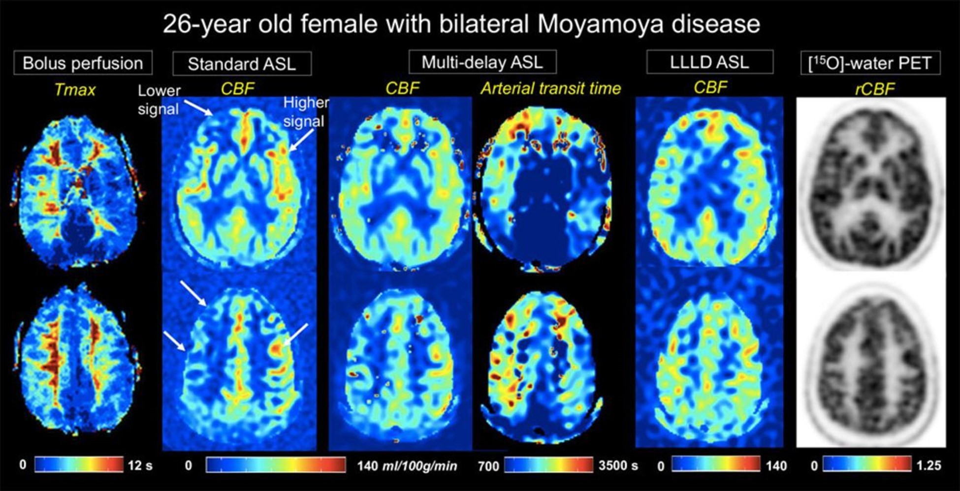
Image source: Stroke
Image source: Stroke
References:
https://www.ahajournals.org/doi/full/10.1161/STROKEAHA.117.017773
“The views, opinions, and positions expressed within this blog are those of the author(s) alone and do not represent those of the American Heart Association. The accuracy, completeness, and validity of any statements made within this article are not guaranteed. We accept no liability for any errors, omissions, or representations. The copyright of this content belongs to the author and any liability with regards to infringement of intellectual property rights remains with them. The Early Career Voice blog is not intended to provide medical advice or treatment. Only your healthcare provider can provide that. The American Heart Association recommends that you consult your healthcare provider regarding your health matters. If you think you are having a heart attack, stroke, or another emergency, please call 911 immediately.”