hidden

Join the race against the clock: Controlling for age in cardiovascular disease

The race against aging has already started. People who want part of this race see the limitless opportunities humans will have if aging is taken out of the equation. Humans will be able to live longer with their loved ones, go back to University when they are 70 years old to study this long-dreamed profession they always wanted to try or take the time to go through every single item on their bucket list, no matter how long the list is. Others are worried about what awaits them at the finish line of the race and what sacrifices will have to be made along the way. They argue that aging is the core of our existence and the reason why we make the choices we make every day. If humans don’t age, will they still find a reason to live and would this type of life be worth living? Regardless of people’s scattered opinions, the remaining question to be answered in our race against the clock is: Can we age disease-free?

As humans began to live longer dying less of problems such as hunger, wars and infections, they were faced with a new type of problem: chronic diseases. As we age and get exposed to different environmental and lifestyle factors, a set of biological and functional changes in our bodies lead to the development of chronic diseases such as cardiovascular diseases (CVD), diabetes, cancer, dementia, arthritis, and the list goes on. Notorious for being the ‘number 1 killer in the world’, preventing CVD has been one of the top priorities in our fight against aging. Age is the best predictor of CVD death, and despite years of research and large amounts of funding spent on biomarker discovery, there are currently no better predictors of CVD death than the age of a person and these are some of the reasons why.

Large blood vessels tend to become stiffer over time as they accumulate more collagen (due to an increase in TGF-b activity leading to collagen synthesis from smooth muscle cells) and lose their elastin content (because of higher metalloproteases and cathepsin activity). This leads to a chronic increase in systolic blood pressure which is worsened by the rise of catecholamines levels usually seen during aging. Both phenomena contribute to left ventricular dysfunction and hypertrophy due to the increase in myocardial oxygen demand. Calcification is another hallmark of aging that also contributes to vessel stiffness and induces stenosis. As we age, skeletal calcium is released and tends to accumulate in the vascular structures.

Apart from leading to vessel stiffness, aging causes the vascular endothelial cell (EC) barrier to become dysfunctional. ECs play a crucial role in maintaining vessel integrity and homeostasis by balancing vasodilatory and vasoconstricting functions and by aligning the vessels with an anti-thrombotic surface. Disruption of this protective barrier over time is characterized by ECs undergoing oxidative stress, reduced nitric oxide (a potent vasodilator) production, increased expression of adhesion molecules (ICAM and VCAM) and secretion of inflammatory chemokines (CXCL8) and cytokines (IL-1b and IL-6). The initiating event of atherosclerosis development starts with endothelial dysfunction which gives way for monocyte infiltration and subsequent foam cell formation contributing to plaque development.

At the molecular level, changes affecting the genome and epigenome are a fundamental feature of aging. With age, the clonal diversity of hematopoietic stem cells decreases resulting in the predominance of one clone. In recent years, clonal hematopoiesis of indeterminate potential (CHIP), which occurs as a result of mutations in transcriptional regulators (DNMT3A, TET2 and ASXL1), was found as a novel CVD risk factor, thereby linking genetic mutations in hematopoietic stem cells, aging and CVD. The number of endothelial progenitor cells also decreases over time which reduces angiogenesis capacity and capillary density leading to microvascular disease (such as peripheral artery disease).

The shortening of chromosome telomers is another molecular change related to aging. As cells replicate, telomeres get shorter until cellular senescence is triggered. Cellular senescence is a cellular protective mechanism that activates NK cells to remove cells with defective genetic material via apoptosis. It has been shown that patients with reduced leukocyte telomere length have increased risk of atherosclerosis. An atherosclerotic plaque, rich in inflammatory cells and trans-differentiated smooth muscle cells, is a dense hypoxic environment characterized by the presence of reactive oxygen species which also induce DNA damage and senescence.

Current therapies for atherosclerosis target some of the pathways of aging highlighted above. While statins are known to reduce plaque lipid content and inflammation, in parallel, they tend to increase calcification leading to vascular stiffness. On the other hand, anti-hypertensive treatments offer benefits beyond reducing CVD mortality, but also decreasing dementia. Recently, novel therapies targeting aging in CVD have focused on stem cell therapy. However, clinical trials using cell therapy to improve left ventricular dysfunction or to reduce cardiovascular events have shown no or modest benefit. This may be because autologous cell therapy of stem cells that already have an ‘aging’ phenotype is not ideal, and these cells may require ex vivo reprograming to improve treatment efficiency.


Since many age-related diseases have similar underlying molecular mechanisms driving them, the future for treating chronic diseases will rely on targeting the mechanisms of aging rather than treating the disease itself. Some of the best ways to slow down aging is by being active, controlling blood glucose levels, opting for diets rich in antioxidants and fibers and introducing low calorie intake periods during the day. However, this usually requires a substantial effort and serious lifestyle changes on our behalf. But, since research on anti-aging therapies and senolytic drugs is booming, it might be possible to slow down aging by taking one or two pills a day without ever needing to change the routines that we are so comfortable with.

References

  1. Paneni F, Diaz Cañestro C, Libby P, Lüscher TF, Camici GG. The Aging Cardiovascular System: Understanding It at the Cellular and Clinical Levels. J Am Coll Cardiol. 2017 Apr 18;69(15):1952–67.
  2. Quyyumi AA, Vasquez A, Kereiakes DJ, Klapholz M, Schaer GL, Abdel-Latif A, et al. PreSERVE-AMI. Circ Res. 2017 Jan 20;120(2):324–31.
  3. Brouilette SW, Moore JS, McMahon AD, Thompson JR, Ford I, Shepherd J, et al. Telomere length, risk of coronary heart disease, and statin treatment in the West of Scotland Primary Prevention Study: a nested case-control study. The Lancet. 2007 Jan 13;369(9556):107–14.
  4. Koopman JJE, Kuipers RS. From arterial ageing to cardiovascular disease. The Lancet. 2017 Apr 29;389(10080):1676–8.
  5. Jaiswal S, Libby P. Clonal haematopoiesis: connecting ageing and inflammation in cardiovascular disease. Nat Rev Cardiol. 2020 Mar;17(3):137–44.
  6. Antonangeli F, Zingoni A, Soriani A, Santoni A. Senescent cells: Living or dying is a matter of NK cells. J Leukoc Biol. 2019 Jun;105(6):1275–83.
  7. What is the Age of My Heart? – Calculate Your Own Heart Age • MyHeart [Internet]. MyHeart. 2015 [cited 2022 May 16]. Available from: https://myheart.net/articles/what-is-the-age-of-my-heart-calculate-your-own-heart-age/

“The views, opinions, and positions expressed within this blog are those of the author(s) alone and do not represent those of the American Heart Association. The accuracy, completeness, and validity of any statements made within this article are not guaranteed. We accept no liability for any errors, omissions, or representations. The copyright of this content belongs to the author and any liability with regards to infringement of intellectual property rights remains with them. The Early Career Voice blog is not intended to provide medical advice or treatment. Only your healthcare provider can provide that. The American Heart Association recommends that you consult your healthcare provider regarding your health matters. If you think you are having a heart attack, stroke, or another emergency, please call 911 immediately.”

hidden

An Interview with Dr. Ilana Kutinsky- Electrophysiologist and Cardiologist of the Apes

As a resident at Beaumont Hospital, I have the good fortune of working alongside some of the finest cardiologists in the country. I am constantly inspired by talent, innovation, and excellence in patient care. Dr. Ilana Kutinsky, associate professor at Oakland University William Beaumont School of Medicine, takes cardiology and commitment to health equity a step further by providing care to apes throughout the country.

Cardiovascular diseases are the leading cause of mortality not only in humans but also in the great ape species, including gorillas, chimpanzees, orangutans, and bonobos. Established in 2010, the Great Ape Heart Project (GAHP) aims to improve the understanding and management of cardiac disease in our cousin species. A cardiac electrophysiologist by training and profession, Dr. Kutinsky is one of the founders of the Great Ape Heart Project. I sat down with Dr. Kutinsky to learn more about her role as ‘cardiologist of the apes’.

DA: How did you get involved with the Great Ape Heart Project?

Dr. Kutinsky: Around 2000, I was a cardiology fellow at the University of Colorado. During my echo rotation, I picked up a call sitting in the darkroom. “We’re calling from the Denver Zoo. We need someone to come and do TEEs on our gorillas and orangutans. Would you be interested?” and I said “What?”. I talked to my attending and we decided to do it. So, we had Phillips loan us a giant TEE machine and bring it to the zoo. They anesthetized the gorilla and we did the TEE. At the time they were just realizing that captive gorillas die from heart diseases and there was no reference to what is normal. The next month, a female orangutan died from extremis from the anesthesia for the TEE. It was disconcerting to see an animal die for a test we did not even know how to interpret. I got in touch with Dr. Hayley Murphy, who had written a paper reporting echo findings from five gorillas in Boston. We shared our frustration and decided to write a grant to try to collect echos over the country and establish a normal. The grant was rejected but I continued to record readings on my own time by taking freehand measurements on VHS tapes. This is how the Gorilla Heart Project got started. Then it got bigger! We wrote a grant to the Institute of Museum and Library Sciences (IMLS) and got a half a million-dollar grant to set up the Great Ape Heart Project. We were able to hire primatologists, pathologists, veterinarians, and sonographers and set up a database. We have been able to establish normal, identify high-risk animals, and treat heart failure in gorillas. We have consulted on cases not only in the United States but all over the world. It’s a wonderful, although busy hobby for me!

DA: How was your first experience as a cardiologist for the apes, the gorilla in Denver you did the TEE for?

Dr. Kutinsky: It was very similar to taking care of a human once the probe was inside the body. But the animals are enormous, between 350 to 580  pounds. Their chests are like barrels and their skulls are ginormous. To get the TEE probe down their mouths, you have to go past their incisors which are almost the size of my hand.

DA: Were you scared?

Dr. Kutinsky: No, you know they are under anesthesia. And someone is standing in the corner with a big gun. So, if they start to wake up, they will be immobilized. I wasn’t scared because I was so in awe that I was able to do something so amazingly cool. I remember calling my mom afterwards and telling her that every night I spent studying or being on call, all the effort made to get into medical school and cardiology was worth it that day.

DA: Were your co-fellows jealous of you?

Dr. Kutinsky: That’s a good question. Um, I think they thought I was crazy! Doing all that extra work on top of a busy fellowship schedule did not make sense to them.

DA: How is it working with a team of so many disciplines?

Dr. Kutinsky: It is amazing to work with a team of veterinarians, human cardiologists, veterinary cardiologists, sonographers, and pathologists. The vets are honestly some of the smartest people I know. They take care of a 500-pound gorilla with heart disease and then they attend to a tiny bird with an eye injury. Their breadth of knowledge is extraordinary. It has been incredibly rewarding experience working with everyone.

DA: What’s been your most interesting experience being a cardiologist for the apes?

Dr. Kutinsky: Going to Cameroon was one of my most interesting experiences. We were invited by the government of Cameroon to a sanctuary of rescued wild gorillas. We were able to see them up close and interact with them from across a mesh. We were there for 2 weeks, seeing them every day and feeling a connection with them. Usually, I don’t get called when they’re doing well. I’m called when they are sick and about to die, so they’re usually anesthetized. To see them awake, eating and going about their usual life was pretty cool.

DA: Do you have a favorite of the great ape species?

Dr. Kutinsky: Gorillas for sure. I like orangutans too but we don’t have any orangutans in the Detroit zoo so I don’t see them much. Chimpanzees are very naughty and a handful to work with.

DA: Have you become attached to any animal?

Dr. Kutinsky: I was very attached to Sunshine, a gorilla in the Detroit Zoo. He was an old, great animal with a bad heart. He died from influenza a few years ago. I was pretty tight with him. I like Mac in Columbus. He is a very handsome boy. There’s Tatu in Omaha, he’s a good boy. I’ll be sad when they die. Gorillas usually don’t live past 40 years and a lot of the ones with heart disease drop dead early. So we’re hoping to change that.

DA: Do you prefer treating humans or gorillas?

Dr. Kutinsky: Depends on the day! I love animals but I cherish the relationships with my patients. I love my job as an electrophysiologist. Everything I’ve learned as a human doctor I use to treat the gorillas. But if I won the lottery, I would probably devote all my time to the gorillas.

DA: Last question, has taken care of gorillas and other apes affected how you treat your patients? 

Dr. Kutinsky: That’s a great question. Hmm, I guess treating animals has made me a more compassionate doctor and person in general. Since the animals can’t complain or whine, you give them symptoms in your mind. You assume they are hurting or sad, and you try to take care of them. I think it has given me an extra bit of sympathy when I take care of my patients. And it makes me happier in general. When you have a passion, it enriches your whole life.

DA: I guess it was a lucky call that day in the echo lab.

Dr. Kutinsky: Absolutely! I was in the right place at the right time. I also think you have to be open to the universe and do things that are outside the box. I was willing to do the extra work outside of a busy fellowship. I was offered something special and I took it.


“The views, opinions, and positions expressed within this blog are those of the author(s) alone and do not represent those of the American Heart Association. The accuracy, completeness, and validity of any statements made within this article are not guaranteed. We accept no liability for any errors, omissions, or representations. The copyright of this content belongs to the author and any liability with regards to infringement of intellectual property rights remains with them. The Early Career Voice blog is not intended to provide medical advice or treatment. Only your healthcare provider can provide that. The American Heart Association recommends that you consult your healthcare provider regarding your health matters. If you think you are having a heart attack, stroke, or another emergency, please call 911 immediately.”

hidden

Women In Cardiology: A Mother’s Day Tribute to Women, Wives, Weekend Warriors, & Workaholics In Cardiology

*Buzz*. Most of us can relate to that alarm going off way earlier than we would like. Wake up, make coffee, throw the clothes in the washer or dryer, clean up the kitchen, put on your office attire or scrubs, and run out the door to start the day taking care of patients, reading nuclear studies, reviewing echocardiograms, or doing catheterizations. For many, this sort of routine is comforting. Enter motherhood or pregnancy to start. This topic in cardiology, a field dominated by men who make up 85% of the workforce, can be an exciting topic to navigate.

The Pregnant Cardiologist

Pregnancy is supposed to be one of the most joyous celebrations in life. I often reminded myself of this while recently pregnant with my second child as I squeezed in OB appointments between patients while simultaneously reassuring them that I would be back after eight weeks. Women in cardiology face obstacles that their male counterparts do not have to use mental bandwidth on. We finish training in our early 30s keenly aware that our “biological clock” is ticking. We try to decide when the right time is to have children whilst trying to grow our own practice and make our footprint. Many questions zoom through our brains. When is the “right” time to take off anywhere from six to twelve weeks with our employer and partners accepting it, covering call, and caring for our patients? How will pregnancy and motherhood affect my career? Is there a maternity leave policy? What if I have complications during pregnancy? Will my partners assume I am going to scale back after I have a child and give preferential treatment to the men? How will pregnancy impact my salary? We sacrifice some of our professional growth to achieve one of the most joyous life events. Or do we? Dr. Martha Gulati, et al recently surveyed women who were members of the American College of Cardiology Women in Cardiology section to assess real life experiences during pregnancy. 341 responded they had children as a practicing cardiologist. Of these, most notified their chair, chief, or practice that they were pregnant in the late first or early second trimester. The most common reason for waiting to report was due to concern of adverse treatment or perception (37.5%). Maternity leave varied dramatically among respondents. The most common leave time was three months (48.9%) followed by less than six weeks (22.6%). 41.2% of respondents had a salary decrease during pregnancy. 37.2% reported performing extra calls or service while pregnant “making up” for being on maternity leave. 21.5% of women thought that taking extra calls or service contributed to pregnancy complications. I must share that I am fortunate to work with some of the most wonderful, understanding, partners and bosses. This was reflected in my pregnancy and leave experience. Not all of us will have a positive experience, but we should. The lack of consistency in maternity leave and support for childbearing is not only a problem in cardiology. We hear this among colleagues of all specialties. Cardiology just so happens to pose a unique challenge given the historical lack of women in the specialty and the rigorous schedule for many. Efforts to create a more uniform maternity leave policy and welcoming pre and post-natal experience are more than past due.

Wonderwomen in Cardiology

Getting back to work after having a child can be overwhelming. The “routine” has changed to a less “routine” routine. That alarm clock buzz is most likely replaced by a crying baby and cold coffee left on the counter somewhere around the house. You are tired. When the sun comes up it is time to feed the baby, get them ready for whatever care you have arranged, and, if breastfeeding, pumping JUST before you leave so you can maximize work time when you arrive. Pumping while doing charts or on zoom calls has become a norm. The concerns about perception at work continue. You wonder, “will I be perceived as fragile or more interested in home life now?”. Struggling to “prove” that motherhood has not taken away your passion for work is almost inevitable. An inherent bias exists. While we juggle motherhood, being a wife (which for many includes cooking, laundry, nighttime routine), and work, we try to convince our colleagues that we are still the same. But are we? I would argue we return better. Finding a group to work with who thinks the same might be hard, but it is attainable. I can attest to that.  Raising children as a cardiologist is a busy new journey. Working together with your spouse is a must. Finally admitting that maybe you cannot do it all at home is okay. Asking for help is okay.  “Subcontracting” household tasks is sometimes necessary to get back to being you: a woman—no, a Wonder Woman both in cardiology and at home. Happy Mother’s Day to all the moms, soon-to-be moms, and those hoping to be moms one day.

Footnote: This was 50% written while pumping and 50% while the children were napping.

REFERENCE

  1. Gulati M, Korn R, Wood M, et al. Childbearing Among Women Cardiologists. J Am Coll Cardiol. 2022 Mar, 79 (11) 1076–1087. https://doi.org/10.1016/j.jacc.2021.12.034

“The views, opinions, and positions expressed within this blog are those of the author(s) alone and do not represent those of the American Heart Association. The accuracy, completeness, and validity of any statements made within this article are not guaranteed. We accept no liability for any errors, omissions, or representations. The copyright of this content belongs to the author and any liability with regards to infringement of intellectual property rights remains with them. The Early Career Voice blog is not intended to provide medical advice or treatment. Only your healthcare provider can provide that. The American Heart Association recommends that you consult your healthcare provider regarding your health matters. If you think you are having a heart attack, stroke, or another emergency, please call 911 immediately.”

hidden

The Protective Role of Anti-Hypertension Medication Among Patients with Comorbidities for COVID-19 Outcomes

Millions of people around the world take Angiotensin-converting enzyme inhibitors (ACEi) and Angiotensin II receptor blockers (ARB) to manage hypertension, heart failure, and coronary artery disease. Concerns of ACEi and ARB potentially increasing the risk of COVID-19 illness severity and mortality among vulnerable populations heightened once scientists reported that risk factors for developing complications included being older, male, and having cardiovascular comorbidities1. One comprehensive study using over 17,000 primary care records found that chronic heart disease has a hazard ratio of 1.57 for COVID-19 related death and the hazard ratio remained high at 1.17 even when accounting for confounding variables, suggesting that people with heart disease are at increased risk of mortality2. In the same study, high blood pressure or hypertension diagnoses were associated with hazard ratio of 0.89, a lower risk of COVID-19 mortality compared to people with normal blood pressure, but insight into how age, sex, comorbidities, and medications influence outcomes were not directly addressed. Such findings fueled a debate about whether ACEI/ARB should be maintained or withdrawn in patients with COVID-19.

The role of ACEi and ARBS drugs in COVID-19 outcomes among cardiovascular patients also became a point of interest due to their mechanism of action in the human body. ACEi and ARB act on the renin-angiotensin-aldosterone system (RAAS), a hormone system important for regulating blood pressure, fluid balance, and inflammation processes that affect cardiovascular health outcomes. While ACEi and ARB drugs are used as the first line of treatment to manage vasoconstriction, there is a question as to how these medications can alter the RAAS balance. In a previous blog, we discussed how the SARS-CoV-2 virus uses the  of angiotensin-converting enzyme 2 (ACE2) receptor to enter host target cells3. This receptor not only acts as the entry point for the virus, but normally acts as a crucial element for regulating RAAS biochemical processes. The inflammatory, tissue damaging, and vasoconstriction effects of Angiotensin II (Ang II) in the body are mitigated by ACE2 activity, and ARB and ACEi drugs also target the Ang II protein4. COVID-19 related research has provided a new understanding of how underlying disease states, behavioral habits like smoking, or genetics could influence ACE2 activity in the body. The unique collaboration between clinicians and scientists during the COVID-19 pandemic has provided new mechanistic insight about how the complex RAAS pathway and the factors that influence disease progression.

Ongoing population studies such as The International Study of Inflammation in COVID-19 (ISIC) and The Michigan Medicine COVID-19 Cohort (M2C2) make use of detailed medical records bio-banked human samples, and advanced statistical modeling to evaluate the potential benefits and harms of ACEi and ARB medications. Using stored blood samples and electronic medical records from patients hospitalized specifically for COVID-19, researchers were able to assess for an association between ACEi or ARB use and in-hospital patient outcomes, such as requiring mechanical ventilation or admission into intensive care. The research team overseeing the ISIC and M2C2 studies analyzed the health outcomes of about 1,600 people hospitalized for COVID-19 and reported that patients taking ACEi or ARB had about 10% mortality compared to 14% who were not on those medications5. Among those taking medications, 24% of patients required ventilation during hospitalization, compared to 20% of those not any treatment. These results were surprising as people taking medication also had significantly more comorbidities such as diabetes compared with the non-ACEi/ARB group. Knowing that people who use ACE inhibitors or ARB are not more susceptible to severe COVID-19 illness or increased risk of mortality during hospitalization has now led to the widely accepted practice of not discontinuing these drugs in people who are infected with SARS-CoV-2. In fact, people on anti-hypertensive medication had lower levels of inflammation biomarkers during hospital admission compared to those who don’t take ACEi and ARB drugs. This insight suggests that ACEi/ARB drugs could counter the inflammatory effects of COVID-19, which could be an interesting future direction of this research. Large scale studies have been valuable for providing evidence on how to mitigate detrimental outcomes during the COVID-19 pandemic and future findings will continue to influence guidelines for monitoring cardiovascular homeostasis, targeting treatments for vulnerable populations, and managing chronic illnesses.

References:

  1. Patel AB, & Verma A. (2020). COVID-19 and Angiotensin-Converting Enzyme Inhibitors and Angiotensin Receptor Blockers: What Is the Evidence? JAMA. https://doi.org/10.1001/jama.2020.4812
  2. Williamson EJ, Walker AJ, Bhaskaran K, et al. (2020). Factors associated with COVID-19-related death using OpenSAFELY. Nature. 2020;584(7821):430-436. doi:1038/s41586-020-2521-4
  3. Raizada MK, & Ferreira AJ, (2007). ACE2: A New Target for Cardiovascular Disease Therapeutics. Journal of Cardiovascular Pharmacology, 50(2), 112–119. https://doi.org/10.1097/FJC.0b013e3180986219
  4. Monterrosa Mena, J. ACE-2 and Immune System Changes in Smokers May Underlie COVID-19 Vulnerability. https://earlycareervoice.professional.heart.org/ace-2-and-immune-system-changes-in-smokers-may-underlie-covid-19-vulnerability/
  5. Pan N, Hayek S, the ISIC Group, et al. (2021). Angiotensin‐Converting Enzyme Inhibitors, Angiotensin II Receptor Blockers, and Outcomes in Patients Hospitalized for COVID‐ Journal of the American Heart Association, 10(24), e023535. https://doi.org/10.1161/JAHA.121.023535

“The views, opinions, and positions expressed within this blog are those of the author(s) alone and do not represent those of the American Heart Association. The accuracy, completeness, and validity of any statements made within this article are not guaranteed. We accept no liability for any errors, omissions, or representations. The copyright of this content belongs to the author and any liability with regards to infringement of intellectual property rights remains with them. The Early Career Voice blog is not intended to provide medical advice or treatment. Only your healthcare provider can provide that. The American Heart Association recommends that you consult your healthcare provider regarding your health matters. If you think you are having a heart attack, stroke, or another emergency, please call 911 immediately.”

hidden

COVID-19 and Endothelial Cell Dysfunction

Photo by CDC on Unsplash

COVID-19, caused by severe acute respiratory syndrome coronavirus 2 (SARS-CoV-2), represents a global health crisis. Cough, fever, and shortness of breath are the most common reported symptoms; however, neurological and gastroenterological manifestations can also be present1. Angiotensin-converting enzyme 2 (ACE-2) has been shown to act as a co-receptor to facilitate coronavirus entry by efficiently binding to the S1 domain of spike protein, a surface glycoprotein of SARS-CoV2. The pathogenesis of COVID-19 depends on the localization of the coronavirus co-receptors. The epithelium of lungs and intestine is rich in ACE-2 expression, thereby providing a possible route of entry for SARS-CoV-2. Further, ACE-2 is also expressed on Type I and type II pneumocytes providing other entry sites for SARS-CoV-2. Virus entry can cause pathological changes at the alveoli-capillary interface. Additionally, copious expression of ACE-2 on the type II alveolar cells can promote rapid viral expansion resulting in further alveolar damage and hyperinflammation3,4.

ACE-2 is also present on endothelial cells, smooth muscle cells, and perivascular pericytes in all the organs, indicating that if SAR-COV-2 is transmitted to the blood circulation, the virus can quickly spread throughout the body3. The postmortem lung tissues of COVID-19 patients exhibited a higher number of ACE-2 positive endothelial cells and a higher prevalence of endothelial injury (disruption of cell membrane and presence of the intracellular virus) and microthrombi than lung tissues of patients who died from influenza-associated respiratory failure1,5. The most common comorbidities observed in COVID-19 patients are hypertension, diabetes, and obesity, all of which are associated with endothelial dysfunction. Further, COVID-19 patients are projected to be at a higher risk of deep vein thrombosis, systemic vasculitis, and acute pulmonary embolism6,7, possibly due to underlying endothelial cell injury and inflammation. Thrombi can directly affect gas exchange and cause and cause multisystem organ dysfunction in COVID-19 pneumonia8. Upon activation, platelets release polyphosphates, which accelerate the activation of factors V and XI, inhibit tissue factor pathway inhibitor and contribute to thicker fibrin strands synthesis. Further, the cytokine release can activate endothelial cells resulting in a prothrombotic environment1.

Acute respiratory distress syndrome is suggested to be caused by the dissociation between lung mechanics, loss of lung perfusion regulation and hypoxic vasoconstriction, and severe hypoxemia9. The loss of hypoxic vasoconstriction can be due to increased mitochondrial oxidative stress resulting in pulmonary endothelial cell dysfunction10. SARS-CoV-2 elements, accumulation of inflammatory cells, intracellular virus, and disrupted cell membranes are detected in the endothelial cells of COVID-19 patients5,11, further indicating endotheliitis /endothelial cell dysfunction during COVID-19 infection. Endothelial cell dysfunction can cause abnormalities in microcirculation in different vascular beds and contribute to life-threatening complications of COVID-19, including thromboembolism and multiple organ damage.

References:

  1. Huertas A, Montani D, Savale L, et al. Endothelial cell dysfunction: a major player in SARS-CoV-2 infection (COVID-19)? Eur Respir J. 07 2020;56(1)doi:10.1183/13993003.01634-2020
  2. Ziegler CGK, Allon SJ, Nyquist SK, et al. SARS-CoV-2 Receptor ACE2 Is an Interferon-Stimulated Gene in Human Airway Epithelial Cells and Is Detected in Specific Cell Subsets across Tissues. Cell. 05 28 2020;181(5):1016-1035.e19. doi:10.1016/j.cell.2020.04.035
  3. Hamming I, Timens W, Bulthuis ML, Lely AT, Navis G, van Goor H. Tissue distribution of ACE2 protein, the functional receptor for SARS coronavirus. A first step in understanding SARS pathogenesis. J Pathol. Jun 2004;203(2):631-7. doi:10.1002/path.1570
  4. Mehta P, McAuley DF, Brown M, et al. COVID-19: consider cytokine storm syndromes and immunosuppression. Lancet. 03 28 2020;395(10229):1033-1034. doi:10.1016/S0140-6736(20)30628-0
  5. Ackermann M, Verleden SE, Kuehnel M, et al. Pulmonary Vascular Endothelialitis, Thrombosis, and Angiogenesis in Covid-19. N Engl J Med. 07 09 2020;383(2):120-128. doi:10.1056/NEJMoa2015432
  6. Bompard F, Monnier H, Saab I, et al. Pulmonary embolism in patients with COVID-19 pneumonia. Eur Respir J. Jul 2020;56(1)doi:10.1183/13993003.01365-2020
  7. Criel M, Falter M, Jaeken J, et al. Venous thromboembolism in SARS-CoV-2 patients: only a problem in ventilated ICU patients, or is there more to it? Eur Respir J. Jul 2020;56(1)doi:10.1183/13993003.01201-2020
  8. Poor HD, Ventetuolo CE, Tolbert T, et al. COVID-19 critical illness pathophysiology driven by diffuse pulmonary thrombi and pulmonary endothelial dysfunction responsive to thrombolysis. Clin Transl Med. Jun 2020;10(2):e44. doi:10.1002/ctm2.44
  9. Gattinoni L, Coppola S, Cressoni M, Busana M, Rossi S, Chiumello D. COVID-19 Does Not Lead to a “Typical” Acute Respiratory Distress Syndrome. Am J Respir Crit Care Med. 05 15 2020;201(10):1299-1300. doi:10.1164/rccm.202003-0817LE
  10. Guignabert C, Phan C, Seferian A, et al. Dasatinib induces lung vascular toxicity and predisposes to pulmonary hypertension. J Clin Invest. 09 01 2016;126(9):3207-18. doi:10.1172/JCI86249
  11. Varga Z, Flammer AJ, Steiger P, et al. Endothelial cell infection and endotheliitis in COVID-19. Lancet. 05 02 2020;395(10234):1417-1418. doi:10.1016/S0140-6736(20)30937-5

“The views, opinions, and positions expressed within this blog are those of the author(s) alone and do not represent those of the American Heart Association. The accuracy, completeness, and validity of any statements made within this article are not guaranteed. We accept no liability for any errors, omissions, or representations. The copyright of this content belongs to the author and any liability with regards to infringement of intellectual property rights remains with them. The Early Career Voice blog is not intended to provide medical advice or treatment. Only your healthcare provider can provide that. The American Heart Association recommends that you consult your healthcare provider regarding your health matters. If you think you are having a heart attack, stroke, or another emergency, please call 911 immediately.”
hidden

PET or MRI, that is the question – Part 1

In our previous blogs, we discussed that doctors can diagnose Moyamoya disease using medical imaging systems, such as positron emission tomography (PET) and magnetic resonance imaging (MRI). But which one is better?

According to some recent studies performed by Dr. Moss Zhao (AHA Postdoctoral Fellow, 2021) at Stanford University, MRI is better thanks to its accessibility, safety, and affordability.

For decades, PET has been the gold standard technique for diagnosing Moyamoya disease based on imaging the blood flow in the abnormal blood vessels in the brain. However, PET uses radioactive tracers to create the images, making it complex and expensive. Although the amount of radiation is less than a 5-hour flight, doctors have sought for alternative and less invasive techniques to replace PET, especially for children. In recent years, arterial spin labeling (ASL), an advanced MRI technique, has emerged to replace PET imaging to measure blood flow in the brain. Because ASL MRI is more accessible at most hospitals and the procedure is less complex than PET, ASL has gained popularity in many research and clinical institutions for Moyamoya patients.

At Stanford University, Dr. Moss Zhao demonstrated that ASL could replace the conventional PET imaging technique without exposing patients to radiation and causing side effects. The image quality and measurement accuracy of ASL are compatible with PET, implying that ASL can be used to characterize the abnormal blood flow and circulation in Moyamoya patients. Among the different implementations of ASL, Dr. Zhao developed an advanced technique dubbed ‘multi-delay ASL’ that gives the best image quality with the least amount of scanning time. For less than 5 minutes, multi-delay ASL can produce images that require more than 20 minutes for PET imaging. The image in this blog shows the images collected by ASL and PET at the same time from normal and healthy people. Using the latest ASL techniques, doctors can identify patients with a high risk for stroke based on their MRI scans for just under 5 minutes without using any radioactive substances. Dr. Zhao’s team is currently testing this technique on the pediatric population to enable this non-invasive imaging technology to be accessible to patients across the lifespan.

Image source: NeuroImage

References:

https://doi.org/10.1016/j.neuroimage.2021.117955


“The views, opinions, and positions expressed within this blog are those of the author(s) alone and do not represent those of the American Heart Association. The accuracy, completeness, and validity of any statements made within this article are not guaranteed. We accept no liability for any errors, omissions, or representations. The copyright of this content belongs to the author and any liability with regards to infringement of intellectual property rights remains with them. The Early Career Voice blog is not intended to provide medical advice or treatment. Only your healthcare provider can provide that. The American Heart Association recommends that you consult your healthcare provider regarding your health matters. If you think you are having a heart attack, stroke, or another emergency, please call 911 immediately.”

hidden

Is Breakfast Still the Most Important Meal of the Day?

“Breaking-fast,” or breakfast, has been endorsed as the most important meal of the day. Replenishing energy stores from overnight fasting boosts energy levels and cognitive function and helps control weight by minimizing fluctuations in blood glucose and preventing binge eating later in the day. But is this still the case today? Has the evolution in the convenience and accessibility of foods, in particular ultra-processed foods and refined high-carbohydrate breakfast items, changed this dogma? Well, the answer is, it depends.

Intermittent fasting, in particular time-restricted feeding, where anywhere from a 6 to 10-hour eating window is followed by fasting, has been growing in popularity as this is the least restrictive form and has less side effects of irritability, and headaches, and decreased concentration (aka “hangry”). Say, for instance, you only consume food from 12 pm to 8 pm and fast the rest of the day. You are essentially skipping breakfast. Short-term studies have found benefits in bodyweight reduction and improved cardiometabolic health.

However, in contrast, our inherent circadian rhythm favors breakfast. Consuming most of our meals during the active phase or light phase of the day results in peak gastrointestinal emptying/motility, increased insulin sensitivity, and lower appetite (in part due to low levels of the hormone ghrelin, which increases appetite). In theory, if we were all able to use our circadian rhythm to our advantage but also eat during a time-restrictive window (6am-4 pm), we could take advantage of all the pleiotropic benefits of fasting. But for most of us, this isn’t realistic because we are social beings and interact with our community that very much revolves around dinner gatherings with family and friends.

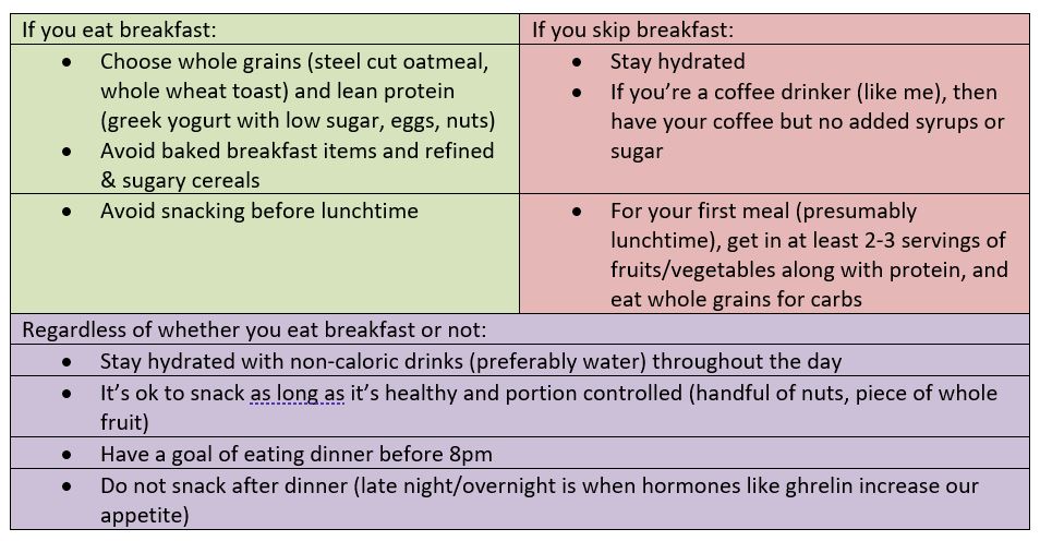
So, what’s the solution? Here are a few tips for using everyday that consider the above:

As a reminder to myself and my patients:

Changing dietary habits is a gradual process with delayed gratification. One small change a day will make all the difference in your lifetime.

Eat well and be well,

References:

  1. Time-Restricted Eating: Benefits, Mechanisms, and Challenges in Translation – PMC (nih.gov)
  2. Nutrition and the Circadian System – PMC (nih.gov)
  3. Effects of Intermittent Fasting on Health, Aging, and Disease | NEJM

“The views, opinions, and positions expressed within this blog are those of the author(s) alone and do not represent those of the American Heart Association. The accuracy, completeness, and validity of any statements made within this article are not guaranteed. We accept no liability for any errors, omissions, or representations. The copyright of this content belongs to the author and any liability with regards to infringement of intellectual property rights remains with them. The Early Career Voice blog is not intended to provide medical advice or treatment. Only your healthcare provider can provide that. The American Heart Association recommends that you consult your healthcare provider regarding your health matters. If you think you are having a heart attack, stroke, or another emergency, please call 911 immediately.”

hidden

What to look forward to at Vascular Discovery 2022

The primary goal of the Vascular Discovery: From Genes to Medicine Scientific Sessions is to provide a forum for the timely exchange of information about new and emerging scientific research in lipids and lipoproteins, arteriosclerosis, thrombosis, vascular biology, genomics, precision medicine, peripheral vascular disease, and vascular surgery.

This meeting is planned to kick-off on Thursday the 12th of May at 7:00 am with an early career round table discussion session moderated by Robert C. Bauer, PhD and Cynthia St. Hilaire PhD. It will be the perfect setting to discuss ideas, including starting your own lab, mentoring, project management and work-life integration with friendly faces around a hot cup of coffee.

After the conference opening, Vascular Discovery 2022 will officially launch with plenary session 1, which includes several concurrent sessions, each covering novel concepts of atherosclerosis. The new discoveries related to inflammation and atherosclerosis will be presented during a packed oral abstract presentations session including the role of autophagy in foam cells as well as the link between plasma cholesterol and the chromatin landscape of bone marrow monocytes. In parallel, another session will be running for those interested in listening to the new research related to the cellular biology of the vessel wall. These topics were launched by a presentation by Jiliang Zhou, MD, PhD about the discovery of long non-coding RNA CARMN and smooth muscle cells. Some of the exciting topics which will be covered during this session are related to the interplay between the endothelium and the inner vasculature. An exciting talk to look forward to is by Paul Cheng, MD, PhD from Stanford University, about the human arterial cell atlas. Discussions on the interplay between COVID-19 and thrombosis will also take place in a concurrent session.

After lunch, the day continues with presentations by the 2022 ATVB Journal Early Career Investigator Award recipients and followed by plenary session 2 with subjects covering lipid metabolism, vascular cells and thrombosis, or hot topics presented by the AHA Career Development Awardees. The day will end with a poster session and reception, which will be a great networking opportunity!

The second day of Vascular Discovery 2022 is also scheduled to start with a special session for Early Career Researchers specifically focused on perfecting your CV and motivation letter. Plenary session three will begin with the stimulating topic of the shapeshifters in the vascular disease, mostly focused on endothelial and smooth muscle cell plasticity, before dividing into three concurrent sessions which will handle various topics from novel therapeutic targets in atherosclerosis to the vascular effects of antithrombotic therapy as well as health disparities in peripheral vascular disease. Plenary session four will include the Page Junior Faculty Research Award Competition featuring exciting topics with a major interest in smooth muscle cells differentiation before splitting into concurrent sessions covering metabolic disorders, platelet production, signaling, and function, or polygenic risk scores for risk stratification. The Hot Off the Bench Oral Abstract Presentations will be a great way to meet the authors of the hottest research currently being done on vascular diseases.

The last day of Vascular Discovery 2022 will be highlighted by the plenary session 6 with a list of invited lecture series covering genomic aging in cardiovascular disease and cerebrovascular disease.

After meeting virtually for the past two years, Vascular Discovery 2022 will be an in-person meeting this year, reuniting us again to discuss our passion for science, form new collaborations, share experiences and finally see each other again after a long break.

Registration for the Vascular Discovery Conference is now open!

“The views, opinions, and positions expressed within this blog are those of the author(s) alone and do not represent those of the American Heart Association. The accuracy, completeness, and validity of any statements made within this article are not guaranteed. We accept no liability for any errors, omissions, or representations. The copyright of this content belongs to the author and any liability with regards to infringement of intellectual property rights remains with them. The Early Career Voice blog is not intended to provide medical advice or treatment. Only your healthcare provider can provide that. The American Heart Association recommends that you consult your healthcare provider regarding your health matters. If you think you are having a heart attack, stroke, or another emergency, please call 911 immediately.”

hidden

Key Takeaways From the 2022 AHA/ACC/HFSA Guideline for the Management of Heart Failure

What’s new in the treatment of heart failure? The 2022 AHA/ACC/HFSA Guideline for the Management was just released in the beginning of April! While much of the ground it covers might not seem particularly groundbreaking to anyone who has been paying attention to discussions on #MedTwitter, #CardioTwitter or the latest clinical trials over the last 2-3 years, it codifies the guideline directed medical therapy (GDMT) that we have all come to know and love for the treatment of heart failure with a reduced ejection fraction (HFrEF). These new guidelines also provide the first-ever guideline recommendations for patients with (heart failure with a preserved ejection fraction) HFpEF and heart failure with mildly reduced ejection fraction (HFmrEF), though the strength of recommendation for these conditions is not as strong as those for HFrEF.

Here are some key takeaways from the new heart failure guideline!

  1. Quadruple therapy GDMT for HFrEF

The latest guideline officially provides class IA recommendations for the use of the following medications in the treatment of HFrEF (defined as LVEF 40% or lower) in patients that have at least NYHA class II symptoms:

  1. Angiotensin-converting enzyme (ACE) inhibitors (ACEi) [i.e. lisinopril] or angiotensin-receptor blockers (ARBs) [i.e. losartan] or angiotensin receptor blocker/neprolysin inhibitor combination (ARNi) [i.e. sacubitril-valsartan]
  2. Beta blockers [i.e. metoprolol, carvedilol]
  3. Mineralocorticoid antagonists [i.e. spironolactone]
  4. SGLT2 inhibitors (SGLTi) [i.e. empagliflozin, dapagliflozin]

The first three classes were previously recommended for the treatment of HFrEF but prior American cardiovascular society guidelines did not include such a strong recommendation for the use of SGLT inhibitors.

  1. We now have HFimpEF

HFimpEF now refers to heart failure in someone who previously had HFrEF but whose LVEF has improved to >40%. The guideline strongly recommends continuing GDMT for patients that fall into this category.

  1. We now have HFmrEF recommendations

The guideline now provides a class 2A recommendation for the use of SGLT2i in the treatment of symptomatic HFmrEF (defined as LVEF 41-49%). It also provides class 2B recommendations for the use of ARNi/ACEi/ARB, beta blockers and MRAs in these patients. These recommendations confirm what some physicians/cardiologists have already begun doing in practice, though the level of evidence to support the use of these medications in HFmrEF as it is for patients with HFrEF.

  1. We also have new HFpEF recommendations

For the first time ever, the guideline recommends medications for the treatment of symptomatic HFpEF (defined as LVEF 50% or greater). Similar to its recommendations for the treatment of HFmrEF, it provides a class IIA recommendation for the use of SGLT2i and class 2B recommendations for beta blockers, ARNi, ACEi, ARB and MRAs, especially if the patient has an LVEF that is closer to 50%. Again, as we know, the level of evidence to support these practices is not as strong as it is for HFrEF. Still, this represents a change from previous guidelines which provided limited options for treatment of HFpEF.

  1. ICD or CRT is still recommended for primary prevention in certain cases

This guideline continues to recommend an implantable cardioverter-defibrillator (ICD)  in a subset of patients, particularly those whose LVEF remains less than or equal to 35% despite being on maximally-tolerated GDMT (there are nuances to this that we will not get into here). Similarly, as before, the guideline also continues to recommend cardiac resynchronization therapy (CRT) for patients who have an LVEF less than or equal to 35%, sinus rhythm, left bundle branch block with a QRS duration of at least 150 ms, NYHA class II-III symptoms.

  1. New recommendations for diagnosis and treatment of cardiac amyloidosis

The new guideline provides class I recommendations for checking serum and urine immunofixation electrophoresis and serum free light chains in patients (which would help diagnose AL amyloidosis) in patients for whom there is clinical suspicion for cardiac amyloidosis. Similarly, there is a class I recommendation for bone scintigraphy to evaluate for transthyretin (TTR) amyloidosis in patients for whom there is sufficient clinical suspicion for amyloidosis (left to the clinician’s judgment). Genetic testing is also recommended if a patient is diagnosed with TTR amyloidosis. For the first time, the guideline provides a class IB recommendation for the use of tafamadis in patients with transthyretin cardiac amyloidosis. And finally, the guideline gives a class IIA recommendation for use of anticoagulation in patients with concurrent atrial fibrillation and cardiac amyloidosis, regardless of CHA2DS2-VASc score.


“The views, opinions, and positions expressed within this blog are those of the author(s) alone and do not represent those of the American Heart Association. The accuracy, completeness, and validity of any statements made within this article are not guaranteed. We accept no liability for any errors, omissions, or representations. The copyright of this content belongs to the author and any liability with regards to infringement of intellectual property rights remains with them. The Early Career Voice blog is not intended to provide medical advice or treatment. Only your healthcare provider can provide that. The American Heart Association recommends that you consult your healthcare provider regarding your health matters. If you think you are having a heart attack, stroke, or another emergency, please call 911 immediately.”

hidden

The PATENS Trial: What does this mean for nitroglycerin in reducing radial artery occlusion?

Written by Aaysha Cader, MD, MRCP and Saidur Rahman Khan, MD, PhD

Ibrahim Cardiac Hospital & Research Institute, Dhaka, Bangladesh

Transradial access (TRA) is gaining increasing popularity for diagnostic and therapeutic interventions globally. Radial Artery Occlusion (RAO) however, is the most frequent complication of transradial access. An occluded radial artery precludes its future use for future catheterization procedures, as a conduit in coronary artery bypass grafting or for arteriovenous fistula creation.1

A number of randomized controlled trials have tested different strategies to reduce RAO, particularly in recent times. Vasodilators such as nitroglycerin have been used prophylactically to reduce radial artery spasms. By virtue of their anti-inflammatory effects and reduction of intimal hyperplasia, they could also enhance regional arterial flow, facilitating patent hemostasis.

A previous randomized trial of intraarterial nitroglycerin vs placebo given through the sheath at the end of the procedure showed a reduction of RAO at 24 hours.2   In the PATENS trial, the investigators used an interesting 2×2 factorial design to evaluate whether nitroglycerin administration either at the beginning or at the end of a TRA procedure, might prevent RAO.3

This trial, recently and published in JACC: Cardiovascular Interventions, was a multicentre, randomized, double-blind, 2-by-2 factorial design trial of all-comer patients undergoing TRA for either diagnostic or therapeutic coronary intervention via either 5 or 6 French sheaths. The trial compared the superiority of 500 µg nitroglycerin (intervention) versus placebo. The 2-by-2 factorial design entailed that the patient be randomized at two-time points:

  • early: immediately after access was obtained and upon sheath insertion; and once again
  • late: after completion of the procedure and just before sheath removal.

Intra-arterial heparin (5,000 U) was given through the radial sheath to all patients. In cases of PCI, additional heparin was used (total 100 IU/kg). Minimal pressure patent arterial hemostasis was achieved by a pneumatic compression device.

The primary outcome was early RAO (at 24 hours): This was evaluated by duplex Doppler Ultrasound (DUS) within 24 hours of removal of the compression device by operators who were blinded to the randomization allocation.

The secondary outcome was late RAO, which was assessed at 30 days by DUS for recanalization among all those patients with early RAO.

The study was powered at 80% to test superiority for the primary endpoint with an estimated 40% reduction of RAO incidence for the intervention, at a significance level of 5%, with an expected incidence of 7.7% of early RAO in the comparator arm. A possible 0.3 interaction was anticipated considering factorial design. The sample size was estimated at 2,040. Accordingly, 2,040 patients underwent transradial catheterization at 3 centers in Brazil, 1020 in each arm at each randomization. The mean age was 61.7 years; 37.9% were women. 75.2% of procedures were diagnostic coronary angiographies. 25.6% of patients had prior radial catheterization. 60.6% had 5 F radial sheaths, while 39.4% had 6 F sheaths.

There were no significant differences in the primary outcome, RAO at 24 hours between nitroglycerin early use and placebo (2.5% vs 2.3%; P = 0.664); RAO was also not different between nitroglycerin late use and placebo (2.3% vs 2.5%; P = 0.664). Furthermore, a preliminary test for interaction among the factors showed no statistical significance (P = 0.665). In the multi-arm analysis, the combined use of nitroglycerin, early and late, did not show benefit compared with placebo at either of the time points examined.

Although not reaching statistical significance, numerically more radial artery spasm was noted among those who did not receive nitroglycerin pre-procedure (early group): 13.4% vs 10.8% (p=0.06). Needless to say, caution must be exercised when interpreting such ‘trends’.

At 30-day follow-up, recanalization by the antegrade flow on DUS was 30.6%, with no difference between groups. Subgroup analysis showed no benefit from nitroglycerin use in any of the evaluated subgroups, which included sex, age, operator experience, procedure type, sheath size, sheath to artery ratio, and spasm. It has not been mentioned if these subgroups were pre-specified.

Among the 24.8% of patients who underwent PCI (11.6% elective, 13.2% ad hoc), only 1.8% had RAO, with no significant difference with nitroglycerin use compared with placebo at either of the 2-time points.

The presence of clinical spasm (OR: 3.53; 95% CI: 1.87-6.66; P < 0.001) and access achieved with more than a single puncture attempt (OR: 2.58; 95% CI: 1.43-4.66; P = 0.002) were significant independent predictors of 24-hour RAO by multivariable logistic regression analysis.

Overall, the results of this factorial design trial indicate that the intraarterial use of nitroglycerin given before or after puncture does not reduce the risk of early RAO after TRA. The investigators included an all-comer population, of whom 24.8% underwent PCI, including ad hoc PCI, lending greater generalizability to the findings. Also, both 5 and 6 F sheaths were allowed, although the majority were indeed 5 F.

Two prior randomized trials have investigated the impact of nitroglycerin in RAO. Dharma et al, randomized 1704 patients 1:1 in 3 global centers to receive either 500µg intra-arterial nitroglycerin or placebo at the end of the radial procedure and before sheath removal. 2 Nitroglycerin reduced the risk of RAO at 24 hours, as compared with placebo (8.3% vs. 11.7%; odds ratio, 0.62; 95% confidence interval (CI), 0.44–0.87; P=0.006).2

In a smaller RCT, Chen et al. randomized 188 patients to receive subcutaneous injection of 0.5 mL 0.1% nitroglycerin or a placebo prior to sheath insertion.4 After sheath insertion, the subjects received an intra-arterial injection of 200 µg nitroglycerin and 3000 IU unfractionated heparin. RAO was substantially lower in the nitroglycerin-treated group versus placebo (5.4% versus 14.4%; P=0.04).

In comparison to both of these trials, very low levels of RAO were recorded in both groups in the PATENS trial, which could be partially explained by the application of contemporary best practices for RAO prevention.1 These include patent hemostasis, the use of intra-arterial heparin in higher doses (5000 U), and potentially, more increased anticoagulation among the PCI (24.8%) patients. Neither of the previous trials mentions a patent hemostasis protocol in their methods,2,4, and indeed the use of a patent hemostasis protocol may have taken away the RAO-prevention advantage of nitroglycerin by improving regional flow.

The percentage of women enrolled in this trial was 37.9%. Particularly as female sex is a risk factor for RAO,1 they should also be enrolled in greater proportions. Furthermore, a pre-procedural DUS with measurements of radial artery diameters may have allowed for further analyses of predictors of RAO and spasm and assessment of nitrates response according to baseline radial artery diameters. Reporting of radial artery spasms in all trials assessing RAO, may also be a useful factor when interpreting trial findings.

So, what are the implications of this trial in clinical practice? In many parts of the world, best practice for RAO prevention is not entirely adhered to, particularly patent hemostasis, and thus, there may still be a role for nitroglycerin role for nitroglycerin in RAO prevention. Also, the fact that spasm was numerically lower in those who received nitroglycerin pre-procedure and that spasm was indeed an independent predictor of RAO is additional food for thought. Furthermore, this trial only looked at the effect of nitroglycerin: there remains scope for randomized evidence in the use of other spasmolytics (e.g. verapamil and diltiazem) as both a strategy to reduce RAO, and to prevent spasms. And finally, any future RAO trial design and planning should also account for the low incidence of RAO in presence of contemporary best-practice.

References

  1. Bernat I, Aminian A, Pancholy S, Mamas M, Gaudino M, Nolan J, et al; RAO International Group. Best Practices for the Prevention of Radial Artery Occlusion After Transradial Diagnostic Angiography and Intervention: An International Consensus Paper. JACC Cardiovasc Interv. 2019;12(22):2235-2246.
  2. Dharma S, Kedev S, Patel T, Kiemeneij F, Gilchrist IC. A novel approach to reduce radial artery occlusion after transradial catheterization: postprocedural/ prehemostasis intra-arterial nitroglycerin. Catheter Cardiovasc Interv. 2015;85:818–825.
  3. da Silva RL, de Andrade PB, Dangas G, Joaquim RM, da Silva TRW, Vieira RG, et al. Randomized clinical trial on prevention of radial occlusion after transradial access using nitroglycerin (PATENS trial). J Am Coll Cardiol Intv. 2022,00(00):000-000.
  4. Chen Y, Ke Z, Xiao J, et al. Subcutaneous injection of nitroglycerin at the radial artery puncture site reduces the risk of early radial artery occlusion after transradial coronary catheterization. Circ Cardiovasc Interv. 2018;11(7):e006571.