hidden

Statins for Chronic Subdural Hemorrhage: Pleiotropy and Pathophysiology

HMG-CoA reductase inhibitors, or statins, are widely used for lipid lowering to risk the risk of cardiovascular disease. Based on the suspected pleiotropic effects of statin medications, such as their anti-inflammatory and endothelial stabilization effects, trials of statin medications for non-cardiovascular indications have proliferated.

Statin medications have been tested for indications ranging from acute respiratory distress syndrome (ARDS) to chronic obstructive pulmonary disease (COPD). Statin therapy was not shown to be beneficial for these indications. So, I was pleasantly surprised to come across a JAMA Neurology publication reporting on a randomized trial of statin therapy for chronic subdural hemorrhage.

Subdural hemorrhage is a common and morbid condition in older individuals. To date, the primary treatment has been neurosurgical. Neurologists are involved in the care of these patients primarily to control seizures.

Jiang and colleagues report the results of a Phase II, randomized, placebo-controlled, double-blind, multi-center trial in which 169 patients with chronic subdural hematoma were randomized to receive atorvastatin or a placebo.1 They followed these patients for up to 24 weeks, and measured hematoma volume, rates of surgery, and clinical outcomes.

Although the size of the study population limits certainty in the results, their results were remarkably consistent across several outcomes, both radiographic and clinical: patients randomized to atorvastatin did better. Remarkably, patients randomized to atorvastatin also less frequently required surgery.

If confirmed, the results of this study speak to the pleiotropic effect of statin medications and inform our understanding of chronic subdural hemorrhage pathophysiology – perhaps further implicating inflammation and endothelial dysfunction. In addition to being clinically useful, these results underscore the value of persistence in clinical investigation.


1Jiang et al. Safety and Efficacy of Atorvastatin for Chronic Subdural Hematoma in Chinese Patients. JAMA Neurology. 2018 [E-pub ahead of print].
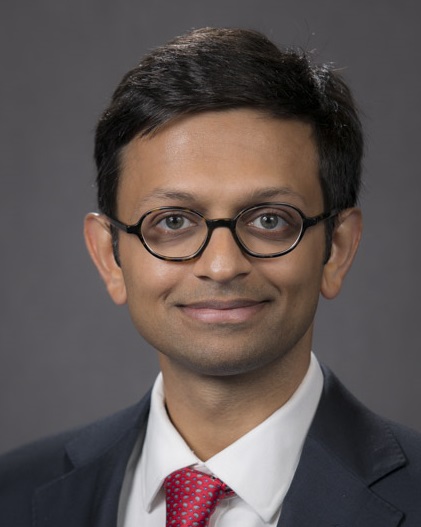
 

Neal Parikh Headshot

Neal S. Parikh, MD, earned his MD from Weill Cornell Medical College and completed residency training in neurology at the same institution. He is now an NIH T32 neuro-epidemiology and vascular neurology fellow at New York-Presbyterian Hospital/Columbia University Medical Center. He tweets @NealSParikhMD and contributes to Blogging Stroke as a blogger.

hidden

Stroke Systems Science: Travel Delays and Access to Care

Advances in science cannot overcome traffic. Patients with strokes due to large vessel occlusions must be taken to hospitals that perform endovascular thrombectomy. Otherwise, these patients do not benefit from the latest and greatest in stroke neurology.

Investigators, including me, have taken interest in measuring the impact of travel delays on stroke care. With my co-investigators, a colleague and I performed a Monte Carlo simulation to model the effects of stroke transfer system configuration on endovascular therapy eligibility and expected outcomes.1 In a recent issue of Stroke, Dr. Regenhardt and colleagues present their analysis of a single hub and spoke system.2

They analyzed 234 patients who were transferred for endovascular therapy. Of these patients, who had a median ASPECTS score of 10 prior to transfer, only 27% of patients ultimately received endovascular therapy.  A median ASPECTS score of 10 correlates with very high eligibility for endovascular therapy prior to transfer.

They found, not surprisingly, that longer transfer time was associated with a decreased odds of undergoing endovascular therapy. The probability of getting endovascular therapy decreased by 1% for each additional minute of transfer time beyond 60 minutes. Being transferred at night was associated with slower transfers (despite less traffic!) and less endovascular therapy.

What does this mean? This means that a patient with a severe stroke who has the misfortune of being taken to a hospital that does not offer endovascular therapy has only a 27% probability of getting this therapy after transfer. At night time, it’s even worse.

Wonderful outcomes can be seen in clinical trials, but they do not benefit society if systems science does not keep up. Infrastructure upgrades and protocol developments may help, along with monitoring and benchmarking of transfer metrics.

References

  1. Parikh, Chatterjee, Diaz, et al. Modeling the Impact of Interhospital Transfer Network Design on Stroke Outcomes in a Large City. Stroke. 2018;49:370-376.
  2. Regenhardt, Mecca, Flavin, et al. Delays in Air or Ground Transfer of Patients for Endovascular Therapy. Stroke. 2018;49:1419-1425.

Neal Parikh Headshot

Neal S. Parikh, MD, earned his MD from Weill Cornell Medical College and completed residency training in neurology at the same institution. He is now an NIH T32 neuro-epidemiology and vascular neurology fellow at New York-Presbyterian Hospital/Columbia University Medical Center. He tweets @NealSParikhMD and contributes to Blogging Stroke as a blogger.

hidden

A different kind of extended window for stroke treatment

To fanfare at International Stroke Conference 2018, the results of the DEFUSE 31 extended window thrombectomy study were announced. The American Heart Association/American Stroke Association acute ischemic stroke guidelines were immediately updated to reflect the practice-changing findings. 

A few months later, Lee Schwamm and colleagues published their findings from MR WITNESS.2 In this study, patients with unwitnessed stroke onset between 4.5 and 24 hours underwent advanced magnetic resonance imaging to identify those individuals with radiographic evidence of hyperacute stroke. Based on prior work, it was known that evolution of imaging characteristic with respect to the fluid-attenuated inversion recovery (FLAIR) sequence correlates with time from onset. Patients who met imaging criteria based on the mismatch between FLAIR signal change and diffusion restriction were given tPA.

The researchers enrolled 80 individuals at multiple centers. Patients were treated at a median of 11 hours from their last known well. The rates of adverse events were very low and within the range of adverse event rates observed in prior stroke treatment trials. 

The standard stroke treatment paradigm allows patients to be treated within 4.5 hours of symptom onset. In general, patients treated beyond this window are at greater risk of brain hemorrhage and poor outcomes. The results of this Phase 2a study challenge the 4.5 hour time window. Like DEFUSE 3, this study uses advanced imaging to personalize acute stroke treatment. A frequent reason for patients to not receive tPA for stroke treatment has been that patients often present to hospitals too late. Expanding the time window for non-large vessel occlusion strokes, which are the vast majority of strokes but nonetheless disabling, has great public health implications. With the rest of the stroke community, I look forward to results of an efficacy trial.

References

  1. Albers GW, Marks MP, Kemp SK, Christensen S, Tsai JP, Santiago O, et al. Thrombectomy for Stroke at 6 to 16 Hours with Selection by Perfusion Imaging. NEJM 2018; 378:708-718.
  2. Schwamm LH, Wu O, Song SS, Ford AL, Hsia AW, Muzikansky A, Betensky RA, et al. Intravenous thrombolysis in unwitnessed stroke onset: MR WITNESS trial results. Ann Neurol 2018 Apr 24 [Epub ahead of print].

Neal Parikh Headshot

Neal S. Parikh, MD, earned his MD from Weill Cornell Medical College and completed residency training in neurology at the same institution. He is now an NIH T32 neuro-epidemiology and vascular neurology fellow at New York-Presbyterian Hospital/Columbia University Medical Center. He tweets @NealSParikhMD and contributes to Blogging Stroke as a blogger.

hidden

Promising Advance In Stroke Thrombolysis Research: Tenecteplase

A recent New York Times article re-surfaced the ‘debate’ regarding alteplase (IV-tPA) for ischemic stroke.1 There are some who continue to argue that the data for IV-tPA are not convincing. In this context, and otherwise, it is worthwhile to discuss a recent study comparing tenecteplase versus alteplase among patients with large vessel occlusion.2

In this study, 202 patients presenting within the IV-tPA treatment window of 4.5 hours and with an ischemic stroke due to large vessel occlusion were randomized to receive IV-tPA versus IV-tenecteplase prior to proceeding with mechanical thrombectomy. The main outcomes relevant for this discussion are the primary outcome of substantial reperfusion (restoration of blood flow in the affected area) and the safety outcome of brain hemorrhage.

Whereas 10% of patients who had received IV-tPA achieved substantial reperfusion prior to undergoing mechanical thrombectomy, 22% achieved substantial reperfusion in the tenecteplase group. The number of brain hemorrhages was the same in both groups (5-6%).

If confirmed, this represents a tremendous advance in thrombolysis because many patients require lengthy transport to reach a center where thrombectomy can be performed. Achieving reperfusion without increased risk of hemorrhage, potentially in the field using stroke ambulances and telemedicine, could dramatically improve population-level care for this otherwise very disabling form of stroke.

Further, these data suggest support the stability of the 6% estimate of brain hemorrhage risk with IV-thrombolysis. The observation that the hemorrhage risk (5 vs 6%) was the same regardless of reperfusion rate (10 vs 22%) is intriguing – if the two are independent, is the risk of hemorrhage from thrombolysis from something other than reperfusion? Further, the results of this study will spur additional study and we will thus have contemporary, high-quality data regarding the efficacy and safety of thrombolysis.

References

  1. https://www.nytimes.com/2018/03/26/health/stroke-clot-buster
  2. Campbell, et al. Tenecteplase versus Alteplase before Thrombectomy for Ischemic Stroke. NEJM. 2018:378:1573-82.

Neal Parikh Headshot

Neal S. Parikh, MD, earned his MD from Weill Cornell Medical College and completed residency training in neurology at the same institution. He is now an NIH T32 neuro-epidemiology and vascular neurology fellow at New York-Presbyterian Hospital/Columbia University Medical Center. He tweets @NealSParikhMD and contributes to Blogging Stroke as a blogger.

hidden

Pharmacological Smoking Cessation Aides And Cardiovascular Safety

Pharmacological smoking cessation therapies have had their challenges. For example, varenicline previously had a US Food and Drug Administration black box warning regarding neuropsychiatric risks.

The EAGLES study, published 2016, was an industry sponsored, randomized, placebo-controlled trial of nicotine, varenicline, and bupropion that sought to address the neuropsychiatric risk profile of these medications.1 They randomized participants with and without known psychiatric comorbidities to these medications and found that these agents were not associated with an increased risk of neuropsychiatric adverse events. Further, the study found varenicline to be more effective than nicotine, bupropion, and placebo for smoking cessation.

The FDA black box warning for varenicline was removed. However, concerns regarding the cardiovascular safety persisted. Apart from abundant observational data on this topic, there have been several randomized trials as well. For example, in 2015, a randomized clinical trial of varenicline versus placebo for patients hospitalized with acute coronary syndrome demonstrated efficacy for cessation and did not raise a safety signal.2

Further, a secondary analysis of the EAGLES study regarding cardiovascular safety was recently published.3 They compared rates of major adverse cardiovascular events, and changes in blood pressure and heart rate, among participants randomized to placebo, varenicline, bupropion, and nicotine replacement. They found very low rates of major cardiovascular events and did not find differences between drugs. Of course, these were not patients with recent or significant cardiovascular comorbidities, so the results do not generalize beyond the general population of smokers.

There is thus mounting evidence for both the psychiatric and cardiovascular safety of pharmacological smoking cessation therapies. While it can be argued that an adequately powered safety trial in patients in acute and/or significant cardiovascular disease has yet to be performed, it may nonetheless be time to create gold standard cessation programs for patients with cardiovascular disease. It may be premature, however, to do the same for patients with cerebrovascular disease – more evidence may be needed.

References:

  1. Anthenelli RM, Benowitz NL, West R, et al. Neuropsychiatric safety and efficacy of varenicline, bupropion, and nicotine patch in smokers with and without psychiatric disorders (EAGLES): a double-blind, randomised, placebo-controlled clinical trial. Lancet. 2016:387;2507-2520.
  2. Eisenberg MJ, Windle SB, Roy N, et al. Varenicline for Smoking Cessation in Hospitalized Patients With Acute Coronary Syndrome. Circulation. 2015:137; https://doi.org/10.1161/CIRCULATIONAHA.115.019634.
  3. Benowitz NL, Pipe A, West R, et al. Cardiovascular Safety of Varenicline, Bupropion, and Nicotine Patch in SmokersA Randomized Clinical Trial. JAMA Internal Medicine. 2018; doi:10.1001/jamainternmed.2018.0397.

Neal Parikh Headshot

Neal S. Parikh, MD, earned his MD from Weill Cornell Medical College and completed residency training in neurology at the same institution. He is now an NIH T32 neuro-epidemiology and vascular neurology fellow at New York-Presbyterian Hospital/Columbia University Medical Center. He tweets @NealSParikhMD and contributes to Blogging Stroke as a blogger.

hidden

Empowering Patients To Treat Hypertension – Self-monitoring

Precision medicine, when applied to prevention, can identify opportunities for an individual to reduce their cardiovascular risk. Resulting interventions are personalized and may take advantage of the latest science, including genetics. Such interventions may presuppose that basic risk factors – such as hypertension and diabetes – have been identified and maximally controlled. Intervening upon more recently identified risk factors may have less or no impact if dominant risk factors such as hypertension are not adequately controlled.  

In contrast, population health interventions could indiscriminately improve health for individuals by targeting well-accepted, high-stakes risk factors. For example, although hypertension is accepted as the leading risk factor for cardiovascular disease, we see patients every day with poorly controlled blood pressure in the clinic and hospital. Controlling such risk factors could raise the bar more uniformly so that more people may benefit from the additional gains of precision prevention. 

Engaging patients in their own health care may be an effective strategy. For example, in the recently published TASMINH4 trial from the United Kingdom, patients empowered to monitor their blood pressure at home and present the data to their doctors had improved blood pressure control, as compared to individuals receiving routine care.1

While such research can be frustratingly complex to perform and interpret, the benefits of standardizing and incorporating sound methods for improving blood pressure control cannot be understated. Translating this into clinical practice may require a new information infrastructure and possibly changes to reimbursement schemes, due to the possible added burden of reviewing additional data. 

In this era of enthusiasm for precision medicine, we should continue to pursue population health and implementation science with equal gusto, especially in countries with high health care disparities such as the United States.  

References:

  1. McManus RJ, Mant J, Franssen M, Nickless A, Schwartz C, Hodgkinson J, et al. Efficacy of self-monitored blood pressure, with or without telemonitoring, for titration of antihypertensive medication (TASMINH4): an unmasked randomized controlled trial. Lancet. 2018. https://doi.org/10.1016/S0140-6736(18)30309-X

Neal Parikh Headshot

Neal S. Parikh, MD, earned his MD from Weill Cornell Medical College and completed residency training in neurology at the same institution. He is now an NIH T32 neuro-epidemiology and vascular neurology fellow at New York-Presbyterian Hospital/Columbia University Medical Center. He tweets @NealSParikhMD and contributes to Blogging Stroke as a blogger.

hidden

Parsing The Updated 2018 Acute Ischemic Stroke Guidelines: Smoking Cessation

The 2018 International Stroke Conference was headlined by the practice-changing results of DEFUSE 3 and related acute stroke care guideline updates. Having returned to our institutions, neurologists are parsing the updated 2018 acute ischemic stroke guidelines1 and wondering how best to operationalize the latest data.
 
Overshadowed by updated guidelines regarding the extended window and buried among changes regarding the utility of indiscriminate use of routine diagnostic testing, was a change regarding smoking cessation.
 
While the guidelines committee did not find any randomized trials of pharmacological smoking cessation aides specifically for stroke patients, they cite a randomized trial in acute coronary patients:2 patients randomized to receive a pharmacological cessation aide had a significant improvement in abstinence. In terms of observational data, a recent study found that patients with stroke who quit smoking had a reduced rate of cardiovascular disease and mortality over 5 years.3 Based partially on such evidence, the updated guidelines provide a IIb recommendation that “for smokers with an acute ischemic stroke, in-hospital initiation of varenicline might be considered”.1
 
A class I recommendation to “strongly advise every patient with acute ischemic stroke who has smoked in the past year to quit” remains in place and is buttressed with a IIb option to consider “interventions that incorporate both pharmacotherapy and behavioral support”.1
 
While Get With the Guidelines-Stroke has seen a substantial improvement in “appropriate” smoking cessation interventions at the time of hospital discharge,4 a distinction between counselling and pharmacotherapy was not made. Therefore, whether effective smoking cessation interventions are being initiated is unknown.
 
Whereas the extended window guidelines influence care for a small group of acute stroke patients, the smoking cessation guidelines apply to every single acute stroke and TIA patient who is an active smoker. Neurologists, particularly stroke neurologists and hospitalists, should familiarize themselves with the updated guidelines, the relevant data, and pharmacological interventions. 
 
References:

  1. Powers WJ, Rabinstein AA, Ackerson T, Adeoye OM, Bambakidis NC, Becker K, et al. 2018 Guidelines for the Early Management of Patients with Acute Ischemic Stroke. Stroke 2018. DOI: 10.1161/STR.0000000000000158.
  2. Eisenberg MJ, Windle SB, Roy N, Old W, Grondin FR, Bata I, et al. Varenicline for Smoking Cessation in Hospitalized Patients with Acute Coronary Syndrome. Circulation. 2016:133;21-30.
  3. Epstein KA, Viscoli CM, Spence JD, Young LH, Inzucchi SE, Gorman M, et al. Smoking cessation and outcome after ischemic stroke or TIA. Neurology. 2017:89;1723-1729. 
  4. Ormseth CH, Sheth KN, Saver JL, Fonarow GC, Schwamm LH. The American Heart Association’s Get With the Guidelines (GWTG)-Stroke development and impact on stroke care. Stroke and Vascular Neurology 2017;2:doi:10.1136/svn-2017-000092

Neal Parikh Headshot

Neal S. Parikh, MD, earned his MD from Weill Cornell Medical College and completed residency training in neurology at the same institution. He is now an NIH T32 neuro-epidemiology and vascular neurology fellow at New York-Presbyterian Hospital/Columbia University Medical Center. He tweets @NealSParikhMD and contributes to Blogging Stroke as a blogger.

hidden

An Early Career Perspective On International Stroke Conference 2018

I have come to look forward to the annual International Stroke Conference each year. Due to the largess of my mentors and support of my residency program, I have had the good fortune of attending the conference each year since my third year of residency. As a third-year resident, I had decided to pursue fellowship training in vascular neurology, and attending the conference amplified my enthusiasm for the field and inspired me to contribute to stroke science. This fueled my passion for stroke research, which ultimately led me to my current fellowship in stroke and neuro-epidemiology at Columbia University. 

First, I want to advocate for resident-level participation in the conference. Exposure to late-breaking science, hearing from leaders in the field, and socializing with members of your institution’s stroke division – these are invaluable opportunities. Now, returning for a third time, I have begun to feel a member of the community. Early attendance may inspire residents to pursue research and provide them with a sense of the scope of current investigation and the priorities of the field. 

This years, as a fellow, I attended the conference with more specific goals. I outlined research areas that I am interested in and scrutinized the program in advance to identify key talks and poster presentations. This allowed me to identify opportunities for future study and to meet individuals to collaborate with. Equally importantly, I had a reunion with friends and classmates from medical school and residency. Speaking with friends 1-3 years ahead in their careers is particularly informative because they provide good guidance. Last, the release of game-changing data created an electric atmosphere that motivated me and surely other early career attendees as well. 

Some of my early career colleagues have reported avoiding the conference when not presenting data. For the reasons outlined above, I encourage residents interested in stroke and stroke fellows to attend and earnestly participate regardless of whether they have data to present. I also encourage program directors and chief residents to encourage resident participation and to make schedule adjustments to permit attendance. Medical students should be similarly included. 

I look forward to attending the conference in Honolulu next year! 

Neal Parikh Headshot

Neal S. Parikh, MD, earned his MD from Weill Cornell Medical College and completed residency training in neurology at the same institution. He is now an NIH T32 neuro-epidemiology and vascular neurology fellow at New York-Presbyterian Hospital/Columbia University Medical Center. He tweets @NealSParikhMD and contributes to Blogging Stroke as a blogger.

hidden

DEFUSE 3 Definitively Expands The Endovascular Therapy Window

Writing from the 2018 International Stroke Conference, it is difficult to report on anything other than the game-changing results of DEFUSE 3. After years of clinical suspicion that endovascular therapy works, definitive evidence establishing the role of endovascular therapy in acute stroke care was first presented at the International Stroke Conference in 2015. Since then, there has been great interest in maximizing the yield of this highly effective therapy. Extending the original window of 0-8 hours has been of particular interest.

Imagine being called the emergency department to find a patient who woke up severely disabled by their stroke only to determine that the last time they were seen well was at dinner the night before. Unable to definitively conclude that their stroke began within the last 6-8 hours, you are unable to provide any therapy beyond standard supportive medical care. The patient worsens while in the hospital and is discharged to a nursing home or subacute rehabilitation facility. 

This is not an uncommon situation. So, learning today that the window for intervention can be extended safely and effectively to 16 hours was moving. The DEFUSE 3 data showed that properly selected patients stand to benefit immensely from endovascular therapy, and these data will arm neurologists with yet another highly impactful intervention to offer patients. By confirming the results of the DAWN trial, which extended the window to a full 24 hours, DEFUSE 3 settles the issue. 

Now, stroke systems of care need to quickly adapt to this new reality so that we can help patients benefit from the remarkable progress of stroke treatment science.
 
Reference:

  1. Nogueira, et al. Thrombectomy 6 to 24 Hours after Stroke with a Mismatch between Deficit and Infarct. NEJM. 2018; 378:11-21.
    Albers et al. Thrombectomy for Stroke at 6 to 16 Hours with Selection by Perfusion Imaging. NEJM. 2018. Ahead of print.

Neal Parikh Headshot

Neal S. Parikh, MD, earned his MD from Weill Cornell Medical College and completed residency training in neurology at the same institution. He is now an NIH T32 neuro-epidemiology and vascular neurology fellow at New York-Presbyterian Hospital/Columbia University Medical Center. He tweets @NealSParikhMD and contributes to Blogging Stroke as a blogger.

hidden

Stroke Advances In 2017: An Overview, Reflections, And A Call To Action

2017 gave us numerous dramatic advances in stroke neurology. We were treated to compelling data regarding the favorability of patent foramen ovale closure in well-selected individuals with cryptogenic stroke.1,2,3 Endovascular therapy matured with the extension of the treatment time window.4 We even saw promising rehabilitation data regarding surgical nerve transfer for chronic spastic arm paralysis.5 Conversely, some widely used therapies such as head positioning6 and oxygen supplementation7 were shown to be ineffective. The list of figurative leaps goes on.
 
As an early career neurologist in a vascular neurology fellowship, I found myself reflecting on the year’s advances, in part to find my place in the field. While thoroughly inspired by the major advances of 2017, I couldn’t help but dwell on the findings of a secondary analysis of the Insulin Resistance Intervention After Stroke (IRIS) trial.
 
The IRIS trial randomized non-diabetic patients with stroke or TIA to pioglitazone or placebo and followed them for several cardiovascular outcomes.8 The primary analysis was published in 2016; patients randomized to pioglitazone had a lower risk of recurrent stroke or heart attack.
 
In a secondary analysis published in Neurology in 2017, Katherine Epstein and colleagues evaluated the association of smoking cessation and recurrent stroke, myocardial infarction, and death.9 In an observational design, they followed individuals who were smoking at the time of their index stroke and quit, and compared them to individuals who did not quit. The 5-year risk of stroke, MI, or death was 16% in quitters versus 23% in non-quitters (adjusted hazard ratio 0.66). Quitters had half the risk of death compared to non-quitters.
 
Granted, this was observational data. Individuals who were motivated to quit smoking may have made other healthy decisions. And, these results are not ground breaking either – we know that smoking cessation is “the most important thing one can do for one’s health” (as we are taught to tell patients in medical school).
 
Regardless, the results are memorable. While advances in acute stroke care, surgical interventions, and novel pharmacotherapies are a testament to scientific ingenuity, we must not neglect the low-hanging fruit. Are neurologists trained to effectively aide in smoking cessation? What are the best tools for this purpose? Are such services adequately incentivized? Some argue that advances in stroke systems of care may now yield more public health gains than scientific advances. If we accept this notion, we must acknowledge that it does not apply exclusively to the acute stroke treatment arena.
 
Included in the AHA/ASA’s Life’s Simple 7 paradigm, and a focus of the FDA’s newest public education campaign entitled “Every Try Counts”,10 smoking cessation deserves our fullest attention. To support these programs and to empower our patients to quit, we must identify and incorporate the best tools available into our practice.  

References

  1. Mas JL, Derumeaux G, Guillon B, Massardier E, Hosseini H, Mechtouff L, et al. Patent Foramen Ovale Closure or Anticoagulation vs Antiplatelets after Stroke. NEJM. 2017:377;1011-1021.
  2. Saver JL, Carroll JD, Thaler DE, Smalling RW, MacDonald LA, Marks DS, et al. Long-Term Outcomes of Patent Foramen Ovale Closure or Medical Therapy after Stroke. NEJM. 2017:377;1022-1032.
  3. Søndergaard LKasner SERhodes JFAndersen GIversen HKNielsen-Kudsk JE, et al. Patent Foramen Ovale Closure or Antiplatelet Therapy for Cryptogenic Stroke. NEJM. 2017:377;1033-1042.
  4. Nogueira RG, Jadhav AP, Haussen DC, Bonafe A, Budzik RF, Bhuva P, et al. Thrombectomy 6 to 24 Hours After Stroke with a Mismatch between Deficit and Infarct. NEJM. 2018:378;11-21.
  5. Zheng MX, Hua XY, Feng JT, Li T, Lu YC, Shen YD, et al.
  6. Anderson CS, Arima H, Lavados P, Billot L, Hacket ML, Olavarria VV, et al. Cluster-Randomized, Crossover Trial of Head Positioning in Acute Stroke. NEJM. 2017:376;2437-2447.
  7. Roffe C, Nevatte T, Sim J, Bishop J, Ives N, Ferdinand P, et al. Effect of Routine Low-Dose Oxygen Supplementation on Death and Disability in Adults With Acute Stroke: The Stroke Oxygen Study Randomized Clinical Trial. JAMA. 2017:318;1125-1135.
  8. Kernan WN, Viscoli CM, Furie KL, Young LH, Inzucchi SE, Gorman M, et al. Pioglitazone after Ischemic Stroke or Transient Ischemic Attack. NEJM. 2016:374;1321-31.
  9. Epstein KA, Viscoli CM, Spence JD, Young LH, Inzucchi SE, Gorman M, et al. Smoking cessation and outcome after ischemic stroke or TIA. Neurology. 2017:89;1723-1729.
  10. Every Try Counts Campaign. Food and Drug Administration. https://www.fda.gov/tobaccoproducts/publichealtheducation/publiceducationcampaigns/everytrycountscampaign/default.htm

Neal Parikh Headshot

Neal S. Parikh, MD, earned his MD from Weill Cornell Medical College and completed residency training in neurology at the same institution. He is now an NIH T32 neuro-epidemiology and vascular neurology fellow at New York-Presbyterian Hospital/Columbia University Medical Center. He tweets @ NealSParikhMD and contributes to Blogging Stroke as a blogger.